More Related Content
Similar to NAPP-DR6 poster
Similar to NAPP-DR6 poster (20)
NAPP-DR6 poster
- 1. TEMPLATE DESIGN © 2007
www.PosterPresentations.com
Identification of novel peptide inhibitors of the DR6-NAPP protein-protein
interaction using a virtual screening approach
Kelly Considine, Dr. Joseph Audie, and Dr. Edward Caliguri
Sacred Heart University, 5151 Park Avenue, Fairfield, CT 06825
Abstract
Introduction
Future Work
References
Acknowledgements
ConclusionMethods
0.33-X0.089-0.00089X-
0.86X-0.65X-X0.075+X-0.79=G
torgap
hbsbc/s-/+empiricalbind,
Results and Discussion (continued)
Nikolaev and co-workers recently described a new apoptotic pathway
that underlies neuronal development and axonal pruning. According to
Nikolaev and co-workers, in response to nerve growth factor (NGF)
withdrawal, the pathway is engaged by binding between the death
receptor six (DR6) ectodomain and an N-terminal fragment of amyloid
precursor protein (NAPP). Nikolaev and co-workers hypothesize that the
DR6-NAPP apoptotic pathway might play a role in the pathophysiology of
Alzheimer's Disease (AD). Thus, inhibitors of the DR6-NAPP interaction
could potentially serve as a new class of drugs for the treatment of AD.
Importantly, a theoretical model of the DR6-NAPP interaction has been
proposed in a recent study. The model implicates a lone NAPP α-helix-
loop motif as crucial to DR6 binding and recognition. Given all of this,
we targeted the NAPP α-helix using structure-based peptide design. In
particular, we virtually screened a focused peptide library against the
NAPP α-helix. Select peptide screening hits were subsequently docked
to the NAPP helix and their binding modes optimized. Final scoring and
ranking was achieved using a well-validated empirical method for
estimating protein-peptide binding affinities. While preliminary, our
results suggest that focused peptide virtual screening, followed by
peptide docking and optimization, and final empirical free energy
scoring, provides an efficient work-flow for identifying novel and viable
peptide inhibitors of the DR6-NAPP interaction.
Alzheimer’s disease (AD) debilitates many individuals, yet there is
no known treatment. A recently described apoptotic pathway involving
the interaction between NAPP and DR6 may become hijacked in the AD
brain and is a potential source for new therapies (3).
The identification of the NAPP-DR6 interaction gives rise to a new
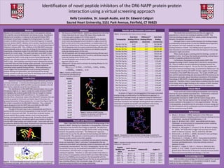
therapeutic target (3; 4). Figure 1 shows the crystal structure of NAPP
and highlights key residues (Lys66-Gln81) that are thought to mediate
the interaction between NAPP and DR6 (5; 6). The residues were
identified from a theoretical model of the DR6-NAPP interaction that was
recently proposed by Ponomarev and Audie; Figure 2 illustrates the
proposed interaction of these proteins (5). It is clear from Figure 2 that
the α-helix is essential to the DR6-NAPP interaction. Thus, molecular
agents designed to bind the NAPP helix and inhibit the DR6-NAPP
interaction hold promise as novel treatments for AD.
The goal of this work was to use a computational work-flow to
quickly identify novel and promising peptides to bind the NAPP helix and
prevent its interaction with DR6. The work-flow combined iterative
focused peptide library design, peptide virtual screening, peptide
docking, and empirical free energy scoring. During the virtual screening
and docking experiments, the peptide ligands were treated as flexible
and the NAPP target protein was treated as rigid. Future work will focus
on molecular dynamics (MD) based refinement and rescoring of peptide-
NAPP interactions and in vitro testing of select peptides.
Figure 1. Secondary structure depiction of NAPP (PDB ID: 1MWP). Residues 66-81
have previously been reported as the region essential to NAPP’s interaction with
DR6 (5).
1. The structure of NAPP (PDB ID: 1MWP) was obtained from the RCSB
Protein Data Bank (6) and Swiss-PDB Viewer (http://spdbv.vital-
it.ch/) was used to prepare it for further study (2).
2. A complete dipeptide library was constructed using the standard
amino acids (excluding glycine) and docked to a region around the
NAPP α-helix (10 GA runs) using AutoDock, as implemented in the
Molecular Docking Server (http://www.dockingserver.com/web) (1).
3. The 20 dipeptides with the lowest predicted binding affinities were
extended into tripeptides at the C-terminus and docked using the
molecular docking server (10 GA runs).
4. The 20 tripeptides with the lowest predicted binding affinities were
extended into tetrapeptides at the C-terminus and docked using the
molecular docking server (10 GA runs).
5. The top 60 peptides were docked to NAPP using a more exhaustive
approach (100 GA runs).
6. CMDescoresm (5) was used to estimate the binding affinities for the
best tetra-peptides:
Table 1. Residues of NAPP involved
in its interaction with DR6 that are
targeted for peptide inhibition (5)
Residues
Lys66
Glu67
Gly68
Ile69
Leu70
Gln71
Tyr72
Cys73
Gln74
Glu75
Val76
Tyr77
Pro78
Glu79
Leu80
Gln81
Figure 3. Backbone of NAPP residues
involved in its interaction with DR6 that are
targeted for peptide inhibition (5)
Table 2. Tetrapeptides with lowest minimum energy
Tetrapeptide
Auto Dock
Binding Affinity
(kcal/mol)
CMDescoresm
Binding Affinity
(kcal/mol)
Auto Dock
Binding
Constant (Ki)
Trp-Asn-Trp-Trp -9.27 -3.5 161.38 nM
Phe-Trp-Lys-Trp -9.02 -2.7 245.87 nM
Pro-Lys-Trp-Trp -8.50 -6.0 586.72 nM
Trp-Phe-Trp-Phe -8.46 -2.9 625.80 nM
Pro-Lys-Trp-Phe -8.37 -2.3 729.20 nM
Trp-Lys-Trp-Trp -8.33 -3.3 748.81 nM
Pro-Trp-Trp-Phe -8.20 -2.3 972.88 nM
Phe-Trp-Trp-Ile -8.06 -2.3 1.25 µM
Trp-Phe-Trp-Trp -8.04 -3.8 1.27 µM
Trp-Trp-Trp-Ile -8.02 -3.0 1.32 µM
Trp-Lys-Pro-Phe -7.92 -2.8 1.57 µM
Trp-Trp-Trp-Cys -7.89 -3.4 1.65 µM
Trp-Lys-Phe-Asp -7.86 -1.6 1.72 µM
Trp-Lys-Phe-Cys -7.83 -1.1 1.82 µM
Pro-Trp-Trp-Leu -7.58 -3.7 2.76 µM
Phe-Trp-Trp-Asn -7.54 -2.7 2.99 µM
Phe-Trp-Lys-Lys -7.48 -2.9 3.30 µM
Phe-Trp-Lys-Leu -7.44 -1.7 3.54 µM
Trp-Trp-Trp-Arg -7.36 -3.2 4.00 µM
Phe-Trp-Trp-Ala -7.34 -1.6 4.18 µM
Figure 2. Theoretical DR6-NAPP interaction model, as taken from the recent paper
by Ponomarev and Audie. NAPP is shown in blue and DR6 is shown in yellow (5).
Kelly Considine acknowledges the financial support from Sacred
Heart University in Fairfield, CT and would like to thank Dr. Joseph
Audie for his help and guidance on this project.
1. Bikadi, Z., & Hazai, E. (2009). Application of the PM6 semi-
empirical method to modeling proteins enhances docking accuracy
of AutoDock. Journal of Cheminformatics, 1, 15.
2. Guex, N., & Peitsch, M.C. (1997). Swiss Model and the Swiss-
PdbViewer: An environment for comparative protein modeling.
Electrophoresis, 18, 2714-2723.
3. Nikolaev, A., McLaughlin, T., O’Leary, D. D. M., & Tessier-Lavigne,
M. (2009). APP binds DR6 to trigger axon pruning and neuron
death via distinct caspases. Nature, 457(7232), 981-989.
4. Osherovich, L., & Writer, S. (2009). Genentech’s new parADigm.
Science-Business eXchange, 2(8), 1-5.
5. Ponomarev, S. Y., & Audie, J. (2011). Computational prediction
and analysis of the DR6-NAPP interaction. Proteins: Structure,
Function, and Bioinformatics, Early View (Articles online in
advance of print).
6. Rossjohn, J. , Cappai, R., Feil, S. C., Henry, A., McKinstry, W.J.,
Galatis, D., et al. (1999). Crystal structure of the N-terminal,
growth factor-like domain of Alzheimer amyloid precursor protein.
Nature Structural and Molecular Biology, 6, 327-331.
Future work will include building pentapeptides and analyzing
them by the same methods described here. Furthermore, molecular
dynamics (MD) simulations of the top tetrapeptides and
pentapeptides that bind to 1MWP with the lowest energy will be
performed. Then, the next step will involve moving from the in silico
process outlined in this project to in vitro experimentation. The top
10 tetrapeptides, as predicted by the estimated minimum energy and
the MD simulations, will be synthesized, and their binding with NAPP
will be monitored using 2D NMR.
Table 2 describes the estimated minimum binding energy of the
peptide to NAPP. Moving from dipeptides to tetrapeptides, there was
a decrease in the minimum free energy as expected. The data from
Docking Server shows very low energies which predict strong binding
between the peptides and protein. The CMDescoresm approach
predicts that Pro-Lys-Trp-Trp will have the best binding with NAPP, at -
6.0 kcal/mol (5). This approach is more focused on the interactions
between protein-protein and protein-peptide.
The binding constants, as predicted and calculated by Docking
Server are reported in Table 2, and show 7 tetrapeptides that are
predicted to bind NAPP and potentially inhibit its interaction with DR6
at nanomolar concentrations. The first 2 tetrapeptides, Trp-Asn-Trp-
Trp and Phe-Trp-Lys-Trp, are predicted to bind NAPP at low nanomolar
concentrations, and then there is a large increase in Ki for the
subsequent peptides. Therefore, in silico data predicts that either or
both of these 2 tetrapeptides will bind NAPP and inhibit DR6 binding.
Based on the CMDBioscience approach, the tetrapeptide with the
lowest minimum binding energy (Pro-Lys-Trp-Trp), is only predicted to
have a binding affinity in the micromolar range, 25.13 µM, as
calculated using the Gibbs Free-Energy relationship.
Results and Discussion
The results here are preliminary but they do suggest the
potential for in vitro/in vivo binding of peptides to NAPP. The
minimum binding energies and the Ki values, predict strong binding
between the peptide and protein.
Although there is some conflict between the binding energies
as predicted by Docking Server (1) and the CMDBioscience approach
(5), utilization of in vitro methods can help compare
binding/inhibition of NAPP. The CMDBioscience approach assumes
vdw interactions cancel which is where the discrepancy in the energy
calculation might come from.
These results do give a starting point for docking a number of
peptides in vitro. As of now, the results are promising, and we have a
number of peptides predicted to bind NAPP.
Furthermore, Ponomarev and Audie predict NAPP-DR6
hydrogen bonding at NAPP residues Gln71 and Gln74, among others
(5). These 2 residues are also predicted to hydrogen bond to the
tetrapeptide shown in Figure 4. Pro-Lys-Trp-Trp is the peptide with
the lowest minimum binding energy, as predicted by CMDescoresm (5),
and its hydrogen bonding with NAPP strongly suggests the possibility
of inhibition of the NAPP-DR6 interaction. The model of NAPP used
here and the selected binding site were based off of the work done by
Ponomarev and Audie, whose theoretical calculations of the binding
free energy of DR6-NAPP is in good agreement with experimental
results (5). Therefore, there is sound reason to believe that the results
of this study, which suggest binding to the residues of NAPP involved
in DR6-NAPP binding, will result in inhibition of this interaction.
Figure 4. Tetrapeptide of lowest estimated binding energy as predicted by
CMDescoresm approach(5), Pro-Lys-Trp-Trp (blue), hydrogen bonding with NAPP
(pink)
As shown in Figure 4, the tetrapeptide of the lowest estimated
binding energy, as predicted by the CMDBioscience approach, is
stabilized by 4 hydrogen bonds, as predicted using the CMDescoresm
hydrogen bond detection method. Table 3 summarizes the residues
and atoms involved in the hydrogen bonding which help stabilize the
interaction of peptide and protein and contribute to the strong
interaction predicted between Pro-Lys-Trp-Trp and NAPP.
Peptide
Residue and
Atom
NAPP Residue
and Atom
Distance (Å) Angles (°)
Trp4 – N Gln71 – O 3.49 119.90 119.0
Trp4 – NE1 Gln 71 – OE1 3.48 147.47 116.0
Lys2 – NZ Gln74 – OE1 2.67 129.17 160.7
Pro1 – N Pro78 – O 3.49 122.44 144.4
Lys2 – O Gln74 – OE1 3.03 117.04 141.1
Table 3. Hydrogen bonding between Pro-Lys-Trp-Trp and NAPP as predicted by
CMDescoresm approach (5)