2. subjected to multiple rounds of selective binding to increase the
prevalence of high affinity aptamers. To further enhance
specificity, we interposed rounds of negative selection against
the closely related AMPA and kainate receptors subtypes,
selecting out aptamers which show high affinity binding for
these subtypes. The resulting candidate aptamers were
characterized using radio-ligand binding and electrophysiology.
We have isolated an aptamer with a high affinity for the
GluN2A containing NMDA receptors (Kd = 120 ± 15 nM) and
little to no effect on the closely related GluA2 AMPA receptor
and GluK2 kainate receptors. Crucially, this aptamer is
neuroprotective in an oxygen glucose deprivation model of
ischemia.
■ RESULTS AND DISCUSSION
As a starting point in developing an NMDA receptor selective
RNA antagonist, we used a RNA library containing 1015
sequences, each 90 nucleotides long. RNAs were synthesized
using 2′-F-pyrimidines instead of 2′-OH-pyrimidines, as these
are more resistant to nucleases and show improved stability in
the bloodstream.18
This initial RNA pool was incubated with
GluN1/GluN2A NMDA receptors isolated in membrane
fractions of transiently transfected HEK cells. The resulting
bound RNA−protein complexes were separated from unbound
RNA using a nitrocellulose filter, and the RNA bound to
GluN1/GluN2A receptors was eluted with 10 mM glutamate,
collected, and used as a template for RT-PCR amplification for
the next SELEX cycle. After three rounds of this positive
selection, negative selection was interposed by incubating the
RNA pool with GluA2 and GluK2 receptors expressed in HEK
cells. RNA molecules which did not bind GluA2 and GluK2
were then collected and used for subsequent selection.
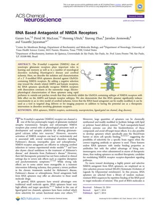
Following three more positive selection rounds and two more
negative selection rounds, the binding affinity of the ninth
round RNA pool was evaluated. Radioligand binding experi-
ments found the ninth round pool had a Kd of 210 ± 10 nM for
GluN1/GluN2A receptors and no saturation binding for
membranes containing GluA2 or GluK2 (Figure 1). The
ninth round RNA pool was cloned and aligned using the
ClustalX program. From sequences of the various cloned
RNAs, a common consensus motif GCGGG was identified in 9
out of 24 clones (Table 1). The saturation binding curve of
clone C26 from among those having the consensus sequence
showed a Kd of 65 ± 3 nM (Figure 2B), considerably higher
affinity than the Kd of 210 ± 10 nM observed for the pool. We
hence used this clone for further analysis.
The secondary structure of the 90-nt RNA of clone C26 was
predicted by Mfold and is shown in Figure 2A.19
Based on this
structure and the consensus motif GCGGG, observed in
position 37−41, two truncated versions of the C26 aptamer,
C26−50 and C26−31 (Figure 2A), were designed with lengths
of 50-nt and 31-nt, respectively. Radioligand binding assays
using membrane preparations of HEK 293T cells expressing
GluN1/GluN2A receptors showed that both aptamers bound
to NMDA receptors with the C26−50 aptamer having a Kd of
120 ± 15 nM while the C26-31 aptamer showed no saturation
binding at concentrations up to 300 nM (Figure 2B).
To evaluate the functional effect of various aptamer we
applied saturating concentrations of glutamate (10 mM) for 1
ms to outside-out patches expressing prototypical glutamate
receptors either alone or in the continuous presence of 1 μM
aptamer. This deactivation jump protocol has the advantage of
closely mimicking the time course of synaptic activation in
native tissue while retaining control over subunit composi-
tion.20
Aptamer C26 robustly inhibited peak GluN1/GluN2A
NMDA receptor responses (25 ± 2% peak responses, n = 7,
Figure 1. Radioligand binding curves before and after SELEX. (A) Binding curves for the starting pool (black circles) and the pool after 9 rounds of
SELEX (gray triangles) on GluN1/GluN2A NMDA receptors. Note saturation binding in the round 9 pool. (B) Similar binding curves on GluA2
AMPA receptors. (C) Binding curves for GluK2 KA receptors.
Table 1. Consensus Motif (bold letters) of Aptamers
aptamer no. sequences
2 GGGGCGGGAGATGGTGTGGTCTCGGGCGGGCGTTAGCGCC
4 AACCTCGCGGTCTGTCAGGCTGGCATAGCGAAGGGGCTGG
6 GGCCCCGTGCGTCGCTCCCCGTGTGGCTGTGGTTCGTATC
7 TACTGCTTCATACCTGTTGCAGCGGTACCTCCGTAGCGAG
9 TCTTCATTTAATCTTCACCCCTGGGCCCTTCCTTTCTCAT
11 AGTTGTCGTGGTAAGGGTACTGTAAAGTGAGATTATTGAG
25 GAGCGCGCAGTTGGAAAGCTGACAACGCATCTTAAAGCGC
26 GTACGTTCATGCGGGGTCTGTCGTCGGTCGCTGGGAGGC
28 GTCGTGGTTGGCGGAGGGTTTTGCGGGTGAAGAGCATGCC
35 AGTACCTGAGCTGGTCGGCGTTGCGGGGGTGATACGGTT
ACS Chemical Neuroscience Research Article
dx.doi.org/10.1021/cn500041k | ACS Chem. Neurosci. 2014, 5, 559−567560
3. Figure 3). Importantly, the same concentration of C26 showed
negligible effects on peak currents from both GluA2 AMPA (83
± 5% peak responses, n = 4) and kainate (KA) receptors (89 ±
2% peak responses, n = 4, Figure 3). We next examined the
minimized constructs C26−50 and C26−31. The strong
inhibition of C26 was preserved in the C26−50 construct
which inhibited peak GluN2A responses 67% (33 ± 3% peak
response, n = 6) while minimally perturbing AMPA (90.2 ±
0.3% peak response, n = 3) and KA receptor responses (96.6 ±
0.5% peak responses, n = 3, Figure 3). Consistent with the loss
of saturation binding at low concentrations (Figure 2B),
GluN2A peak inhibition was substantially reduced with C26−
31 construct (67 ± 2% peak response, n = 6, Figure 3).
Interestingly, selectivity was also lost, with C26−31 showing
stronger inhibition at AMPA receptors (80 ± 3% peak
response, n = 3) and especially KA receptors (73 ± 1% peak
response, n = 3, Figure 3). We therefore selected C26−50 as
our candidate aptamer for further functional characterization.
To determine the inhibitory potency of C26−50 at GluN2A
containing NMDA receptors, we measured the peak current
inhibition of long (2 s) 10 mM glutamate applications by the
continual presence of varying concentrations of aptamer
(Figure 4). As seen in Figure 4A, C26−50 produced a dose-
dependent inhibition of peak responses in outside out patches.
The IC50 for C26−50 calculated from these data on GluN2A
peak responses was 1.3 ± 0.1 μM (nh = 0.88 ± 0.04, n = 4−6
patches per point, Figure 4B). Interestingly, we observed that
C26−50 also inhibited steady-state responses in these patches
(Figure 4A), despite the presence of a super saturating
concentration of glutamate (10 mM glutamate compared to
an EC50 of 4 μM).21
It was expected that C26−50 would be a
competitive antagonist and as such high concentrations of
Figure 2. Truncation of candidate C26 aptamer. (A) Predicted secondary structure of the C26 aptamer showing segments truncated. (B)
Radioligand binding curves for the indicated aptamer on GluN1/GluN2A NMDA receptors. Note the loss of saturating binding in C26−31 aptamer
corresponding to a reduction in binding affinity.
ACS Chemical Neuroscience Research Article
dx.doi.org/10.1021/cn500041k | ACS Chem. Neurosci. 2014, 5, 559−567561
4. glutamate would ultimately surmount inhibition of steady-state
but not peak responses.22,23
Instead, the persistent block by
C26−50 at such high glutamate concentrations suggests that it
acts as a noncompetitive antagonist. To further explore this, we
carried out a competition experiment to determine if C26−50
acts as a competitive or noncompetitive antagonist. Under
whole cell recording conditions with larger equilibrium
response amplitudes, we attempted to surmount inhibition by
C26−50 by increasing glutamate concentration. As seen in
Figure 5, 1 μM C26−50 produces approximately 50%
inhibition of the steady-state response when coapplied with
100 μM glutamate. Notably, this extent of inhibition is similar
to that produced by 1 μM C26−50 for steady-state responses
to 10 mM glutamate in patches, which is consistent with C26−
50 being a noncompetitive antagonist (Figure 4). Increasing
glutamate concentration to 1 mM in the same recording
produced little to no relief of inhibition as would be predicted
for a noncompetitive mechanism (Figure 5). Interestingly, a
prominent tail current was observed at the end of agonist
application (Figure 5A). This tail current likely reflects the
C26−50 dissociating from the receptor while glutamate is still
bound, allowing some activation to occur before glutamate
finally dissociates. To further probe the mechanism of action of
C26−50, we compared the glutamate dose−response curves in
the absence of aptamer and the presence of 1 μM C26−50
which gives roughly 50% inhibition. In the absence of aptamer,
steady-state responses to glutamate exhibited an EC50 of 5.4 ±
0.3 μM (nh = 1.23 ± 0.07, n = 10 cells per point, Figure 5C)
which is in excellent agreement with previous work.20
The
presence of 1 μM C26−50 induced small but statistically
significant shift in the EC50 for glutamate with an EC50 of 9.6 ±
1.1 μM (nh = 0.79 ± 0.07, n = 11, Figure 5C). We also
constructed glutamate dose−response curves with 0.3 and 3
μM C26−50 which showed similar small but statistically
significant right shifts in glutamate EC50. Specifically, the EC50
for glutamate in the presence of 0.3 μM C26−50 was 8.0 ± 0.4
μM (nh = 1.11 ± 0.06, n = 12) and was further shifted to 16 ±
2 μM (nh = 0.76 ± 0.15, n = 12) with 3 μM C26−50. A Schild
plot of these shifts in EC50 as a function of C26−50
concentration is shown in Figure 5D. The slope of this plot
is 0.6 ± 0.1, clearly distinct from the slope of 1 expected for a
pure competitive antagonist. Based on these results, we
conclude that C26−50 is a noncompetitive antagonist of
NMDA receptors with some allosteric effect on glutamate
potency. Having established that C26−50 was selective
noncompetitive inhibitor of NMDA receptor, we next moved
on to assess C26−50′s antagonistic potential in a more
clinically relevant experiment.
To assess what, if any, therapeutic utility the C26−50
aptamer might have we examined the neuroprotective potential
of these aptamers in an oxygen-glucose deprivation (OGD)
model of neuronal cell death. Exposure of rat primary cortical
Figure 3. Glutamate receptor inhibition by truncated C26 aptamers. (A) Example responses to a 1 ms pulse of 10 mM glutamate in outside out
patches containing GluN1/GluN2A NMDA receptors (upper row), GluA2 AMPA receptors (middle row), and GluK2 KA receptors (lower row) in
the presence of 1 μM C26 (left column), C26−50 (middle column), and C26−31 (right column). Note NMDA receptor inhibition is reduced with
C26−31. (B) Summary of inhibition by 1 μM of various truncated aptamers for each receptor type.
Figure 4. Inhibition curve of peak GluN1/GluN2A responses by the C26−50 aptamer. (A) Example responses of an outside-out patch containing
GluN1/GluN2A receptors to a 2 s application of 10 mM glutamate alone or in the continual presence of the indicated concentration of C26−50
aptamer. (B) Summary of peak inhibition data.
ACS Chemical Neuroscience Research Article
dx.doi.org/10.1021/cn500041k | ACS Chem. Neurosci. 2014, 5, 559−567562
5. neurons to C26−50 aptamer or RNA pools (0th and ninth) at
concentrations of up to 1 μM for 24 h did not produce
cytotoxicity, as determined with LDH and MTT assays (Figure
6A). Rat primary cortical neurons incubated with RNA from
pool 0 at concentrations up to 1 μM for 24 h during OGD
showed no effect in both the LDH and MTT assay. However,
cells incubated with 1 μM RNA from pool 9 showed statistically
significant reductions in LDH release and increases in MTT
Figure 5. C26−50 is a noncompetitive inhibitor. (A) Representative whole cell responses to 100 mM glutamate alone (black trace), coapplied with 1
mM C26−50 (gray trace) or 1 mM glutamate coapplied with C26−50 (dark gray trace). Note that increasing glutamate concentration 10-fold does
not relieve steady-state inhibition. (B) Summary of competition experiment showing additional glutamate did not relieve C26−50 inhibition. (C)
Glutamate dose−response curves in the absence (black circles) and presence (red circles) of 1 μM C26−50. (D) Schild plot showing the shift in
glutamate EC50 in the presence of various concentrations of C26−50 aptamer. The slope of the fit is 0.6 ± 0.1, confirming that C26−50 is a
noncompetitive inhibitor.
Figure 6. Neuronal injury and viability measured by LDH and MTT assays. (A) Percentages of neuronal survival in the presence of the indicated
compounds without OGD. Note the absence of toxicity of these compounds. (B) Neuronal survival in the presence of the indicated compounds
following OGD. Note that the C26−50 aptamer was neuroprotective at levels concentrations to memantine.
ACS Chemical Neuroscience Research Article
dx.doi.org/10.1021/cn500041k | ACS Chem. Neurosci. 2014, 5, 559−567563
6. reactivity (Figure 6B), consistent with pool 9′s higher affinity
binding to GluN1/GluN2A receptors (Figure 1A). The C26−
50 aptamer showed even stronger neuroprotection, with
concentrations as low as 250 nM showing robust decreases in
LDH release and increases in MTT reactivity (Figure 6B).
Interestingly, no additional neuroprotection was observed at
concentrations higher than 250 nM and this concentration also
corresponded to the cell viability observed with the non-
competitive NMDA receptors inhibitor memantine (Figure
6B). Taken together, these results demonstrate the selective
isolation, characterization, and evaluation of C26−50, a novel
RNA based antagonist of NMDA receptors with possible
therapeutic applications.
Here we have used a modified SELEX procedure to develop
a potent and selective RNA based antagonist of NMDA
receptors. This alternating pattern of positive and negative
selection produces an RNA pool with high affinity binding to
the target subtype of the NMDA receptor and minimal
detectable binding to the closely related AMPA and KA
receptors (Figure 1). Subsequent cloning and screening isolated
C26, an aptamer with high affinity for the NMDA receptor
(Figure 2). Electrophysiological characterization, using brief 1
ms pulses of 10 mM glutamate to mimic synaptic transmission,
revealed that this aptamer strongly inhibited NMDA receptors
(Figure 3) with less of an effect on AMPA or KA receptors.
Truncation experiments reduced the size of this aptamer to
C26−50, a 50 base pair aptamer which retained potent NMDA
receptor antagonism (IC50 = 1.3 ± 0.1 μM) and selectivity
(Figures 3 and 4). Interestingly, both kinetic analysis and
competition experiments suggest this aptamer acts via a
noncompetitive mechanism (Figure 5). Crucially, low concen-
trations of C26−50 reduce OGD induced neuronal damage, as
measured by LDH release and MTT reactivity (Figure 6B), just
as well as the noncompetitive NMDA receptor antagonist
memantine (Figure 6B).
There are several competitive and noncompetitive antago-
nists of the NMDA receptors that are currently available.
However, the type of aptamer reported here offers several
advantages over these traditional small molecule antagonists.
First, the selectivity can be engineered. In the present case, we
set out to deliberately evolve an antagonist to NMDA receptors
with little to no inhibitory potency at AMPA or KA receptors.
Such a goal is very time-consuming and expensive using
classical methods of structure−activity studies and chemistry.
Even in silico structure based design involves detailed structural
information and computational resources. In contrast, our
alternating negative and positive selection method was robust,
efficient, and relatively inexpensive. Importantly, no prior
structural information is required. Second, our aptamer can be
easily truncated and optimized through simple cloning and in
vitro translation instead of expensive and specialized chemical
synthesis. Aptamers are also more amenable to chemical
alternations and can be packaged together with modern drug
delivery methods such as immunoliposomes.10−12
This
combination of deliberately evolved selectivity and tissue-
specific, and perhaps even brain region specific, delivery may
open up new avenues of glutamatergic interventions in various
disease states.
Previous RNA aptamers developed to inhibit ionotropic
glutamate receptors have all focused on inhibiting the AMPA
class of these receptors.14,24−26
These studies have isolated
RNA sequences capable of blocking the AMPA receptor with
high affinity24,26
and also show some degree of state-dependent
inhibition14
as well as therapeutic potential.27
However, from a
drug developmental perspective, AMPA receptors are difficult
targets as their signaling is so prevalent in the CNS and the
subunits are highly related, making subtype selectivity a
persistent problem. Moreover, AMPA receptors are often
accompanied by several auxiliary proteins which can regulate
their function and alter drug responses.28
In contrast, NMDA
receptors make more attractive targets as there are fewer
auxiliary proteins which alter pharmacology, more room for
subtype specificity, and less frequent signaling. This advantage
is further evidenced by the number of NMDA antagonists that
have been involved in clinical trials compared to AMPA
receptor antagonists.1
Here we report the first description of an
aptamer evolved specifically for GluN1/GluN2A NMDA
receptors. We evolved this aptamer with a combination of
positive selection to extract sequences with high affinity and
negative selection to suppress sequences which show poor
selectivity. Our result was the C26−50 aptamer which binds
with nanomolar affinity to the GluN1/GluN2A NMDA
receptor (Kd = 120 ± 15 nM, Figure 2) and inhibits activation
of NMDA receptors by high concentrations of glutamate with
an IC50 of 1.3 ± 0.1 μM (Figure 3). Most importantly, this
aptamer shows neuroprotective effects at levels comparable to
memantine, the only NMDA antagonist to be approved for
clinical use to date.1
Combining this candidate aptamer with
specific delivery methods may open up new avenues of
therapeutic intervention.
■ METHODS
Chemicals. Glutamate was purchased from Sigma (St. Louis, MO),
[α-32
P]-ATP (25 Ci/mmol), and L-[α-3
H]-glutamic acid (49.0 Ci/
mmol) were from MP Bio (Solon, OH) and PerkinElmer (Boston,
MA). 2′-Fluoro-2′-deoxyuridine-5′-triphosphate (2′-F-dUTP) and 2′-
fluoro-2′-deoxycytidine-5′-triphosphate (2′-F-dCTP) were purchased
from Trilink Technologies (San Diego, CA), and ATP and GTP were
from Ambion (Austin, TX). Memantine was obtained from Tocris
(Bristol, U.K.).
Cell Culture and Protein Expression. HEK 293T cells (ATCC,
CRL-11268) were maintained in DMEM supplemented with 10% FBS
and 5% Pen/Strep at 37 °C and 5% CO2. Cells were transfected using
Lipofectamine 2000 following manufacturer’s instructions (Invitrogen,
Carlsbad, CA). The cDNAs for GluN1 and GluN2A were obtained
from Dr. Nakanishi (Kyoto University, Japan), GluA2-flip was
obtained from Dr. Peter Seeburg (Max Planck Institute, Germany),
and GluK2 was provided by Dr. Kathyrn Partin (Colorado State
University, Fort Collins, CO). For SELEX and radioactive ligand
binding experiments, transfected cells were harvested 48 h after
transfection as described previously.29
For membrane enrichment, cells
were resuspended in 20 mM HEPES buffer (pH 7.4) containing 250
mM sucrose, 1 mM EDTA, and EDTA-free Complete Protease
Inhibitor Mixture (Roche Diagnostics). Then 0.1 M NaCl and 0.4 mM
MgSO4 were added to the buffer, and cells were centrifuged for 1 h at
100 000g and 4 °C. The pellet was resuspended in 20 mM HEPES
(pH 7.4) and 1 mM EDTA buffer and ultracentrifuged again and then
resuspended in SELEX buffer, which contained 145 mM NaCl, 5.3
mM KCl, 1.8 mM CaCl2, 1.7 mM MgCl, and 25 mM HEPES (pH 7.4)
for expression verifying using western analysis.
Preparation of 2′-F-Pyrimidine RNA Pool. The PAGE-purified
108-nt single-stranded (ss) DNA library and two HPLC-purified
primers were obtained from Sigma-Genosys (The Woodlands, TX).
The synthetic ssDNA template contained a 40-nt central randomized
region flanked by two constant regions (5′-ACC GAG TCC AGA
AGC TTG TAG TAC T-N40-GCC TAG ATG GCA GTT GAA TTC
TCC CTA TAG TGA GTC GTA TTA C-3′). The two primers used
for the generation of double-stranded (ds) DNA template pool were:
5′-GTA ATA CGA CTC ACT ATA GGG AGA ATT CAA CTG
ACS Chemical Neuroscience Research Article
dx.doi.org/10.1021/cn500041k | ACS Chem. Neurosci. 2014, 5, 559−567564
7. CCA TCT A-3′ (P-40), and 5′-ACC GAG TCC AGA AGC TTG
TAG T-3′ (P-22). For generating the 1015
library, error-prone PCR
amplification was performed.30,31
The initial nuclease resistant 2′-F-
pyrimidine RNA pool (RNA Pool 0) was obtained by in vitro
transcription of the 108-nt dsDNA template pool using T7 RNA
polymerase (Ambion, Austin, TX), ATP, GTP, and 2′-fluoro-modified
pyrimidine nucleotide triphosphates.32
The template DNA was
removed using RNase-free DNase I (2 U/μL) (Ambion). The RNA
thus obtained was heat denatured at 65 °C and renatured at room
temperature to allow the formation of secondary structures, and its
integrity verified using an 8% denaturing polyacrylamide gel.
Displacement SELEX. GluN1/GluN2A membranous protein was
used as a target for the SELEX experiments. The molar ratio of the
RNA pool to protein started from 1:1 and was gradually increased to a
final ratio of 100:1 in round 9. In each round, RNA pool−protein
complex was incubated in SELEX buffer supplemented with 0.3 μg/μL
yeast t-RNA (pH 7.4), for 40 min. Bound and unbound RNAs were
filter separated using a nitrocellulose membrane (Millipore, Billerica,
MA).31,33
After washing with SELEX buffer, the bound RNA was
displaced with 10 mM glutamate. The collected supernatant
containing the displaced RNA was phenol- and chloroform-extracted,
and the purified RNA was used as a template for reverse-transcription
(RT) PCR amplification to obtain the DNA pool for the initiating next
SELEX round. For counter SELEX or negative selection, GluA2 and
GluK2 membranous protein was used, and unbound RNA passing
through the membrane was collected during successive rounds.
Following 9 cycles of in vitro selection, the RNA pool was cloned into
the pGEM3Z vector (Promega, Madison, WI) for subsequent DNA
sequencing and aptamer characterization.
Radioligand Binding. The binding affinities of the RNA aptamer
pools and individual clones were determined by saturation binding
analysis using HEK 293 cell membranes transiently expressing GluN1/
GluN2A, GluA2, or GluK2. Constant amounts of membrane protein
(10 μg) were incubated with increasing concentrations of the [32
P]-
labeled individual RNA aptamer (1−300 nM) in SELEX buffer for 1h
at room temperature and passed through nitrocellulose membranes
using a vacuum manifold (Millipore). Bound radioligand−protein
complex was washed three times and radioactivity determined using a
liquid scintillation counter. Nonspecific binding of [32
P]-RNA was
determined by including 10 mM glutamate in the binding assay. The
Kd values for the ligands were determined using eq 1:
=
+
y
xB
x K
( )
( )
max
d (1)
where y is the concentration of bound radioactive ligand given x
concentration of free ligand, Bmax is the maximum amount of bound
radioactive ligand, and Kd is the dissociation constant.
Electrophysiology. Outside-out patch recordings were performed
on HEK 293T cells transfected with either GluN1 and GluN2A (1:3
μg cDNA/10 mL media), GluA2 (5 μg cDNA), or GluK2 (2 μg
cDNA) and eGFP as a marker (1 μg cDNA) using Lipofectamine
2000 at a 1:1.5 ratio of cDNA to lipofectamine. Following 24 to 48 h
of transfection, outside-out patches were pulled from eGFP expressing
cells using thick walled borosilicate glass pipettes of 3−5 MΩ, coated
with beeswax, fire-polished, and filled with a solution which contained
(in mM) 135 CsF, 33 CsOH, 11 EGTA, 10 HEPES, 2 MgCl2, 1 CaCl2,
pH 7.4. External solutions were made in DEPC treated water and
composed of (in mM) 150 NaCl, 10 HEPES, 1 CaCl2, and 0.1 glycine
and adjusted to pH 7.4 with 5 N NaOH. All recordings were
performed with a holding potential of −60 mV using an Axopatch
200B amplifier (Molecular Devices, Sunnyvale, CA) acquired at 40
kHz and filtered at 10 kHz (8-pole Bessel) under the control of
pCLAMP 10 software. Series resistances (3−10 MΩ) were routinely
compensated by >95% where the amplitude exceeded 100 pA. Rapid
application was performed using home-built theta (Warner Instru-
ments, Hamden, CT) or multibarrel (Vitrocom, Mountain Lake, NJ)
glass application pipettes, pulled to 100−150 μm, and translated using
a piezoelectric microstage (Burleigh Instruments). Solution exchange
as estimated from open tip potentials was 100−300 μs (10−90% rise-
time). For whole cell experiments, a custom built multibarrel perfusion
system was driven by solenoid valves (Warner Instruments). Inhibition
curves were fit with eq 2:
=
+⎜ ⎟
⎛
⎝
⎞
⎠( )
y
I
1
x
n
max
IC50
h
(2)
where y is the peak current at aptamer concentration x, Imax is the
maximal peak current in the absence of aptamer, IC50 is the
concentration of aptamer producing half-maximal inhibition, and nh
is the Hill coefficient. Dose−response curves were fit with eq 3:
=
+( )( )
y
I
1 x
n
max
EC50
h
(3)
where y is the peak current at aptamer concentration x, Imax is the
maximal peak current in the absence of aptamer, EC50 is the
concentration of glutamate producing half-maximal response, and nh is
the Hill coefficient. Statistical significance was evaluated using the two-
tailed Student’s t test.
Primary Cortical Neuron Cultures. Primary neuron cultures
were prepared from E15 to E16 day rat embryos as described by Zhao
and co-workers.34
Neocortices were dissected and dissociated by
trituration. The dissociated cells were then plated onto poly-L-lysine
coated culture plates in Neurobasal Medium with B27 at a density of
700/mm2
and cultured in a 5% CO2 and 21% O2 incubator at 37.0 ±
0.5 °C. The culture medium was changed every 3 days. After a total of
10 days in culture, the cells were used for experiments.
Oxygen-Glucose-Deprivation Injury. The cell injury was
induced by subjecting the cultured cells to oxygen-glucose-deprivation
(OGD) for 1 h as previously described.35
In a gastight humidified
chamber, 5% CO2/95% N2 gas mixture was flushed for 10 min and
then cells were cultured at 37.0 ± 0.5 °C for 1 h. For OGD, the media
was changed to a glucose-free mixture (Invitrogen, Carlsbad, CA), and
oxygen was removed for 1 h at 37 °C. At the end of OGD injury,
glucose was added into the cultures, and the culture plates were
returned to the original culture incubator for reoxygenation. The cell
survival/death was measured 24 h after OGD. For testing RNA
aptamers, 0.025, 0.25, 1, and 5 μM of the C26−50 aptamer or the 0.25
and 1 μM of zeroth (initial RNA pool) and ninth round RNA pools
were added directly to the culture medium 15 min before OGD. The
RNA aptamer remained in the media throughout the experiment. The
initial RNA pool 0 was used as negative control while the NMDA
receptor antagonist memantine (5 μM) was included as positive
control.
LDH Assay and MTT Assay. Assessment of neuronal injury was
performed by measuring the release of lactate dehydrogenase (LDH)
into the culture media using an LDH assay kit (Promega, Madison,
WI). A volume of 50 μL of culture media was collected from each
culture well and incubated with 50 μL of the LDH assay reagent for 30
min in a 96-well plate. The colorimetric value (OD) was determined
using an ELISA Reader (Bio-Rad) at 490 nm. The data were expressed
as percentage over control. The neuronal viability after OGD was
measured using a 3-(4,5-dimethylthiazol-2-yl)-2,5-diphenyltetrazolium
bromide (MTT) kit (Promega, Madison, WI). The soluble tetrazolium
was added into the neuron culture media at 20 h after the onset of
OGD and incubated for 4 h. The insoluble formazan that formed in
the metabolically viable neurons was dissolved with a mixture of
isopropanol/formic acids (95/5), and the optical densities were
measured at 545 nm.
Statistical Analysis. All data were expressed as mean ± SEM. Two
independent OGD experiments were performed with each condition
having five duplicates (5 culture wells each time). The data were
analyzed using Prism and InStat (GraphPad Software Inc., San Diego,
CA). One-way analysis of variance (ANOVA) followed by the
Newman-Keuls post-test was used to evaluate the difference among
the groups. P ≤ 0.05 (represented by *) and P ≤ 0.001 (represented
by **) were considered significant.
ACS Chemical Neuroscience Research Article
dx.doi.org/10.1021/cn500041k | ACS Chem. Neurosci. 2014, 5, 559−567565
8. ■ AUTHOR INFORMATION
Corresponding Author
*Mailing address: MSB 6.174, 6431 Fannin, Department of
Biochemistry and Molecular Biology, University of Texas
Health Science Center, Houston, Texas 77030, USA. E-mail:
vasanthi.jayaraman@uth.tmc.edu.
Author Contributions
∥
G.L. and D.M.M. contributed equally to the manuscript. G.L.
performed aptamer development and radioligand binding
studies. D.M.M. carried out electrophysiology experiments.
X.Z. performed the OGD assay and analysis. H.U., J.A., and V.J.
designed the study. G.L., D.M.M., and V.J. drafted the
manuscript which was approved by all authors.
Funding
This work was supported by a Muscular Dystrophy Association
Grant MDA199041 (V.J.) and in part by National Institutes of
Health Grant GM094246 (V.J.), and an American Heart
Association postdoctoral fellowship (D.M.M). H.U. acknowl-
edges grant support by Fundação de Amparo à Pesquisa do
Estado de São Paulo (FAPESP), Conselho Nacional de
Desenvolvimento Cient ıfico e Tecnol ógico (CNPq), and
the Provost’s Office for Research of the University of São Paulo,
Grant Number: 2011.1.9333.1.3 (NAPNAUSP), Brazil.
Notes
The authors declare no competing financial interest.
■ ACKNOWLEDGMENTS
We wish to thank members of the Jayaraman lab for critical
reading of the manuscript.
■ REFERENCES
(1) Traynelis, S. F., Wollmuth, L. P., McBain, C. J., Menniti, F. S.,
Vance, K. M., Ogden, K. K., Hansen, K. B., Yuan, H., Myers, S. J., and
Dingledine, R. (2010) Glutamate receptor ion channels: structure,
regulation, and function. Pharmacol. Rev. 62, 405−496.
(2) Kalia, L. V., Kalia, S. K., and Salter, M. W. (2008) NMDA
receptors in clinical neurology: excitatory times ahead. Lancet Neurol.
7, 742−755.
(3) Calabresi, P., Centonze, D., Cupini, L. M., Costa, C., Pisani, F.,
and Bernardi, G. (2003) Ionotropic glutamate receptors: still a target
for neuroprotection in brain ischemia? Insights from in vitro studies.
Neurobiol. Dis. 12, 82−88.
(4) Caplan, L. R. (1998) Stroke treatment: promising but still
struggling. JAMA, J. Am. Med. Assoc. 279, 1304−1306.
(5) Martinez-Vila, E., and Sieira, P. I. (2001) Current status and
perspectives of neuroprotection in ischemic stroke treatment.
Cerebrovasc. Dis. 11 (Suppl 1), 60−70.
(6) Plum, F. (2001) Neuroprotection in acute ischemic stroke.
JAMA, J. Am. Med. Assoc. 285, 1760−1761.
(7) Winblad, B., Jones, R. W., Wirth, Y., Stoffler, A., and Mobius, H. J.
(2007) Memantine in moderate to severe Alzheimer’s disease: a meta-
analysis of randomised clinical trials. Dementia Geriatr. Cognit. Disord.
24, 20−27.
(8) Aarsland, D., Ballard, C., Walker, Z., Bostrom, F., Alves, G.,
Kossakowski, K., Leroi, I., Pozo-Rodriguez, F., Minthon, L., and
Londos, E. (2009) Memantine in patients with Parkinson’s disease
dementia or dementia with Lewy bodies: a double-blind, placebo-
controlled, multicentre trial. Lancet Neurol. 8, 613−618.
(9) De Keyser, J., Van de Velde, V., Schellens, R. L., Hantson, L.,
Tritsmans, L., Gheuens, J., Van Peer, A., Woestenborghs, R., Franke,
C. L., and van Gorp, J. (1997) Safety and pharmacokinetics of the
neuroprotective drug lubeluzole in patients with ischemic stroke. Clin.
Ther. 19, 1340−1351.
(10) Dua, P., Kim, S., and Lee, D. K. (2011) Nucleic acid aptamers
targeting cell-surface proteins. Methods (San Diego, CA, U.S.) 54, 215−
225.
(11) Ruigrok, V. J., Levisson, M., Eppink, M. H., Smidt, H., and van
der Oost, J. (2011) Alternative affinity tools: more attractive than
antibodies? Biochem. J. 436, 1−13.
(12) Tan, W., Wang, H., Chen, Y., Zhang, X., Zhu, H., Yang, C.,
Yang, R., and Liu, C. (2011) Molecular aptamers for drug delivery.
Trends Biotechnol. 29, 634−640.
(13) Smuc, T., Ahn, I. Y., and Ulrich, H. (2013) Nucleic acid
aptamers as high affinity ligands in biotechnology and biosensorics. J.
Pharm. Biomed. Anal. 81−82, 210−217.
(14) Huang, Z., Han, Y., Wang, C., and Niu, L. (2010) Potent and
selective inhibition of the open-channel conformation of AMPA
receptors by an RNA aptamer. Biochemistry 49, 5790−5798.
(15) Hess, G. P., Ulrich, H., Breitinger, H. G., Niu, L., Gameiro, A.
M., Grewer, C., Srivastava, S., Ippolito, J. E., Lee, S. M., Jayaraman, V.,
and Coombs, S. E. (2000) Mechanism-based discovery of ligands that
counteract inhibition of the nicotinic acetylcholine receptor by cocaine
and MK-801. Proc. Natl. Acad. Sci. U.S.A. 97, 13895−13900.
(16) Blank, M., Weinschenk, T., Priemer, M., and Schluesener, H.
(2001) Systematic evolution of a DNA aptamer binding to rat brain
tumor microvessels. selective targeting of endothelial regulatory
protein pigpen. J. Biol. Chem. 276, 16464−16468.
(17) Cheng, C., Chen, Y. H., Lennox, K. A., Behlke, M. A., and
Davidson, B. L. (2013) In vivo SELEX for Identification of Brain-
penetrating Aptamers. Mol. Ther. 2, e67.
(18) Ulrich, H., Martins, A. H., and Pesquero, J. B. (2004) RNA and
DNA aptamers in cytomics analysis. Cytometry, Part A 59, 220−231.
(19) Zuker, M. (2003) Mfold web server for nucleic acid folding and
hybridization prediction. Nucleic Acids Res. 31, 3406−3415.
(20) Clements, J. D., Lester, R. A., Tong, G., Jahr, C. E., and
Westbrook, G. L. (1992) The time course of glutamate in the synaptic
cleft. Science 258, 1498−1501.
(21) Paoletti, P., Bellone, C., and Zhou, Q. (2013) NMDA receptor
subunit diversity: impact on receptor properties, synaptic plasticity and
disease. Nat. Rev. Neurosci. 14, 383−400.
(22) Maclean, D. M., and Bowie, D. (2011) Transmembrane AMPA
receptor regulatory protein regulation of competitive antagonism: a
problem of interpretation. J. Physiol. 589, 5383−5390.
(23) Wyllie, D. J., and Chen, P. E. (2007) Taking the time to study
competitive antagonism. Br. J. Pharmacol. 150, 541−551.
(24) Du, M., Reid, S. A., and Jayaraman, V. (2005) Conformational
changes in the ligand-binding domain of a functional ionotropic
glutamate receptor. J. Biol. Chem. 280, 8633−8636.
(25) Huang, Z., Pei, W., Jayaseelan, S., Shi, H., and Niu, L. (2007)
RNA aptamers selected against the GluR2 glutamate receptor channel.
Biochemistry 46, 12648−12655.
(26) Park, J. S., Wang, C., Han, Y., Huang, Z., and Niu, L. (2011)
Potent and selective inhibition of a single alpha-amino-3-hydroxy-5-
methyl-4-isoxazolepropionic acid (AMPA) receptor subunit by an
RNA aptamer. J. Biol. Chem. 286, 15608−15617.
(27) Du, M., Ulrich, H., Zhao, X., Aronowski, J., and Jayaraman, V.
(2007) Water soluble RNA based antagonist of AMPA receptors.
Neuropharmacology 53, 242−251.
(28) Yan, D., and Tomita, S. (2012) Defined criteria for auxiliary
subunits of glutamate receptors. J. Physiol. 590, 21−31.
(29) Sirrieh, R. E., MacLean, D. M., and Jayaraman, V. (2013)
Amino-terminal domain tetramer organization and structural effects of
zinc binding in the N-methyl-D-aspartate (NMDA) receptor. J. Biol.
Chem. 288, 22555−22564.
(30) Ulrich, H., Martins, A. H., and Pesquero, J. B. (2005) RNA and
DNA aptamers in Cytomics Analysis. In Current Protocols in Cytometry
(Robinson, J. R., Darzynkiewic, Z., Hyu,W., Orfa, A., Rabinovitch, P.,
Eds.), unit 7.28, pp 1−39, Wiley, New York.
(31) Ulrich, H., Ippolito, J. E., Pagan, O. R., Eterovic, V. A., Hann, R.
M., Shi, H., Lis, J. T., Eldefrawi, M. E., and Hess, G. P. (1998) In vitro
selection of RNA molecules that displace cocaine from the membrane-
ACS Chemical Neuroscience Research Article
dx.doi.org/10.1021/cn500041k | ACS Chem. Neurosci. 2014, 5, 559−567566
9. bound nicotinic acetylcholine receptor. Proc. Natl. Acad. Sci. U.S.A. 95,
14051−14056.
(32) Ulrich, H., Magdesian, M. H., Alves, M. J., and Colli, W. (2002)
In vitro selection of RNA aptamers that bind to cell adhesion receptors
of Trypanosoma cruzi and inhibit cell invasion. J. Biol. Chem. 277,
20756−20762.
(33) Ulrich, H., and Wrenger, C. (2013) Identification of aptamers as
specific binders and modulators of cell-surface receptor activity.
Methods Mol. Biol. 986, 17−39.
(34) Zhao, X., Ou, Z., Grotta, J. C., Waxham, N., and Aronowski, J.
(2006) Peroxisome-proliferator-activated receptor-gamma (PPARgam-
ma) activation protects neurons from NMDA excitotoxicity. Brain Res.
1073−1074, 460−469.
(35) Kim, D. H., Zhao, X., Tu, C. H., Casaccia-Bonnefil, P., and
Chao, M. V. (2004) Prevention of apoptotic but not necrotic cell
death following neuronal injury by neurotrophins signaling through
the tyrosine kinase receptor. J. Neurosurg. 100, 79−87.
ACS Chemical Neuroscience Research Article
dx.doi.org/10.1021/cn500041k | ACS Chem. Neurosci. 2014, 5, 559−567567