
Recommended
More Related Content
What's hot
What's hot (20)
Similar to Assessment of the Musculoskeletal System Review
Similar to Assessment of the Musculoskeletal System Review (20)
More from shaheen khowaja
Recently uploaded
Recently uploaded (20)
Assessment of the Musculoskeletal System Review
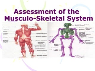
- 1. Assessment of the Musculo-Skeletal System
- 2. Review of Anatomy and Physiology • The musculo-skeletal system consists of the muscles, tendons, bones and cartilage together with the joints • The primary function of which is to produce skeletal movements
- 3. Muscles Three types of muscles exist in the body • 1. Skeletal Muscles – Voluntary and Striated • 2. Cardiac muscles – Involuntary and Striated • 3. Smooth/Visceral muscles – Involuntary and NON-striated
- 4. TENDONS •Bands of fibrous connective tissue that tie bones to muscles
- 5. LIGAMENTS •Strong, dense and flexible bands of fibrous tissue connecting bones to another bone
- 6. BONES • Variously classified according to shape, location and size • Functions 1. Locomotion 2. Protection 3. Support and lever 4. Blood production 5. Mineral deposition
- 7. JOINTS The part of the Skeleton where two or more bones are connected
- 8. CARTILAGES •A dense connective tissue that consists of fibers embedded in a strong gel-like substance
- 9. BURSAE Sac containing fluid that are located around the joints to prevent friction
- 10. • CHECK YOUR EQUIPMENT PRIOR TO ENTERING THE PATIENT’S ROOM. • MAKE SURE YOU HAVE EVERYTHING YOU NEED TO COMPLETE YOUR ASSESSMENT PRIOR TO ENTERING THE PATIENT’S ROOM
- 11. Make the Patient Comfortable • Showing concern for privacy and patient modesty must become ingrained in your professional behavior • Be sure to close nearby doors or examination room PRIOR to beginning physical examination • Your goal is to visualize one area of the body at a time
- 12. Make the Patient Comfortable • Be sensitive to the patient’s feelings and physical comfort • When you have completed the examination, show your attentiveness, by rearranging the patients pillows, or adding blankets for warmth; make sure their immediate environment is to their satisfaction • Be sure to lower the bed completely, and make sure side rails are up and call bell is in the patient’s reach • As you leave be sure to WASH YOUR HANDS
- 15. Cardinal Techniques of Examination • Inspection • Palpation • Percussion • Auscultation
- 16. Outlines 1. Review of Anatomy and physiology of musculoskeletal system 2. Physical Exam 3. Inspection 4. Palpation 5. ROM (Rang of motion)
- 17. Objectives • Apply knowledge of Anatomy and physiology of musculoskeletal system • Differentiate between normal and abnormal • Implement physical assessment
- 18. Musculoskeletal • Muscle or joint pain • Stiffness • Arthritis • Gout • Backache • If present, describe location or affected joints or muscles, any swelling, redness, pain, tenderness, stiffness, weakness, or limitation of motion or activity; include timing of symptoms duration, and any history of trauma • Neck or low back pain • Joint pain with systemic features such as fever, chills, rash, anorexia, weight loss, or weakness
- 19. Skin • Rashes • Lumps • Sores • Itching • Dryness • Changes in color • Changes in hair or nails
- 21. What do muscles do ? • Muscles simply move you! • Without muscles you couldn't open your mouth, speak, shake hands, walk, talk, or move your food through your digestive system. • There would be no exploring, running, climbing, smiling, blinking, breathing. You couldn't move anything inside or outside you. The fact is, without muscles, you wouldn't be alive for very long
- 23. The skeleton is the name given to the collection of bones that holds our body up. Our skeleton is very important to us. It does three major jobs. 1. It protects our vital organs such as the brain, the heart, and the lungs. 2. It gives us the shape that we have. Without our skeleton we would just be a blob of blood and tissue on the floor. 3. It allows us to move. Because our muscles are attached to our bones, when our muscles move, they move the bones, and we move
- 24. Physical Exam 1. Inspection • Observe any lack of symmetry and any evidence of trauma or disease. • Look for muscle wasting; • Inspect the joint contour (shape) and observe any evidence of swelling, deformity or inflammation.
- 25. • Ask the client to point to, or otherwise identify, any painful areas, including sites of radiation of pain. Screening questions for musculoskeletal disorders 1. Do you have any pain or stiffness in your arms, legs or back? 2. Can you walk up and down stairs without difficulty? 3. Can you dress yourself in everyday clothes without any difficulty?
- 26. • Assessment of Gait • Ask the patient to walk back and forth across the room . • Observe for equality of arm swing , balance and rapidity and ease of turning . • Next, ask the patient to walk on his tiptoes ,then on heels . • Ask the patient to tandem walk . • Test patient's ability to stand with feet together with eyes open and then closed. (Romberg's test .)Reassure patient that you will support him, in case he becomes unsteady . • Normal :Person can walk in balance with the arms swinging at sides and can turn smoothly. Person should be able to stand with feet together without falling with eyes open or closed. tiptoes heels tandem
- 27. Upper Extremity Muscles • Inspect the muscles of the shoulder, arm, forearm and hand. • Note muscle size (bulk). • Look for asymmetry, atrophy and fasciculation. • Look for tremor and other abnormal movement at rest and with arms outstretched.
- 28. Determine muscle power by • Gently trying to overpower contraction of each group of muscles. – Shoulder: Abduction (Deltoid) –, Adduction –, (Trapezius) Abduction Adduction Trapezius)
- 29. – Elbow: flexion (Biceps) – Elbow extension (Triceps) – Wrist: Flexion ( )and extension().
- 30. – Hand: Grip – opposition of thumb and index finger – opposition of thumb and little finger and – finger abduction and adduction. Grip
- 31. • Determine limb tone (resistance to passive stretch). • With the patient relaxed • Gently move the limb at the shoulder, elbow and wrist joints and note whether tone is normal, increased or decreased
- 32. Normal findings • Muscles are symmetrical in size with no involuntary movements. • In some, muscles may be slightly larger on the dominant side. • Muscle power obviously varies. You should not be able to overpower with reasonable resistance. • You have to learn to appreciate the normal tone from practice.
- 33. Neck: Range of Motion of • Fix the head with one hand while you examine neck • Inspection – Note the normal concavity of cervical spine – Identify Transverse process of C7 – Observe Trapezius and Sternomastoid muscles • Palpation – Feel each spinous process looking for focal areas of tenderness – Joint • Feel for crepitus during passive motion – Para spinal muscles • Range of motion – Active • Touch chin for flexion • Throw head back for extension Touch chin Throw head back
- 34. • Touch each shoulder with ears for lateral flexion • Touch each shoulder with chin for lateral rotation – Passive • Feel for crepitus during passive motion • Normal: – 30 degree rotation, able to touch chest with chin, 55 degree extension and 40 degree lateral bend. – No resistance during the range of motion.
- 35. Muscles of Lower Extremity Inspect the muscles of the hip, knee and ankle . • Note muscle size( bulk .) • Look for asymmetry, atrophy and fasciculation . • Look for abnormal movement . • Determine muscle power by gently trying to overpower contraction of each group of muscles . – Hip :Flexion( Iliopsoas), Extension (Gluteus maximus), Abduction, Adduction . Hip flexion
- 36. Assessment of the Musculoskeletal System Muscle Strengthscale 0 No detection of muscular contraction 1 A barely detectable flicker or trace of contraction with observation or palpation. 2 Active movement of body part with elimination of gravity. 3 Active movement against gravity only and not against resistance 4 Active movement against gravity & some resistance 5 Active movement against full resistance without evident fatigue (Normal muscle strength)
- 37. The Knee Exam • Inspection • Make sure that both knees are fully exposed. The patient should be in either a gown or shorts. Rolled up pant legs do not provide good exposure ! • Watch the patient walk. • Do they limp or appear to be in pain? • When standing, is there evidence of bowing (varus) or knock- kneed (valgus) deformity? There is a predilection for degenerative joint disease to affect the medical aspect of the knee, a common cause of bowing . varus Knee deormity,more marked on the left leg
- 38. • Is there evidence of atrophy of the quadriceps, hamstring, or calf muscle groups? Knee problems/pain can limit the use of the affected leg, leading to wasting of the muscles . While both legs have well developed musculature, the left calf and hamstring are bulkier than the right
- 39. – Knee : Flexion( Hamstrings ), Extension( Quadriceps ) – Ankle : Dorsiflexion( Tibialis anterior), Plantar flexion (Gastronemius .) • Determine limb tone resistance to passive stretch. With the patient relaxed, gently move the limb at the hip, knee and ankle and note whether tone is normal, increased or dicreased. Flex the hip and knee. • Support the knee, dorsiflex the ankle sharply and hold the foot in this position checking for clonus . Dorsiflexion Knee extension Knee flexion
- 40. Spine (Bone) • The examiner should stand behind the patient and observe the alignment of the spine in the flexed position to determine scoliosis . • View the spine from the side to determine kyphosis . • Ask the patient if he is aware of sore spots. Palpate the spinous process and be gentle with the sore spots .Percuss one vertebra at a time, starting from head . • .
- 41. • Assess range of motion of spine by having patient bend down to pick up an object without bending his legs while you hold his hips . • Normal : • Gentle concavities in cervical and lumbar regions and a convexity in the thorax . • Vertebral line and gluteal cleft align
- 42. Posture •Normal - Comfortably erect Look for straight lines across body parts
- 43. Lordosis - Increased Curvature of the Spine
- 44. Kyphosis is a curving of the spine that causes a bowing of the back, which leads to a hunchback or slouching posture.
- 45. Scoliosis – curvature of the spine away from middle or sideways