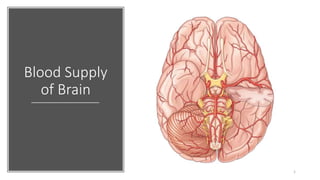
Blood supply of brain
•Download as PPTX, PDF•
4 likes•288 views

The origin, course, branches, and distribution of internal carotid artery. The origin, course, branches, and distribution of basilar artery. Describe the formation, branches and distribution of circulus arteriosus. Outline the venous drainage of the brain.

Recommended
More Related Content
What's hot
What's hot (20)
Similar to Blood supply of brain
Similar to Blood supply of brain (20)
Recently uploaded
Recently uploaded (20)
Blood supply of brain
- 2. Contents • The origin, course, branches and distribution of internal carotid artery. • The origin , course , branches and distribution of basilar artery. • Describe the formation, branches and distribution of circulus arteriosus. • Outline the venous drainage of brain. 2
- 3. Blood supply of Brain-Introduction The brain is one of the most metabolically active organs of the body as it depends on aerobic metabolism of glucose. Although the brain constitutes only 2% (1/50) of the total body weight, it receives 20% (1/5) of the total cardiac output and consumes 20% of the total O2 used by the body. The cerebrovascular diseases (thrombosis, embolism, and hemorrhage) are the third most common cause of death and the neurological signs depend on the site of lesion. An adequate knowledge of the blood supply of the brain is essential for proper diagnosis and treatment of these diseases. 3
- 4. The brain The brain Composed of three parts: - Cerebrum - Cerebellum - Brainstem The cerebrum is divided into four lobes: 1.Frontal 2.Parietal 3.Temporal 4.Occipital 4
- 5. Arteries of the brain Two pairs of arteries, the carotid and vertebral. ■ The internal carotid system, 80% of the brain’s blood supply (anterior circulation). ■ The vertebral basilar system from subclavian artery(20 %) (posterior circulation) Anastomose in the arterial circle of Willis. 5
- 6. Arteries of the brain Internal carotid artery: Origin: ■ One of the two terminal branches of common carotid artery. ■ Begins in the neck at the upper border of thyroid cartilage. 6
- 7. Arteries of the brain Internal carotid artery: Course in the cranial cavity- • Enters the base of skull & passes thro’ the carotid canal in the petrous portion of the temporal bone. Its course here is extradural. • Passes horizontally forward through cavernous sinus in an S-shaped curve (the carotid siphon). • Emerges on medial side of anterior clinoid process by piercing the dura & arachnoid mater. • Enters the subarachnoid space. • Then it runs upwards to the anterior perforated substance where it terminates by dividing into the anterior and middle cerebral arteries. 7
- 8. Arteries of the brain Parts of the internal carotid artery: • Cervical part – neck • Petrous part – carotid canal • Cavernous part – cavernous sinus • Cerebral part – base of the brain Branches of the cerebral part : • Ophthalmic • Posterior communicating • Anterior choroidal • Anterior cerebral • Middle cerebral 8
- 9. 9
- 10. Anterior cerebral artery Origin : Smaller terminal branch of the internal carotid artery Course : Runs above the optic nerve to the medial surface of the frontal lobe of the cerebral hemisphere . • Communicate with the ACA of opposite side by the anterior communicating artery • Curves round the genu of the corpus callosum. • Enters anterior part of median longitudinal fissure • Curves backwards above the corpus callosum. • It terminates near the splenium by anastomosing with the posterior cerebral artery. 10
- 11. Anterior cerebral artery Branches: • Cortical branches • Central branches • Recurrent artery of Heubner . Cortical branches - ▪ Orbital, frontal, parietal branches Supply - orbital surface of the frontal lobe, all medial surface of the cerebral cortex as far back as the parieto-occipital sulcus and adjacent strip of the suprolateral surface of the cerebral hemisphere. 11
- 13. Anterior cerebral artery : Functional areas supplied: Upper part of primary motor area(4) Upper part of somatosensory area (3,1,2) Upper part of premotor area (6) Upper part of prefrontal cortex (9,10,11,12) 13
- 14. Anterior cerebral artery : Central branches: • Anteromedial group of central arteries. • Joins the fellow of the opposite side by the anterior communicating artery. Recurrent artery of Heubner: • Pierces the anterior perforated substance. • Supplies: basal ganglia, Internal capsule – anterior limb 14
- 15. Middle cerebral artery Origin: Larger terminal branch and main continuation of internal carotid artery .Together the middle and anterior cerebral arteries are referred to as the anterior circulation of the brain. The middle cerebral artery is the artery most often occluded in stroke. Course ▪ Runs laterally in the stem of lateral sulcus between the orbital and tentorial surfaces to the insula ▪ Terminates on the insula by dividing into branches Branches : • Cortical branches • Central branches (anterolateral group) 15
- 17. Middle cerebral artery Cortical branches: • Orbital, frontal, parietal and temporal branches Supply the cerebral cortex Supply: ▪ Most of superolateral surface (except narrow strip along the superomedial border from the frontal pole to parieto-occipital sulcus, Occipital lobe, Inferior temporal gyrus) & insular lobe. ▪ Lateral part of orbital surface. ▪ Anterior part of tentorial surface. 17
- 18. Middle cerebral artery Central branches (lateral striate /lenticulostriate arteries) anterolateral group 18
- 19. Middle cerebral artery • Functional areas supplied: Lower parts of Primary motor area (area 4) Premotor area (area 6) Somatosensory area (area 3 1 2) Prefrontal area (area 9,10,11,12) Broca’s motor speech area (45,44) Auditory area (41,42) Supramarginal gyrus (40) Angular gyrus (39) Superior temporal gyrus (22) 19
- 21. Distribution of middle cerebral arteryDistribution of middle cerebral artery Arteries of the brain
- 22. Anterior choroidal artery: Origin : Branch of internal carotid artery Course : Passes backward, enters inferior horn of lateral ventricle, and ends in choroid plexus. Distribution: Supplies: • Choroid plexus of the inferior horn of the lateral ventricle. • Lateral geniculate body. • Posterior limb & retrolentiform part of internal capsule. • Middle 3/5 of crus cerebri. • Basal ganglia – globus pallidus. 22
- 23. Vertebral artery It has 4 parts. Origin - Arise from the I part of subclavian artery. Course - • I Part - Ascends towards the foramen transversarium of C6 vertebra. • II Part - Ascend through the transverse foramen of the upper six cervical vertebrae (C6-C1). • III Part - Emerges from the transverse foramen of the atlas (C1) runs on the groove on the upper surface of the post arch of the atlas pierces the atlanto-occipital membrane and enters the foramen magnum. • IV Part - Lies within cranium, the two arteries run on the anterior aspect of the medulla oblongata then unite at the lower border of the pons to form the basilar artery. 23
- 24. Vertebral artery • Branches of 4th part: 1. Meningeal 2. Medullary 3. Anterior spinal 4. Posterior spinal 5. Posterior inferior cerebellar 24
- 25. Basilar artery • It’s formed by the union of 2 vertebral arteries. Ascends in a groove on the anterior surface of the pons. • At the upper border of the pons/ ponto-midbrain junction, divides into 2 posterior cerebral arteries. 25
- 26. Basilar artery Branches from the basilar artery: 1. Anterior inferior cerebellar artery. 2.The labyrinthine artery. 3.The pontine branches (multiple vessels that penetrate the pons as paramedian, short circumferential, and long circumferential arteries). 4.The superior cerebellar arteries. 5. The basilar artery terminates by dividing into the posterior cerebral arteries at the upper border of the pons. 26
- 27. 27
- 28. Posterior cerebral artery Posterior cerebral artery: Terminal branch of basilar artery. Course: • Ascends beside the midbrain to reach the splenium of the corpus callosum on the medial side of the cerebral hemisphere and terminates by dividing into branches • Connected to the internal carotid artery by the posterior communicating artery Branches: - Cortical - Central branches - Posterior choroidal artery 28
- 29. Posterior cerebral artery Cortical branches: ▪ Inferior surfaces of temporal lobe by temporal branches. ▪ Medial surface of the occipital lobe by calcarine branches. ▪ Medial surface of the post part of the parietal & part of the occipital lobes by parieto occipital branches. Functional area supplied: ▪ Visual area (17,18,19). 29
- 30. Posterior cerebral artery Posterior choroidal artery supply • Choroid plexus of III ventricle. • Choroid plexus of central part of lateral ventricle. • Ventral part of thalamus. • Fornix and superior colliculus of midbrain. • Central branches: • Posteromedial group, posterolateral group • Many small perforating arteries arise from PCA to supply the midbrain, thalamus, hypothalamus and medial and lateral geniculate bodies. 30
- 31. Circulus arteriosus-Circle of Willis Location: • Lies in the interpeduncular fossa at the base of brain around optic chiasma, tuber cinereum and mamillary bodies. Formation: Anterior part: Anterior cerebral arteries (Rt & Lt) Anterior communicating artery Posterior part: Basilar artery Posterior cerebral arteries (Rt & Lt) Lateral part: Internal carotid artery (Rt & Lt) Posterior communicating arteries (Rt & Lt) 31
- 32. Circulus arteriosus-Circle of Willis 32
- 33. Circulus arteriosus-Circle of Willis Distribution of central arteries: • Central arteries, arise from the arterial circle and proximal portion of the principal arteries, penetrate the substance of the brain and supply the deep structures. • Branches of the circle of Willis : • Central arteries 6 groups: ▪ Anteromedial(AM) group ▪ Posteromedial(PM) group ▪ 2 Anterolateral(AL) groups ▪ 2 Posterolateral (PL)groups 33
- 34. Circulus arteriosus-Circle of Willis 34 Anteromedial group of central arteries: Collectively they are referred to as the medial striate arteries.The largest and most lateral of these to enter the brain is the medial striate or recurrent artery of Huebner which arises from the anterior cerebral artery. Origin: • Anterior cerebral and anterior communicating arteries Distribution: • Anterior part of hypothalamus • Head of caudate and lentiform nuclei • Anterior limb of the internal capsule.
- 35. Circulus arteriosus-Circle of Willis Anterolateral group of central arteries: They are also called the lateral striate or lenticulostriate arteries. Origin: • Middle cerebral artery Distribution: • Internal capsule ( Genu, anterior & posterior limbs) • Caudate and lentiform nuclei • Lateral hypothalamus The most common sites of spontaneous hemorrhage in individuals with long-standing hypertension and is termed the artery of cerebral hemorrhage. 35
- 36. Circulus arteriosus-Circle of Willis Posteromedial group of central arteries: (Thalamoperforate arteries) Origin- • Posterior cerebral artery (Thalamoperforate arterirs) • Posterior communicating arteries Distribution- • midbrain • Anterior and medial parts of thalamus • Posterior part of hypothalamus • Subthalamus Posterolateral group of central arteries: (Thalamogeniculate arteries) Origin- • Posterior cerebral artery) Distribution- • Posterior and lateral parts of thalamus • Geniculate bodies • Colliculi of midbrain and pineal gland 36
- 37. Arteries of the brainstem and cerebellum • The cerebellum is supplied by three pairs of cerebellar arteries. • Superior cerebellar artery: supplies the superior surface of the cerebellum. • Anterior inferior cerebellar and posterior inferior cerebellar arteries: supplies the inferior surface of the cerebellum. 37
- 38. Arteries of the brainstem and cerebellum The medulla is supplied by • 1. Two vertebral arteries. • 2. Anterior and posterior spinal arteries. • 3. Anterior and posterior inferior cerebellar arteries. • 4. Basilar artery. The pons is supplied by 1. Numerous (pontine) branches from the basilar artery. 2. Anterior inferior cerebellar artery. The midbrain is supplied by 1. Basilar artery through its posterior cerebral and superior cerebellar arteries. Basilar artery also supplies themidbrain through direct branches. 2. Branches of posterior communicating and anterior choroidal arteries. 38
- 39. Arterial supply of the internal capsule • Anterior limb: lenticulostriate branches of middle cerebral artery (superior half) & recurrent artery of Heubner of the anterior cerebral artery(inferior half). • Genu: lenticulostriate branches of middle cerebral artery • Posterior limb: lenticulostriate branches of middle cerebral artery (superior half) & anterior choroidal artery off of the internal carotid artery (inferior half). • Retrolentiform + sublentiform parts = supplied by anterior choroidal. 39
- 40. Veins of brain • Veins of the brain have no muscular tissue in the wall and no valves. • Veins – Located in the subarachnoid space. • Venous sinuses – located between 2 layers of the dura mater. 40
- 41. Veins of brain Cerebral veins are divided into: 1.Superficial cerebral veins: • Superior cerebral veins. • Inferior cerebral veins. • Superficial middle cerebral vein. 2. Deep cerebral veins: • Internal cerebral vein. • Basal vein (Rosenthal). • Great cerebral vein (of Galen). 41
- 42. Cerebral veins • Superior cerebral veins drain the upper parts of the superolateral and medial surface and end in the superior sagittal sinus. • Inferior cerebral veins drain the lower part of the hemisphere. • Superficial middle cerebral vein drains superolateral surface and posterior end of this vein is connected to the superior sagittal sinus by the superior anastomotic vein and to the transverse sinus by the inferior anastomotic vein. superficial middle cerebral vein terminates in the cavernous sinus. 42
- 43. Cerebral veins Internal cerebral vein: • Situated in the tela choroidea of third ventricle, in the roof of third ventricle. • Formation: At the interventricular foramen by the union of thalamostriate vein and choroidal veins. • Termination: Below the splenium two Internal cerebral vein joins to form the great cerebral vein. 43
- 44. Cerebral veins Great cerebral vein (vein of Galen): • Situated below the splenium, in the cystern of great cerebral vein. • Formation: By the union of two internal cerebral veins. • Termination: Joins the inferior sagittal sinus to form straight sinus. • Tributaries: Right and left basal veins. 44
- 45. Cerebral veins •Internal cerebral veins joins to form the great cerebral vein and the two basal veins. • The deep cerebral veins drains the thalamus, hypothalamus, the corpus striatum, the internal capsule, copus callosum and the choroid plexuses. The corpus striatum and internal capsule are drained by superior and inferior striate veins. 45
- 46. Veins of cerebellum & brain stem • The veins from the upper surface of the cerebellum drain into the straight, transverse, and superior petrosal venous sinuses. • The veins from the lower surface of cerebellum drain into right and left sigmoid , inferior petrosal, occipital and straight sinus. • The veins of the midbrain drain into the great cerebral vein. The pons and medulla drain into the superior and inferior petrosal sinuses, transverse and occipital sinuses. • Inferiorly, the veins of the medulla are continuous with the veins of the spinal cord. 46
- 47. Middle meningeal veins • These veins accompany the branches of middle menigeal artery and consist of : 1.Frontal (Anterior) trunk. 2.Parietal (Posterior) trunk. • Both trunks communicate above with superior sagittal sinus through the venous lacunae. • The frontal trunk terminates into pterygoid venous plexus through the foramen ovale, or into the spheno- parietal or cavernous sinus. • The parietal trunk ends in the pterygoid venous plexus through the foramen spinosum. 47
- 48. Blood Supply of Brain - Clinical Importance 48
- 49. Clinical importance Berry aneurysms(Congenital cerebral aneurysms): A brain aneurysm (AN-yoo-riz-um) is a bulge or ballooning in a blood vessel in the brain. It often looks like a berry hanging on a stem. These occur mostly at the sites where two arteries join in the formation of the circle of Willis. The basic abnormality at these points is the congenital deficiency of the tunica media (elastic tissue) in the arterial wall. A brain aneurysm can leak or rupture, causing bleeding into the brain (hemorrhagic stroke). 49
- 50. Clinical importance Subarachnoid hemorrhage: • A subarachnoid hemorrhage is bleeding in the subarachnoid space. • Bleeding usually results from the rupture of an abnormal bulge in a blood vessel (aneurysm) in brain. • Sometimes bleeding is caused by trauma, an abnormal tangle of blood vessels in brain (arteriovenous malformation), or other blood vessel or health problems. • The primary symptom is a sudden, severe headache. 50
- 51. Clinical importance Occlusion of middle cerebral artery: The occlusion of middle cerebral artery occurs commonly. The most common cause of MCA occlusion is embolism. Signs and symptoms: Contralateral hemiplegia and hemianesthesia involving mainly the face and arm, due to involvement of most of the primary motor and sensory areas. Aphasia, if left dominant hemisphere is involved—due to involvement of motor and sensory speech areas. Cerebral thrombosis most commonly affects the middle cerebral artery or its main branches because it is a direct continuation of the internal carotid artery. 51
- 52. Clinical importance • Subdural hemorrhage: • It occurs due to rupture of cerebral veins in the subdural space. • The superior cerebral veins are most commonly torn, where they enter the superior sagittal sinus. • The cause is usually a blow on the front or back of the head, resulting in excessive anteroposterior displacement of the brain within the skull. 52
- 53. Summary Arteries of the brain Brain: Anterior , posterior , middle cerebral arteries and branches of circle of Willis. Cerebellum: Superior , anterior inferior and posterior inferior cerebellar arteries. Medulla: • Anterior and posterior spinal arteries • Vertebral arteries • Posterior inferior cerebellar arteries. Pons: • Paramedian branches of basilar artery (pontine arteries) • Short circumferential branches of basilar artery • Superior cerebellar artery and anterior inferior cerebellar arteries. Midbrain: Branches from basilar a. • Posterior cerebral artery and superior cerebellar artery (sup. & inf. colliculi) • Anterior choroidal a. and Posterior communicating artery. 53
- 54. Summary Cerebral veins 1.Superficial cerebral veins: Superior cerebral veins, inferior cerebral veins and superficial middle cerebral vein. 2. Deep cerebral veins: Internal cerebral vein, basal vein (Rosenthal) and Great cerebral vein (of Galen). • Veins draining the brain open into the dural venous sinuses. These are the superior sagittal , inferior sagittal, straight, transverse, sigmoid, cavernous, sphenoparietal, petrosal and occipital sinuses. • The blood from all these sinuses reaches the sigmoid sinus which becomes continuous with internal jugular vein. • The intracranial venous sinuses communicate with veins outside the skull through emissary veins. 54
- 55. Summary • Veins of cerebellum & brain stem • The veins from upper surface of the cerebellum drain into the straight, transverse, and superior petrosal venous sinuses. • The veins from lower surface of cerebellum drain into right and left sigmoid , inferior petrosal, occipital and straight sinus. • The veins of the midbrain drain into the great cerebral vein. The pons and medulla drain into the superior and inferior petrosal sinuses, transverse and occipital sinuses. • Inferiorly, the veins of the medulla are continuous with the veins of the spinal cord. 55
- 56. THANK YOU 56
Editor's Notes
- 21