
Recommended
More Related Content
Similar to Psoriasis
Similar to Psoriasis (20)
Recently uploaded
Recently uploaded (20)
Psoriasis
- 2. PORIASIS Psoriasis is non-contagious chronic autoimmune disease that causes skin inflammation
- 3. PATHOPHYSIOLOGY • Thickness of epidermis is intact due to regulated turn over of keratinocytes • Microbes live on skin enter the skin when any break in skin • The dendritic cells (Stratum spinosum) catch microbes and process and present antigen to T Cells • T cells release Cytokines ( IL 12, IL23,Interferon γ, TNF, IL 17) • Release of cytokines leads to proliferation of keratinocytes and recruit immune cells (neutrophils) • When microbes are killed the immune response returns to normal
- 4. PSORIASIS •In Psoriasis due to etiological factors (Genetic, Environmental- trauma or infection •The immune response doesn't shut off leads to excess inflammation and chronic skin damage •Blood vessels in Basale increases which increases immune cells neutrophils in epidermis •The neutrophils collect in stratum corneum, Keratinocytes proliferate excessively and immature abnormally •Thins out stratum Basale but thickens other layers ( Spinosm and corneum) •Piling of keratinocytes and defects of keratinocytes In stratum corneum •Produce more keratin –thicker •Retain Nuclei – Parakeratosis •Do not adhere properly breaks in epidermis – Scaly appearance •Scales Picked off leads to localized bleeding called Auspitz Sign
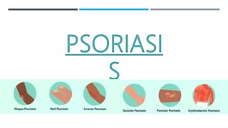
- 5. TYPES AND SYMPTOMS 1) Plaque Psoriasis – Most common Flattened areas of epidermal elevation Inflamed and red due to dilated blood vessels White silver scales which are itchy Present on scalp and tensor regions, groin, lower back and knees
- 7. 2) Gluttate Psoriasis Small red individual spots on trunk and limbs Starts in childhood and triggered by infection 3) Inverse Psoriasis Smooth and shiny red lesions lack scales within skin folds like genital region, underarms and under breasts 4) Pustular psoriasis Red skin, white elevated of clouded pus which are tender present on hands and feet
- 11. 5) Erythrodermic Psoriasis Fire red scales can cover any large area of skin Extremely itchy and painful, scales fall off in sheets 6) Psoriatic Arthritis Inflammation in joints Nail Pitting and holes in nails
- 14. DIAGNOSIS AND TREATMENT Based on distribution of the skin damage Tissue biopsy – Confirm the diagnosis and see classic changes in epidermal layers Treatment – Moisturizers and emollients Clear plaques and minimize itching Topical and systemic immunosuppressive therapies UV phototherapy – Induce DNA Damage in keratinocytes
- 15. New Research – Stress management Dietary interventions Immunotherapies Treatment of Depression and anxiety- Psychological counseling Psychodermatology- Anti-anxiety medication Biofeedback Allergy and immune function testing Cognitive behavioral therapy
- 16. MEDICAL MANAGEMENT Goal to reduce inflammation and suppress rapid turnover of epidermal cells. No cure, but control is possible Topical treatments: Corticosteroids, tazarotene, calcipotriene, anthralin, calcineurin inhibitors (tacrolimus) Intralesional injection of corticosteroids for chronic plaques Systemic treatments: Natural or artificial UVB. PUVA (UVA with topical or systemic photosensitizer [psoralen]) Traditional and oral therapies: antimetabolite (methotrexate), retinoid, apremilast, immunosuppressant (cyclosporine) Biologic therapies: adalimumab, etanercept, infliximab, ustekinumab, brodalumab, certolizumab pegol, secukinumab, golimumab, guselkumab, ixekizumab for moderate to severe plaque disease