SlideShare a Scribd company logo
1 of 46
RADIOGRAPHIC
TECHNIQUE OF
SKULL
PRESENTER:- Saru Gosain
B.Sc. MIT 2017 Batch
MODERATOR:- RANJIT KUMAR JHA
Assistant Professor
Contoso Ltd.
ANATOMY OF SKULL
The skull consists of the 28 bones; calvaria
consists of 14 bones including 3 pair of
ossicles:
1. Parietal-2
2. Temporal-2
3. Malleus-2
4. Incus-2
5. Stapes-2
6. Frontal 1
7. Occipital-1
8. Sphenoid-1
9. Ethmoid-1
Add a footer
2
Contoso Ltd.
Anatomical Terminology
LANDMARKS
Outer canthus of the eye: the point where the upper and
lower eyelids meet laterally.
Infra-orbital margin/point: the inferior rim of the orbit, with
the point being located at its lowest point.
Nasion: the articulation between the nasal and frontal bones.
Glabella: a bony prominence found on the frontal bone
immediately superior to the nasion.
 Vertex: the highest point of the skull in the median sagittal
plane (MSP).
External occipital protuberance (inion): a bony prominence
found on the occipital bone, usually coincident with the
median sagittal plane.
External auditory meatus: the opening within the ear that
leads into the external auditory canal. 3
Contoso Ltd.
• Inter-orbital (inter-pupillary) line: joins the
centre of the two orbits or the centre of the
two pupils when the eyes are looking straight
forward.
• Infra-orbital line: joints the two infra-orbital
points.
• Anthropological baseline: passes from the
infra-orbital point to the upper border of the
external auditory meatus (also known as the
Frankfurter line).
• Orbito-meatal base line (radiographic
baseline): extends from the outer canthus of
the eye to the centre of the external auditory
meatus. This line is angled approximately 10
degrees to the anthropological baseline.
• Reid’s baseline: line drawn from the inferior
margin of the orbit (orbital point) to the
centre of the orifice of the external auditory
meatus.
Add a footer
4
Lines
Contoso Ltd.
○ Median sagittal plane: divides the skull into
right and left halves.
○ Coronal planes: these are at right-angles to
the median sagittal plane and divide the
head into anterior and posterior parts.
○ Anthropological plane: a horizontal plane
containing the two anthropological
baselines and the infra-orbital line. It is an
example of an axial plane.
○ Auricular plane: perpendicular to the
anthropological plane. Passes through the
centre of the two external auditory
meatuses. It is an example of a coronal
plane.
5
Planes
Basic Projections
o Occipito-Frontal
o Lateral
6
OtherProjectionsfor cranium
○ Towne’s Projection
○ Submento-vertical
○ Sella turcica: lateral
○ Optic foramina postero-anterior oblique
○ Jugular foramina: submento-vertical 20 degrees cauded
○ Temporal bones: fronto-occipital 35 degrees cauded
○ Mastoid: lateral oblique 25 degrees cauded
○ Petrous bone: anterior oblique (Stenver’s)
Contoso Ltd.
Indications
○ Different types of fractures such as
basal, blowout, contre-coup,
depressed, Leforte, linear, tripod)
○ Mastoiditis
○ Metastases
○ Osteomyelitis
○ Osteopetrosis
○ Osteoporosis
○ Paget’s disease
○ Polyp
○ Sinusitis
○ Tumor
○ All metal objects are removed from
the patient, e.g. hair clips and
hairpins.
○ Bunches of hair often produce
artefacts and thus should be
untied.
○ If the area of interest includes the
mouth, then false teeth containing
metal and metal dental bridges
should be removed.
○ The patient should be provided
with a clear explanation of any
movements and film positions
associated with the normal
operation of the skull unit.
1. Cassette size: 24 x 30 cm
2. FFD: 100cm
3. kVp: 70-85
4. mAs: 20-25
5. Focal spot: small
6. Grid: yes
Patientpreparation Technicalparameters
Contoso Ltd.
Position of patient and cassette
○ Patient stands erect or lies prone (prone uncomfortable).
○ For the erect position, patient stands facing the vertical erect Bucky
○ MSP coincident with the midline of Bucky and perpendicular to it
○ Neck is flexed so that the orbito-meatal baseline is perpendicular to the Bucky.
○ Mid-part of frontal bone is positioned to the centre of the Bucky.
Direction and centering of x-ray beam
○ Directed perpendicular to the Bucky along the MSP
Essential image characteristics
○ All the cranial bones should be included within the image, including the skin margins
○ Ensure that the skull is not rotated.
○ For – Occipito- frontal: the petrous ridges should be completely superimposed within the orbit, with their upper
borders coincident with the upper third of the orbit.
○ -OF 10°↓: the petrous ridges appear in the middle third of the orbit.
○ -OF 15°↓ (Caldwell method): the petrous ridges appear in the lower third of the orbit.
○ -OF 20 °↓: the petrous ridges appear just below the inferior orbital margin 8
Occipito-frontal
9
o for the demonstration of
frontal bone, OF projection is
done without giving any
angulation
o the petrous ridges should be
completely superimposed
within the orbit
the petrous ridges appear in the
middle third of the orbit.
o the petrous ridges appear just
below the inferior orbital
margin
o for the demonstration of the
superior orbital fissures, the
central ray is directed through
the midorbits at an angle of
20-25 degrees cauded
Contoso Ltd.
Position of patient and cassette
○ Patient sits facing the erect Bucky
○ Head is rotated, such that the MSP is parallel to the Bucky and the inter-
orbital line is perpendicular to it.
○ Cassette placed transversely in the erect Bucky, such that its upper border is
5 cm above the vertex of the skull.
Direction and centering of the X-ray beam
○ Centre midway between the glabella and the external occipital
protuberance to a point approximately 5 cm superior to the external
auditory meatus.
Essential image characteristics
○ The image should contain all of the cranial bones and the first cervical
vertebra. Both the inner and outer skull tables should be included.
○ A true lateral will result in perfect superimposition of the lateral portions of
the floors of the anterior cranial fossa and those of the posterior cranial
fossa. The clinoid processes of the sella turcica should also be
superimposed.
10
Lateral
Contoso Ltd.
INDICATIONS: skull fractures, neoplastic processes, Paget’s diseases,
chronic suppurative otitis media.
Position of patient and cassette
○ Patient lies supine, with the posterior aspect of the skull resting on
a grid cassette.
○ MSP right angles to the cassette and coincident with its midline.
○ The orbito-meatal base line perpendicular to the film.
Direction and centering of the X-ray beam
○ Angled caudally so it makes an angle of 30 degrees to the orbito-
meatal plane.
○ Centre in the midline such that the beam passes midway between
the external auditory meatuses. This is to a point
○ approximately 5 cm above the glabella.
○ The top of the cassette positioned adjacent to the vertex of the
skull to ensure that the beam angulation does not project the area
of interest off the bottom of the image.
11
Towne’sprojection(Fronto-occipital30° cauded)
Contoso Ltd.
Essential image characteristics
o The sella turcica of the sphenoid bone is projected
within the foramen magnum.
o The image must include all of the occipital bone
and the posterior parts of the parietal bone, and
the lambdoidal suture should be visualized clearly.
o The skull should not be rotated. This can also be
assessed by ensuring that the sella turcica appears
in the middle of the foramen magnum.
12
Add a footer
NOTE: Reverse Towne’s projections (Hass method)
can also be performed with occipito-frontal 30°
cranial angulation on obese patient or who cannot
be correctly adjusted for the Towne’s projection
which will carry a lower radiation dose to sensitive
structures than the equivalent antero-posterior
projection.
Contoso Ltd.
Position of patient and cassette
○ Patient erect or supine. Patients placed in the supine position may have
increased intracranial pressure, therefore erect position is preferred.
Erect
○ The patient sits a short distance away from a vertical Bucky. The neck is
hyperextended to allow the head to fall back until the vertex of the skull makes
contact with the centre of the vertical Bucky.
○ Head adjusted to bring the external auditory meatuses equidistant from the
cassette.
○ The MSP at right-angles to the cassette along its midline.
○ The orbito-meatal plane should be as near as possible parallel to the cassette.
Direction and centering of the X-ray beam
○ Directed right-angles to the orbito-meatal plane and centred midway between
the external auditory meatuses.
Essential image characteristics
○ A correct projection will show the angles of the mandible clear of the petrous
portions of the temporal bone.
○ The foramina of the middle cranial fossa should be seen symmetrically either
side of the midline.
13
Submento-vertical
Sella turcica:
lateral
INDICATIONS: To see the sella expansion by a large lesion.
Position of patient and cassette
• Patient sits facing the erect Bucky, head rotated, such that the
MSP parallel to the Bucky and the inter-orbital line
perpendicular to the Bucky.
• Shoulders may be rotated slightly to allow the correct position
to be attained. Patient may grip the Bucky for stability.
• The head and Bucky heights are adjusted so that the centre of
the Bucky is 2.5 cm vertically above a point 2.5 cm along the
baseline from the external auditory meatus
Direction and centering of the X-ray beam
• Centred to a point 2.5 cm vertically above a point 2.5 cm along
the baseline from the auditory meatus nearer the X-ray tube.
14
Contoso Ltd.
OpticForamina:postero-anterior
oblique(Rhesemethod)
15
INDICATIONS: detection of optic nerve glioma
Position of patient and cassette
• Patient lies prone or, more commonly, erect with the nose, cheek
and chin of the side being examined in contact
• with the Bucky or cassette table.
• The centre of the orbit of the side under examination should
coincide with the centre of the Bucky or cassette table.
• The MSP adjusted to make an angle of 35 degrees to the vertical (55
degrees to the table).
• The orbito-meatal base line is raised 35 degrees from the horizontal.
Direction and centering of the X-ray beam
• The horizontal central ray centred to a point 7.5 cm above and 7.5
cm behind the uppermost external auditory meatus, so that the
central ray emerges from the centre of the orbit in contact with the
table.
Jugular foramina: submento-
vertical 20° cauded
INDICATIONS: detection of glomus jugulare tumour
Position of patient and cassette
Patient erect or supine. If the patient is unsteady, then a supine technique is
advisable.
Supine
• Patient’s shoulders are raised and the neck is hyperextended to bring the
vertex of the skull in contact with the grid cassette or table.
• Head adjusted to bring the external auditory meatuses equidistant from the
cassette.
• The MSP at right-angles to the cassette along its midline.
• The orbito-meatal plane should be as near as possible parallel to the cassette.
Direction and centering of the X-ray beam
Angled caudally so that it makes an angle of 70 degrees to the orbitomeatal
plane and centred in the midline to pass midway between the external auditory
meatuses.
Contoso Ltd.
Temporal bone: fronto-occipital35° cauded
Patient lies supine or erect
• Patient may be supine or erect with their back to an erect Bucky.
Head adjusted to bring the external auditory meatuses equidistant
from the table, so that the MSP is at right-angles to, and in the
midline of, the table.
• Chin is depressed so that the orbito-meatal line is at right-angles to
the table.
• A cassette is placed transversely and is centred to coincide with the
angled central ray.
Direction and centering of the X-ray beam
• A caudal angulation is employed, such that it makes an angle of 35
degrees to the orbito-meatal plane.
• The beam is centred midway between the external auditory
meatuses.
Essential image characteristics
• The sella turcica of the sphenoid bone should be projected within
the foramen magnum and include mastoid and petrous part of the
temporal bone supra-inferiorly.
• The skull should not be rotated. This can also be assessed by ensuring
that the sella turcica appears in the middle of the foramen magnum.
Mastoid- lateral
oblique 25° cauded
Position of patient and cassette
• Patient sits facing the erect Bucky.
• Head is rotated, such that the MSP is parallel to the Bucky and the inter-
orbital line is perpendicular to the Bucky.
• The auricle of the ear adjacent to the table is folded forward to ensure that its
soft-tissue outline is not superimposed over the region of interest.
• Cassette positioned longitudinally in the Bucky and is centred to coincide with
the central ray and mastoid process.
Direction and centering of the X-ray beam
• A 25-degree caudal angulation is employed and centred 5 cm above and 2.5
cm behind the external auditory meatus remote from the cassette.
Essential image characteristics
• Ensure that all of the mastoid air cells have been included within the image.
18
Add a footer
Mastoid-profile
Position of patient and cassette
• Patient lies supine on the table, with the orbito-meatal baseline
perpendicular to the table top.
• From a position with the MSP perpendicular to the table, the
head is rotated through an angle of 35 degrees away from the
side under examination, such that the MSP now makes an angle
of 55 degrees to the table.
• The vertical tangent to the skull should now be at the level of the
middle of the mastoid process under examination, so that the
mastoid process is in profile.
• Finally, the head is moved transversely across the table so that
the mastoid process being examined is in the midline of the table.
Direction and centering of the X-ray beam
• Angled caudally so that it makes an angle of 25 degrees to the
orbito-meatal plane and is centred to the middle of the mastoid
process on the side under examination.
19
Petrous bone: anterior oblique
(Stenver’s projection)
Position of patient and cassette
• Patient may be prone or erect facing a vertical Bucky.
• The middle of the supra-orbital margin on the side being examined is
centred to the middle of the Bucky.
• Neck flexed so that the nose and forehead are in contact with the
table and the orbito-meatal line is perpendicular to the table.
• From a position where the MSP is perpendicular to the table, the
head is rotated toward the side under examination, such that the
MSP is now at an angle of 45 degrees to the table. This brings the
petrous part of the temporal bone parallel to the cassette.
• The neck is extended so that the orbito-meatal line is raised five
degrees from horizontal.
• Cassette placed transversely in the Bucky and is centred at a level to
coincide with the central ray.
20
Contoso Ltd.
Direction and
centering of
the X-ray beam
• A 12-degree cephalad beam angulation is
employed, i.e. at an angle of seven degrees
to the orbito-meatal plane, to separate the
occiput from the petrous bone.
• Centre midway between the external
occipital protuberance and the external
auditory meatus furthest from the cassette.
21
Styloid process: PA axial ( Cahoon method)
o The styloid process is a needle like projection of varying lengths averaging 2-3 cm which projects from the inferior part
of the petrous temporal bone.
22
Add a footer
Position of patient and cassette
• Patient in the seated-upright or prone position.
• Adjust the forehead and nose so that the MSP is perpendicular
to the midline o f the Bucky.
• Center the IR to the nasion.
• Adjust the flexion of the patient's neck so that the OML is
perpendicular to the plane of the IR.
Direction and centering of x-ray beam
• Directed to the nasion at an angle of 25 degrees cephalad.
Essential image characteristics
• Temporal styloid processes free of superimposition from
frontal bone, occipital bone, and coronoid processes of the
mandible.
• Styloid processes projected within or just above the maxillary
sinuses.
Facialbones and sinuses
Facial skeleton consists of 14 bones.
1. Maxilla-2
2. Zygomatic-2
3. Nasal-2
4. Lacrimal-2
5. Palatine-2
6. Inferior nasal concha-2
7. Mandible-1
8. Vomer -1
Within these bones, there are series of air-filled cavities known as the paranasal sinuses. There are 4
paranasal sinuses;
1. Maxillary sinuses
2. Frontal sinuses
3. Sphenoid sinuses
4. Ethmoid sinuses
23
Add a footer
Contoso Ltd.
1. Occipito-mental
2. Occipito-mental 30° cauded
3. Lateral
4. Zygomatic arches: infero-superior
5. Orbits: occipito-mental (modified)
6. Nasal bones: lateral
7. Mandible: lateral 30° cephalad
: postero-anterior
: postero-anterior oblique
8. Temporal-mandibular joints: lateral 25°
cauded
24
Add a footer
Projectionsfor paranasalsinuses
1. Occipito-mental with open mouth (Water’s view)
2. Occipito-frontal 15° cauded
3. Lateral
Projectionsfor facial bones
Contoso Ltd.
Occipito-mental for facial bones
25
Position of patient and cassette
• Patient seated facing the vertical erect Bucky.
• Patient’s nose and chin are placed in contact with the midline of
the cassette holder. The head is then adjusted so that the orbito-
meatal baseline makes 45-degree angle to the cassette holder.
• The horizontal central line of the Bucky/cassette holder should be
at the level of the lower orbital margins.
• Ensure that the MSP is at right-angles to the Bucky/cassette
holder by checking the outer canthi of the eyes and that the
external auditory meatuses are equidistant.
Direction and centering of the X-ray beam
• Perpendicular and centred to the middle of the cassette holder.
• To check that the beam is centred properly, the cross-lines on the
Bucky or cassette holder should coincide with the patient’s anterior
nasal spine.
Essential image characteristics
• The petrous ridges must appear below the
floors of the maxillary sinuses.
• There should be no rotation. This can be
checked by ensuring that the distance from
the lateral orbital wall to the outer skull
margins is equidistant on both sides.
26
Add a footer
Contoso Ltd.
Position of patient and cassette
○ Patient seated facing the vertical Bucky.
○ The patient’s nose and chin are placed in contact with the
midline of the cassette holder. The head then is adjusted to bring
the orbito-meatal baseline to a 45-degree angle to the cassette
holder.
○ The horizontal central line of the Bucky or cassette holder should
be at the level of the symphysis menti.
○ Ensure that the MSP is at right-angles to the Bucky by checking
that the outer canthi of the eyes and the external auditory
meatuses are equidistant.
Direction and centering of the X-ray beam
○ The tube is angled 30 degrees caudally and centred along the
midline, such that the central ray exits at the level of the lower
orbital margins.
○ To check that the beam is centred properly, the cross-lines on
the Bucky or cassette holder should coincide approximately with
the upper symphysis menti region.
27
Occipito-mental 30° cauded
Contoso Ltd.
Essential image characteristics
o The floors of the orbit will be clearly visible
through the maxillary sinuses, and the lower
orbital margin should be demonstrated clearly.
o There should be no rotation. This can be checked
by ensuring that the distance from the lateral
orbital wall to the outer skull margins is
equidistant on both sides.
28
Add a footer
Contoso Ltd.
Position of patient and cassette: In cases of injury, this projection should be
taken using a horizontal beam in order to demonstrate any fluid levels in the
paranasal sinuses.
Erect
○ Patient sits facing the vertical Bucky. The head is rotated, such that the side
under examination is in contact with the Bucky or cassette holder.
○ The Bucky height is altered, such that its centre is 2.5 cm inferior to the outer
canthus of the eye.
○ The MSP is brought parallel to the cassette by ensuring that the inter-orbital
line is at right angles to the cassette and the nasion and external occipital
protuberance are equidistant from it.
Direction and centering of the X-ray beam
○ Centre the horizontal central ray to a point 2.5 cm inferior to the outer
canthus of the eye.
Essential image characteristics
○ The image should contain all of the facial bones sinuses, including the frontal
sinus and posteriorly to the anterior border of the cervical spine.
○ A true lateral will have been obtained if the lateral portions of the floor of the
anterior cranial fossa are superimposed. 29
Lateral for facial bones
Contoso Ltd.
Zygomatic arches: infero-superior (jug-
handle projection) Position of patient and cassette
INDICATION: Fracture of the zygomatic bones.
This projection is essentially a modified submento-vertical
(SMV) projection. This method is tangential projection.
o Patient lies supine, with one or two pillows under the
shoulders to allow the neck to be extended fully.
o Cassette is placed against the vertex of the skull, such
that its long axis is parallel with the axial plane of the
body. It should be supported in this position with foam
pads and sandbags.
o The flexion of the neck is now adjusted to bring the
long axis of the zygomatic arch parallel to the cassette.
o The head in now tilted five to ten degrees away from
the side under examination. This allows the zygomatic
arch under examination to be projected on to the film
without superimposition of the skull vault or facial
bones.
30
Contoso Ltd.
Direction and centering of the X-ray beam
○ Perpendicular to the cassette and long axis of the zygomatic arch.
○ A centering point should be located such that the central ray passes
through the space between the midpoint of the zygomatic arch and
the lateral border of the facial bones.
Essential image characteristics
○ The whole length of the zygomatic arch should be demonstrated
clear of the skull. If this has not been achieved, then it may be
necessary to repeat the examination and alter the degree of head tilt
to try and bring the zygomatic arch clear of the skull.
31
Add a footer
Contoso Ltd.
Position of patient and cassette
○ Patient seated facing the vertical Bucky or cassette holder.
○ The patient’s nose and chin are placed in contact with the midline
of the cassette holder. The head is then adjusted to bring the
orbito-meatal baseline to a 35-degree angle to the cassette holder.
○ The horizontal central line of the vertical Bucky or cassette holder
should be at the level of the midpoint of the orbits.
○ Ensure that the MSP is at right-angles to the Bucky or cassette
holder by checking that the outer canthi of the eyes and the
external auditory meatuses are equidistant.
Direction and centering of the X-ray beam
○ Perpendicular to the cassette holder or Bucky and by design will be
centred to the middle of the image receptor. If this is the case and
the above positioning is performed accurately, then the beam will
already be centred.
○ To check that the beam is centred properly, the cross-lines on the
Bucky or cassette holder should coincide with the midline at the
level of the mid-orbital region.
32
Orbits: occipito-mental(modified)
Contoso Ltd.
Essential image characteristics
o The orbits should be roughly circular in
appearance (they will be more oval in the
occipito-mental projection).
o The petrous ridges should appear in the lower
third of the maxillary sinuses.
o There should be no rotation. This can be checked
by ensuring that the distance from the lateral
orbital wall to the outer skull margins is
equidistant on both sides.
33
Add a footer
Contoso Ltd.
Nasal bones:
lateral
34
Position of patient and cassette
○ Patient sits facing the cassette stand or a vertical Bucky.
○ The head is turned so that MSP is parallel with the cassette and
the inter-pupillary line is perpendicular to the cassette.
○ The nose should be roughly coincident with the centre of the
cassette.
Direction and centering of the X-ray beam
○ A horizontal central ray is directed through the centre of the
nasal bones and collimated to include the nose.
Contoso Ltd.
Position of patient and cassette
○ Patient lies in the supine position. The trunk is rotated
slightly and then supported with pads to allow the side
of the face being examined to come into contact with
the cassette, which will be lying on the tabletop.
○ The MSP should be parallel with the cassette and the
inter-pupillary line perpendicular.
○ The neck may be flexed slightly to clear the mandible
from the spine.
○ The long axis of the cassette should be parallel with the
long axis of the mandible and the lower border
positioned 2 cm below the lower border of the
mandible.
35
Mandible: lateral 30° cephalad
Contoso Ltd.
Direction and centering of the X-ray beam
○ The central ray is angled 30 degrees cranially at an angle of 60
degrees to the cassette and is centred 5 cm inferior to the angle
of the mandible remote from the cassette.
○ Collimate to include the whole of the mandible and temporo-
mandibular joint (TMJ) (include the external auditory meatus
(EAM) at the edge of the collimation field).
Essential image characteristics
○ The body and ramus of each side of the mandible should not be
superimposed.
○ The image should include the whole of the mandible, from the
TMJ to the symphysis menti.
36
Add a footer
Contoso Ltd.
Position of patient and cassette
○ Patient sits facing the vertical Bucky.
○ MSP should be coincident with the midline of the Bucky or cassette holder.
The head is then adjusted to bring the orbito-meatal baseline
perpendicular to the Bucky or cassette holder.
○ The MSP should be perpendicular to the cassette. Check that the external
auditory meatuses are equidistant from the cassette.
○ The cassette should be positioned such that the middle of the cassette,
when placed longitudinally in the Bucky or cassette holder, is centred at
the level of the angles of the mandible.
Direction and centering of the X-ray beam
○ Directed perpendicular to the cassette and centred in the midline at the
levels of the angles of the mandible.
Essential image characteristics
○ The whole of the mandible from the lower portions of the TMJs to the
symphysis menti should be included in the image.
○ There should be no rotation evident.
37
Mandible: postero-anterior
Contoso Ltd.
Position of patient and cassette
○ Patient sits facing the vertical erect Bucky.
○ MSP should be coincident with the midline of the Bucky or
cassette holder. The head is then adjusted to bring the
orbito-meatal baseline perpendicular to the Bucky or
cassette holder.
○ From a position with the MSP perpendicular to the cassette,
the head is rotated 20 degrees to either side, so that the
cervical vertebra will be projected clear of the symphysis
menti.
○ The head is now repositioned so the region of the symphysis
menti is coincident with the middle of the cassette.
○ The cassette should be positioned such that the middle of an
18 24-cm cassette, when placed longitudinally in the Bucky
or cassette holder, is centred at the level of the angles of the
mandible.
38
Mandible: postero-anterioroblique
Contoso Ltd.
39
Add a footer
Direction and centering of the X-ray beam
○ Directed perpendicular to the cassette and centred 5 cm from
the midline, away from the side being examined, at the level of
the angles of the mandible.
Essential image characteristics
○ The symphysis menti should demonstrated without any
superimposition of the cervical vertebra.
Contoso Ltd.
Position of patient and cassette
○ Patient sits facing the vertical Bucky or lies prone on the Bucky table.
The head is rotated to bring the side of the head under examination in
contact with the table.
○ The head and Bucky or cassette holder level is adjusted so the centre
cross-lines are positioned to coincide with a point 1 cm along the
orbito-meatal baseline anterior to the external auditory meatus.
○ The MSP is brought parallel to the cassette by ensuring that the inter-
pupillary line is at right-angles to the table top and the nasion and
external occipital protuberance are equidistant from it.
Direction and centering of the X-ray beam
○ Angled 25 degrees caudally and will be centred to a point 5 cm
superior to the joint remote from the cassette so the central ray
passes through the joint nearer the cassette.
Essential image characteristics
○ Condyle lying in mandibular fossa in the closed-mouth examination
○ Condyle lying inferior to articular tubercle in the open-mouth
examination if the patient is normal and able to open the mouth wide 40
Temporal-mandibularjoints:lateral 25° cauded(Schuller method)
Contoso Ltd.
41
Position of patient and cassette
○ Patient seated facing the vertical Bucky.
○ The patient’s nose and chin are placed in contact with the midline
of the cassette holder. The head is then adjusted to bring the
orbito-meatal baseline to a 45-degree angle to the cassette holder.
○ The horizontal central line of the Bucky or cassette holder should
be at the level of the lower orbital margins.
○ Ensure that the MSP is at right-angles to the Bucky or cassette
holder by checking that the outer canthi of the eyes and the
external auditory meatuses are equidistant.
○ The patient should open the mouth as wide as possible before
exposure. This will allow the posterior part of the sphenoid sinuses
to be projected through the mouth.
Paranasal sinuses occipito-
mental (Water’s view)
Contoso Ltd.
Direction and centering of the X-ray beam
○ Perpendicular to the cassette holder or Bucky and by design will be
centred to the middle of the image receptor. If this is the case and the
above positioning is performed accurately, then the beam will already
be centred.
○ To check the beam is centred properly, the cross-lines on the Bucky or
cassette holder should coincide with the patient’s anterior nasal spine.
○ Collimate to include all of the sinuses.
Essential image characteristics
○ The petrous ridges must appear below the floors of the maxillary
sinuses.
○ There should be no rotation. This can be checked by ensuring that the
distance from the lateral orbital wall to the outer skull margins is
equidistant on both sides.
NOTES: This projection is designed to project the petrous part of the
temporal bone below the floor of the maxillary sinuses so that fluid levels
and pathological changes in the lower part of the sinuses can be visualized
clearly.
-To distinguish a fluid level from mucosal thickening, an additional
projections may be undertaken with the head tilted, such that a transverse
plane makes an angle of about 20 degrees to the floor.
42
Add a footer
Contoso Ltd.
This projection is used to demonstrate the frontal and
ethmoid sinuses
Position of patient and cassette
○ Patient seated facing the vertical Bucky or skull unit
cassette holder so the MSP is coincident with the
midline of the Bucky and is also perpendicular to it.
○ The head is positioned so that the orbito-meatal
baseline is raised 15 degrees to the horizontal.
○ Ensure that the nasion is positioned in the centre of
the Bucky.
Direction and centering of the X-ray beam
○ Directed perpendicular to the vertical Bucky along
the MSP so the beam exits at the nasion.
○ A collimation field or extension cone should be set to
include the ethmoidal and frontal sinuses.
43
Occipito-frontal15°cauded
(Caldwell Method)
Essential image characteristics
o All the relevant sinuses should be included
within the image.
o The petrous ridges should be projected just
above the lower orbital margin.
o It is important to ensure that the skull is not
rotated. This can be assessed by measuring
the distance from a point in the midline of
the skull to the lateral orbital margins. If this
is the same on both sides of the skull, then it
is not rotated.
Add a footer
44
Contoso Ltd.
Position of patient and cassette
○ Patient sits facing the vertical Bucky or skull unit cassette holder. The head
is then rotated, such that the MSP is parallel to the Bucky and the inter-
orbital line is perpendicular to the Bucky.
○ The head and Bucky heights are adjusted so that the centre of the Bucky is
2.5 cm along the orbito-meatal line from the outer canthus of the eye.
○ A cassette is positioned longitudinally in the erect Bucky, such that its lower
border is 2.5 cm below the level of the upper teeth.
Direction and centering of the X-ray beam
○ A horizontal central ray should be employed to demonstrate fluid levels.
○ The tube should have been centred previously to the Bucky, such that the
central ray will now be centred to a point 2.5cm posterior to the outer
canthus of the eye.
Essential image characteristics
○ A true lateral will have been achieved if the lateral portions of the floors of
the anterior cranial fossa are superimposed.
45
Add a footer
Paranasal sinus: lateral
Contoso Ltd.

More Related Content

What's hot

Radiography Positioning Spine
Radiography Positioning SpineRadiography Positioning Spine
Radiography Positioning SpineDeepak Prasath
 
Radiograpic views for shoulder joint
Radiograpic views  for shoulder jointRadiograpic views  for shoulder joint
Radiograpic views for shoulder jointGanesan Yogananthem
 
Skull anatomy and positioning
Skull anatomy and positioningSkull anatomy and positioning
Skull anatomy and positioningAmit Rauniyar
 
Projections of carpal tunnel and wrist
Projections of  carpal tunnel and wristProjections of  carpal tunnel and wrist
Projections of carpal tunnel and wristDonBenny2
 
Chest radiography ppt
Chest radiography pptChest radiography ppt
Chest radiography pptNiranjan Jha
 
Mammography positioning technique for Cranio Caudal (CC)
Mammography positioning technique for Cranio Caudal (CC) Mammography positioning technique for Cranio Caudal (CC)
Mammography positioning technique for Cranio Caudal (CC) Selin Prasad
 
X ray imaging of forearm and elbow
X ray imaging of forearm and elbowX ray imaging of forearm and elbow
X ray imaging of forearm and elbowMaajid Mohi ud din
 
SKULL: Radiologic Positioning (Non-contrast)
SKULL: Radiologic Positioning (Non-contrast)SKULL: Radiologic Positioning (Non-contrast)
SKULL: Radiologic Positioning (Non-contrast)Marona Ysabel Julabar
 
CT procedure of neck
CT procedure of neckCT procedure of neck
CT procedure of neckSabitaMandal1
 
Skull x ray plain evaluations
Skull  x ray  plain evaluations Skull  x ray  plain evaluations
Skull x ray plain evaluations Tarek Mansour
 
tomography tomography - Presentation Transcript 1. PRINCIPLE OFTOMOGRAPHY...
tomography tomography - Presentation Transcript     1. PRINCIPLE OFTOMOGRAPHY...tomography tomography - Presentation Transcript     1. PRINCIPLE OFTOMOGRAPHY...
tomography tomography - Presentation Transcript 1. PRINCIPLE OFTOMOGRAPHY...Prem Murti
 
Radiographic Film and Its processing
Radiographic Film and Its processingRadiographic Film and Its processing
Radiographic Film and Its processingTarique Ajij
 
Shoulder girdle presentation
Shoulder girdle presentationShoulder girdle presentation
Shoulder girdle presentationEmmie Macabiog
 
Mandible and maxilla oblique radiography
Mandible and maxilla oblique radiographyMandible and maxilla oblique radiography
Mandible and maxilla oblique radiographyDinesh Darshana
 
Radiographic views of lumbar spine
Radiographic views of lumbar spineRadiographic views of lumbar spine
Radiographic views of lumbar spineChandan Prasad
 
Xray beam restrictors 1436711280968
Xray beam restrictors 1436711280968Xray beam restrictors 1436711280968
Xray beam restrictors 1436711280968Sarin Vidya
 

What's hot (20)

Skull, pns views.pot
Skull, pns views.potSkull, pns views.pot
Skull, pns views.pot
 
Radiography Positioning Spine
Radiography Positioning SpineRadiography Positioning Spine
Radiography Positioning Spine
 
Radiograpic views for shoulder joint
Radiograpic views  for shoulder jointRadiograpic views  for shoulder joint
Radiograpic views for shoulder joint
 
Skull anatomy and positioning
Skull anatomy and positioningSkull anatomy and positioning
Skull anatomy and positioning
 
Projections of carpal tunnel and wrist
Projections of  carpal tunnel and wristProjections of  carpal tunnel and wrist
Projections of carpal tunnel and wrist
 
Mammography technique
Mammography techniqueMammography technique
Mammography technique
 
Chest radiography ppt
Chest radiography pptChest radiography ppt
Chest radiography ppt
 
Mammography positioning technique for Cranio Caudal (CC)
Mammography positioning technique for Cranio Caudal (CC) Mammography positioning technique for Cranio Caudal (CC)
Mammography positioning technique for Cranio Caudal (CC)
 
X ray imaging of forearm and elbow
X ray imaging of forearm and elbowX ray imaging of forearm and elbow
X ray imaging of forearm and elbow
 
SKULL: Radiologic Positioning (Non-contrast)
SKULL: Radiologic Positioning (Non-contrast)SKULL: Radiologic Positioning (Non-contrast)
SKULL: Radiologic Positioning (Non-contrast)
 
the lower limb positioning
the lower limb positioningthe lower limb positioning
the lower limb positioning
 
CT procedure of neck
CT procedure of neckCT procedure of neck
CT procedure of neck
 
Skull x ray plain evaluations
Skull  x ray  plain evaluations Skull  x ray  plain evaluations
Skull x ray plain evaluations
 
tomography tomography - Presentation Transcript 1. PRINCIPLE OFTOMOGRAPHY...
tomography tomography - Presentation Transcript     1. PRINCIPLE OFTOMOGRAPHY...tomography tomography - Presentation Transcript     1. PRINCIPLE OFTOMOGRAPHY...
tomography tomography - Presentation Transcript 1. PRINCIPLE OFTOMOGRAPHY...
 
Radiographic Film and Its processing
Radiographic Film and Its processingRadiographic Film and Its processing
Radiographic Film and Its processing
 
Shoulder girdle presentation
Shoulder girdle presentationShoulder girdle presentation
Shoulder girdle presentation
 
Mandible and maxilla oblique radiography
Mandible and maxilla oblique radiographyMandible and maxilla oblique radiography
Mandible and maxilla oblique radiography
 
Radiographic views of lumbar spine
Radiographic views of lumbar spineRadiographic views of lumbar spine
Radiographic views of lumbar spine
 
Xray beam restrictors 1436711280968
Xray beam restrictors 1436711280968Xray beam restrictors 1436711280968
Xray beam restrictors 1436711280968
 
Trauma Radiography
Trauma RadiographyTrauma Radiography
Trauma Radiography
 

Similar to Radiographic technique of skull

positioningofskull-130812100608-phpapp01.pdf
positioningofskull-130812100608-phpapp01.pdfpositioningofskull-130812100608-phpapp01.pdf
positioningofskull-130812100608-phpapp01.pdfEmmanuelOluseyi1
 
Oral and Maxillofacial Radiology
Oral and Maxillofacial RadiologyOral and Maxillofacial Radiology
Oral and Maxillofacial RadiologyTahmasub Tayyab
 
Exraoral and intraoral radiography
Exraoral and intraoral radiographyExraoral and intraoral radiography
Exraoral and intraoral radiographyRahma Mohammed
 
Radiological anatomy of the Head and Neck
Radiological anatomy of the Head and NeckRadiological anatomy of the Head and Neck
Radiological anatomy of the Head and NeckRAVINDRANATH GANDRAKOTA
 
Radiography and Anatomy of orbit
Radiography and Anatomy of orbit Radiography and Anatomy of orbit
Radiography and Anatomy of orbit Pankaj Kaira
 
Imaging in Skull base
Imaging in Skull baseImaging in Skull base
Imaging in Skull baseRakesh Ca
 
ca-skullbase-141003082027-phpapp02 2.pptx
ca-skullbase-141003082027-phpapp02 2.pptxca-skullbase-141003082027-phpapp02 2.pptx
ca-skullbase-141003082027-phpapp02 2.pptxShivanjanGoyal
 
Mandible lateral oblique
Mandible lateral obliqueMandible lateral oblique
Mandible lateral obliqueAmila Abeyweera
 
SKULL_RADIOGRAPHY.G.pptx
SKULL_RADIOGRAPHY.G.pptxSKULL_RADIOGRAPHY.G.pptx
SKULL_RADIOGRAPHY.G.pptxDavidKimunyan
 
SKULL_RADIOGRAPHY.G.pptx
SKULL_RADIOGRAPHY.G.pptxSKULL_RADIOGRAPHY.G.pptx
SKULL_RADIOGRAPHY.G.pptxDavidKimunyan
 
Exta oral radiographs by dr nwahajioke
Exta oral radiographs by dr nwahajiokeExta oral radiographs by dr nwahajioke
Exta oral radiographs by dr nwahajiokeChinonso Nwahajioke
 
Radiographic Techniques in Pediatric Dentistry - Part Two.......
Radiographic Techniques in Pediatric Dentistry - Part Two.......Radiographic Techniques in Pediatric Dentistry - Part Two.......
Radiographic Techniques in Pediatric Dentistry - Part Two.......Dr Simran Deepak Vangani
 
Extra oral radiograph
Extra oral radiographExtra oral radiograph
Extra oral radiographislam kassem
 
ELBOW Orthopedic Surgical Approaches
ELBOW Orthopedic Surgical Approaches ELBOW Orthopedic Surgical Approaches
ELBOW Orthopedic Surgical Approaches Abdallah El-Azanki
 
(4-M) 301 - A Shoulder to Clavicle.pptx
(4-M) 301 - A  Shoulder to Clavicle.pptx(4-M) 301 - A  Shoulder to Clavicle.pptx
(4-M) 301 - A Shoulder to Clavicle.pptxShiennaRoseAnnManalo1
 
(4-M) 301 - A Shoulder to Clavicle.pptx
(4-M) 301 - A  Shoulder to Clavicle.pptx(4-M) 301 - A  Shoulder to Clavicle.pptx
(4-M) 301 - A Shoulder to Clavicle.pptxShiennaRoseAnnManalo1
 

Similar to Radiographic technique of skull (20)

positioningofskull-130812100608-phpapp01.pdf
positioningofskull-130812100608-phpapp01.pdfpositioningofskull-130812100608-phpapp01.pdf
positioningofskull-130812100608-phpapp01.pdf
 
Oral and Maxillofacial Radiology
Oral and Maxillofacial RadiologyOral and Maxillofacial Radiology
Oral and Maxillofacial Radiology
 
Exraoral and intraoral radiography
Exraoral and intraoral radiographyExraoral and intraoral radiography
Exraoral and intraoral radiography
 
Radiological anatomy of the Head and Neck
Radiological anatomy of the Head and NeckRadiological anatomy of the Head and Neck
Radiological anatomy of the Head and Neck
 
Radiography and Anatomy of orbit
Radiography and Anatomy of orbit Radiography and Anatomy of orbit
Radiography and Anatomy of orbit
 
Imaging in Skull base
Imaging in Skull baseImaging in Skull base
Imaging in Skull base
 
RADIO.pptx
RADIO.pptxRADIO.pptx
RADIO.pptx
 
ca-skullbase-141003082027-phpapp02 2.pptx
ca-skullbase-141003082027-phpapp02 2.pptxca-skullbase-141003082027-phpapp02 2.pptx
ca-skullbase-141003082027-phpapp02 2.pptx
 
Mandible lateral oblique
Mandible lateral obliqueMandible lateral oblique
Mandible lateral oblique
 
SKULL_RADIOGRAPHY.G.pptx
SKULL_RADIOGRAPHY.G.pptxSKULL_RADIOGRAPHY.G.pptx
SKULL_RADIOGRAPHY.G.pptx
 
SKULL_RADIOGRAPHY.G.pptx
SKULL_RADIOGRAPHY.G.pptxSKULL_RADIOGRAPHY.G.pptx
SKULL_RADIOGRAPHY.G.pptx
 
Extraoral radiography
Extraoral radiographyExtraoral radiography
Extraoral radiography
 
Exta oral radiographs by dr nwahajioke
Exta oral radiographs by dr nwahajiokeExta oral radiographs by dr nwahajioke
Exta oral radiographs by dr nwahajioke
 
Radiographic Techniques in Pediatric Dentistry - Part Two.......
Radiographic Techniques in Pediatric Dentistry - Part Two.......Radiographic Techniques in Pediatric Dentistry - Part Two.......
Radiographic Techniques in Pediatric Dentistry - Part Two.......
 
Seminar new
Seminar newSeminar new
Seminar new
 
Imaging in ent
Imaging in entImaging in ent
Imaging in ent
 
Extra oral radiograph
Extra oral radiographExtra oral radiograph
Extra oral radiograph
 
ELBOW Orthopedic Surgical Approaches
ELBOW Orthopedic Surgical Approaches ELBOW Orthopedic Surgical Approaches
ELBOW Orthopedic Surgical Approaches
 
(4-M) 301 - A Shoulder to Clavicle.pptx
(4-M) 301 - A  Shoulder to Clavicle.pptx(4-M) 301 - A  Shoulder to Clavicle.pptx
(4-M) 301 - A Shoulder to Clavicle.pptx
 
(4-M) 301 - A Shoulder to Clavicle.pptx
(4-M) 301 - A  Shoulder to Clavicle.pptx(4-M) 301 - A  Shoulder to Clavicle.pptx
(4-M) 301 - A Shoulder to Clavicle.pptx
 

More from SaruGosain

Jugular venography
Jugular venographyJugular venography
Jugular venographySaruGosain
 
Frontal venography
 Frontal venography Frontal venography
Frontal venographySaruGosain
 
Abdomen & Pelvis II
Abdomen & Pelvis IIAbdomen & Pelvis II
Abdomen & Pelvis IISaruGosain
 
Abdomen & pelvis part I
Abdomen & pelvis part IAbdomen & pelvis part I
Abdomen & pelvis part ISaruGosain
 
Oral cholecystography
Oral cholecystographyOral cholecystography
Oral cholecystographySaruGosain
 
Faults in x ray tube and its care
Faults in x ray tube and its careFaults in x ray tube and its care
Faults in x ray tube and its careSaruGosain
 

More from SaruGosain (7)

Jugular venography
Jugular venographyJugular venography
Jugular venography
 
Frontal venography
 Frontal venography Frontal venography
Frontal venography
 
Abdomen & Pelvis II
Abdomen & Pelvis IIAbdomen & Pelvis II
Abdomen & Pelvis II
 
Abdomen & pelvis part I
Abdomen & pelvis part IAbdomen & pelvis part I
Abdomen & pelvis part I
 
Oral cholecystography
Oral cholecystographyOral cholecystography
Oral cholecystography
 
Faults in x ray tube and its care
Faults in x ray tube and its careFaults in x ray tube and its care
Faults in x ray tube and its care
 
X ray filters
X ray filtersX ray filters
X ray filters
 

Recently uploaded

VIP Mumbai Call Girls Hiranandani Gardens Just Call 9920874524 with A/C Room ...
VIP Mumbai Call Girls Hiranandani Gardens Just Call 9920874524 with A/C Room ...VIP Mumbai Call Girls Hiranandani Gardens Just Call 9920874524 with A/C Room ...
VIP Mumbai Call Girls Hiranandani Gardens Just Call 9920874524 with A/C Room ...Garima Khatri
 
Call Girls Service Noida Maya 9711199012 Independent Escort Service Noida
Call Girls Service Noida Maya 9711199012 Independent Escort Service NoidaCall Girls Service Noida Maya 9711199012 Independent Escort Service Noida
Call Girls Service Noida Maya 9711199012 Independent Escort Service NoidaPooja Gupta
 
College Call Girls Pune Mira 9907093804 Short 1500 Night 6000 Best call girls...
College Call Girls Pune Mira 9907093804 Short 1500 Night 6000 Best call girls...College Call Girls Pune Mira 9907093804 Short 1500 Night 6000 Best call girls...
College Call Girls Pune Mira 9907093804 Short 1500 Night 6000 Best call girls...Miss joya
 
Bangalore Call Girls Marathahalli 📞 9907093804 High Profile Service 100% Safe
Bangalore Call Girls Marathahalli 📞 9907093804 High Profile Service 100% SafeBangalore Call Girls Marathahalli 📞 9907093804 High Profile Service 100% Safe
Bangalore Call Girls Marathahalli 📞 9907093804 High Profile Service 100% Safenarwatsonia7
 
VIP Call Girls Pune Vani 9907093804 Short 1500 Night 6000 Best call girls Ser...
VIP Call Girls Pune Vani 9907093804 Short 1500 Night 6000 Best call girls Ser...VIP Call Girls Pune Vani 9907093804 Short 1500 Night 6000 Best call girls Ser...
VIP Call Girls Pune Vani 9907093804 Short 1500 Night 6000 Best call girls Ser...Miss joya
 
Russian Call Girls in Pune Tanvi 9907093804 Short 1500 Night 6000 Best call g...
Russian Call Girls in Pune Tanvi 9907093804 Short 1500 Night 6000 Best call g...Russian Call Girls in Pune Tanvi 9907093804 Short 1500 Night 6000 Best call g...
Russian Call Girls in Pune Tanvi 9907093804 Short 1500 Night 6000 Best call g...Miss joya
 
Call Girls Horamavu WhatsApp Number 7001035870 Meeting With Bangalore Escorts
Call Girls Horamavu WhatsApp Number 7001035870 Meeting With Bangalore EscortsCall Girls Horamavu WhatsApp Number 7001035870 Meeting With Bangalore Escorts
Call Girls Horamavu WhatsApp Number 7001035870 Meeting With Bangalore Escortsvidya singh
 
Call Girls In Andheri East Call 9920874524 Book Hot And Sexy Girls
Call Girls In Andheri East Call 9920874524 Book Hot And Sexy GirlsCall Girls In Andheri East Call 9920874524 Book Hot And Sexy Girls
Call Girls In Andheri East Call 9920874524 Book Hot And Sexy Girlsnehamumbai
 
Low Rate Call Girls Ambattur Anika 8250192130 Independent Escort Service Amba...
Low Rate Call Girls Ambattur Anika 8250192130 Independent Escort Service Amba...Low Rate Call Girls Ambattur Anika 8250192130 Independent Escort Service Amba...
Low Rate Call Girls Ambattur Anika 8250192130 Independent Escort Service Amba...narwatsonia7
 
Russian Call Girls Chickpet - 7001305949 Booking and charges genuine rate for...
Russian Call Girls Chickpet - 7001305949 Booking and charges genuine rate for...Russian Call Girls Chickpet - 7001305949 Booking and charges genuine rate for...
Russian Call Girls Chickpet - 7001305949 Booking and charges genuine rate for...narwatsonia7
 
VIP Call Girls Tirunelveli Aaradhya 8250192130 Independent Escort Service Tir...
VIP Call Girls Tirunelveli Aaradhya 8250192130 Independent Escort Service Tir...VIP Call Girls Tirunelveli Aaradhya 8250192130 Independent Escort Service Tir...
VIP Call Girls Tirunelveli Aaradhya 8250192130 Independent Escort Service Tir...narwatsonia7
 
Aspirin presentation slides by Dr. Rewas Ali
Aspirin presentation slides by Dr. Rewas AliAspirin presentation slides by Dr. Rewas Ali
Aspirin presentation slides by Dr. Rewas AliRewAs ALI
 
Call Girls Service in Bommanahalli - 7001305949 with real photos and phone nu...
Call Girls Service in Bommanahalli - 7001305949 with real photos and phone nu...Call Girls Service in Bommanahalli - 7001305949 with real photos and phone nu...
Call Girls Service in Bommanahalli - 7001305949 with real photos and phone nu...narwatsonia7
 
Call Girl Service Bidadi - For 7001305949 Cheap & Best with original Photos
Call Girl Service Bidadi - For 7001305949 Cheap & Best with original PhotosCall Girl Service Bidadi - For 7001305949 Cheap & Best with original Photos
Call Girl Service Bidadi - For 7001305949 Cheap & Best with original Photosnarwatsonia7
 
Call Girl Chennai Indira 9907093804 Independent Call Girls Service Chennai
Call Girl Chennai Indira 9907093804 Independent Call Girls Service ChennaiCall Girl Chennai Indira 9907093804 Independent Call Girls Service Chennai
Call Girl Chennai Indira 9907093804 Independent Call Girls Service ChennaiNehru place Escorts
 
Russian Call Girl Brookfield - 7001305949 Escorts Service 50% Off with Cash O...
Russian Call Girl Brookfield - 7001305949 Escorts Service 50% Off with Cash O...Russian Call Girl Brookfield - 7001305949 Escorts Service 50% Off with Cash O...
Russian Call Girl Brookfield - 7001305949 Escorts Service 50% Off with Cash O...narwatsonia7
 
Call Girls Chennai Megha 9907093804 Independent Call Girls Service Chennai
Call Girls Chennai Megha 9907093804 Independent Call Girls Service ChennaiCall Girls Chennai Megha 9907093804 Independent Call Girls Service Chennai
Call Girls Chennai Megha 9907093804 Independent Call Girls Service ChennaiNehru place Escorts
 
VIP Call Girls Indore Kirti 💚😋 9256729539 🚀 Indore Escorts
VIP Call Girls Indore Kirti 💚😋  9256729539 🚀 Indore EscortsVIP Call Girls Indore Kirti 💚😋  9256729539 🚀 Indore Escorts
VIP Call Girls Indore Kirti 💚😋 9256729539 🚀 Indore Escortsaditipandeya
 

Recently uploaded (20)

VIP Mumbai Call Girls Hiranandani Gardens Just Call 9920874524 with A/C Room ...
VIP Mumbai Call Girls Hiranandani Gardens Just Call 9920874524 with A/C Room ...VIP Mumbai Call Girls Hiranandani Gardens Just Call 9920874524 with A/C Room ...
VIP Mumbai Call Girls Hiranandani Gardens Just Call 9920874524 with A/C Room ...
 
Call Girls Service Noida Maya 9711199012 Independent Escort Service Noida
Call Girls Service Noida Maya 9711199012 Independent Escort Service NoidaCall Girls Service Noida Maya 9711199012 Independent Escort Service Noida
Call Girls Service Noida Maya 9711199012 Independent Escort Service Noida
 
College Call Girls Pune Mira 9907093804 Short 1500 Night 6000 Best call girls...
College Call Girls Pune Mira 9907093804 Short 1500 Night 6000 Best call girls...College Call Girls Pune Mira 9907093804 Short 1500 Night 6000 Best call girls...
College Call Girls Pune Mira 9907093804 Short 1500 Night 6000 Best call girls...
 
Escort Service Call Girls In Sarita Vihar,, 99530°56974 Delhi NCR
Escort Service Call Girls In Sarita Vihar,, 99530°56974 Delhi NCREscort Service Call Girls In Sarita Vihar,, 99530°56974 Delhi NCR
Escort Service Call Girls In Sarita Vihar,, 99530°56974 Delhi NCR
 
Bangalore Call Girls Marathahalli 📞 9907093804 High Profile Service 100% Safe
Bangalore Call Girls Marathahalli 📞 9907093804 High Profile Service 100% SafeBangalore Call Girls Marathahalli 📞 9907093804 High Profile Service 100% Safe
Bangalore Call Girls Marathahalli 📞 9907093804 High Profile Service 100% Safe
 
VIP Call Girls Pune Vani 9907093804 Short 1500 Night 6000 Best call girls Ser...
VIP Call Girls Pune Vani 9907093804 Short 1500 Night 6000 Best call girls Ser...VIP Call Girls Pune Vani 9907093804 Short 1500 Night 6000 Best call girls Ser...
VIP Call Girls Pune Vani 9907093804 Short 1500 Night 6000 Best call girls Ser...
 
Russian Call Girls in Pune Tanvi 9907093804 Short 1500 Night 6000 Best call g...
Russian Call Girls in Pune Tanvi 9907093804 Short 1500 Night 6000 Best call g...Russian Call Girls in Pune Tanvi 9907093804 Short 1500 Night 6000 Best call g...
Russian Call Girls in Pune Tanvi 9907093804 Short 1500 Night 6000 Best call g...
 
Call Girls Horamavu WhatsApp Number 7001035870 Meeting With Bangalore Escorts
Call Girls Horamavu WhatsApp Number 7001035870 Meeting With Bangalore EscortsCall Girls Horamavu WhatsApp Number 7001035870 Meeting With Bangalore Escorts
Call Girls Horamavu WhatsApp Number 7001035870 Meeting With Bangalore Escorts
 
Call Girls In Andheri East Call 9920874524 Book Hot And Sexy Girls
Call Girls In Andheri East Call 9920874524 Book Hot And Sexy GirlsCall Girls In Andheri East Call 9920874524 Book Hot And Sexy Girls
Call Girls In Andheri East Call 9920874524 Book Hot And Sexy Girls
 
Low Rate Call Girls Ambattur Anika 8250192130 Independent Escort Service Amba...
Low Rate Call Girls Ambattur Anika 8250192130 Independent Escort Service Amba...Low Rate Call Girls Ambattur Anika 8250192130 Independent Escort Service Amba...
Low Rate Call Girls Ambattur Anika 8250192130 Independent Escort Service Amba...
 
Russian Call Girls Chickpet - 7001305949 Booking and charges genuine rate for...
Russian Call Girls Chickpet - 7001305949 Booking and charges genuine rate for...Russian Call Girls Chickpet - 7001305949 Booking and charges genuine rate for...
Russian Call Girls Chickpet - 7001305949 Booking and charges genuine rate for...
 
sauth delhi call girls in Bhajanpura 🔝 9953056974 🔝 escort Service
sauth delhi call girls in Bhajanpura 🔝 9953056974 🔝 escort Servicesauth delhi call girls in Bhajanpura 🔝 9953056974 🔝 escort Service
sauth delhi call girls in Bhajanpura 🔝 9953056974 🔝 escort Service
 
VIP Call Girls Tirunelveli Aaradhya 8250192130 Independent Escort Service Tir...
VIP Call Girls Tirunelveli Aaradhya 8250192130 Independent Escort Service Tir...VIP Call Girls Tirunelveli Aaradhya 8250192130 Independent Escort Service Tir...
VIP Call Girls Tirunelveli Aaradhya 8250192130 Independent Escort Service Tir...
 
Aspirin presentation slides by Dr. Rewas Ali
Aspirin presentation slides by Dr. Rewas AliAspirin presentation slides by Dr. Rewas Ali
Aspirin presentation slides by Dr. Rewas Ali
 
Call Girls Service in Bommanahalli - 7001305949 with real photos and phone nu...
Call Girls Service in Bommanahalli - 7001305949 with real photos and phone nu...Call Girls Service in Bommanahalli - 7001305949 with real photos and phone nu...
Call Girls Service in Bommanahalli - 7001305949 with real photos and phone nu...
 
Call Girl Service Bidadi - For 7001305949 Cheap & Best with original Photos
Call Girl Service Bidadi - For 7001305949 Cheap & Best with original PhotosCall Girl Service Bidadi - For 7001305949 Cheap & Best with original Photos
Call Girl Service Bidadi - For 7001305949 Cheap & Best with original Photos
 
Call Girl Chennai Indira 9907093804 Independent Call Girls Service Chennai
Call Girl Chennai Indira 9907093804 Independent Call Girls Service ChennaiCall Girl Chennai Indira 9907093804 Independent Call Girls Service Chennai
Call Girl Chennai Indira 9907093804 Independent Call Girls Service Chennai
 
Russian Call Girl Brookfield - 7001305949 Escorts Service 50% Off with Cash O...
Russian Call Girl Brookfield - 7001305949 Escorts Service 50% Off with Cash O...Russian Call Girl Brookfield - 7001305949 Escorts Service 50% Off with Cash O...
Russian Call Girl Brookfield - 7001305949 Escorts Service 50% Off with Cash O...
 
Call Girls Chennai Megha 9907093804 Independent Call Girls Service Chennai
Call Girls Chennai Megha 9907093804 Independent Call Girls Service ChennaiCall Girls Chennai Megha 9907093804 Independent Call Girls Service Chennai
Call Girls Chennai Megha 9907093804 Independent Call Girls Service Chennai
 
VIP Call Girls Indore Kirti 💚😋 9256729539 🚀 Indore Escorts
VIP Call Girls Indore Kirti 💚😋  9256729539 🚀 Indore EscortsVIP Call Girls Indore Kirti 💚😋  9256729539 🚀 Indore Escorts
VIP Call Girls Indore Kirti 💚😋 9256729539 🚀 Indore Escorts
 

Radiographic technique of skull

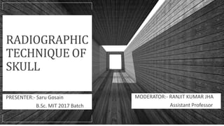
  • 1. RADIOGRAPHIC TECHNIQUE OF SKULL PRESENTER:- Saru Gosain B.Sc. MIT 2017 Batch MODERATOR:- RANJIT KUMAR JHA Assistant Professor
  • 2. Contoso Ltd. ANATOMY OF SKULL The skull consists of the 28 bones; calvaria consists of 14 bones including 3 pair of ossicles: 1. Parietal-2 2. Temporal-2 3. Malleus-2 4. Incus-2 5. Stapes-2 6. Frontal 1 7. Occipital-1 8. Sphenoid-1 9. Ethmoid-1 Add a footer 2
  • 3. Contoso Ltd. Anatomical Terminology LANDMARKS Outer canthus of the eye: the point where the upper and lower eyelids meet laterally. Infra-orbital margin/point: the inferior rim of the orbit, with the point being located at its lowest point. Nasion: the articulation between the nasal and frontal bones. Glabella: a bony prominence found on the frontal bone immediately superior to the nasion.  Vertex: the highest point of the skull in the median sagittal plane (MSP). External occipital protuberance (inion): a bony prominence found on the occipital bone, usually coincident with the median sagittal plane. External auditory meatus: the opening within the ear that leads into the external auditory canal. 3
  • 4. Contoso Ltd. • Inter-orbital (inter-pupillary) line: joins the centre of the two orbits or the centre of the two pupils when the eyes are looking straight forward. • Infra-orbital line: joints the two infra-orbital points. • Anthropological baseline: passes from the infra-orbital point to the upper border of the external auditory meatus (also known as the Frankfurter line). • Orbito-meatal base line (radiographic baseline): extends from the outer canthus of the eye to the centre of the external auditory meatus. This line is angled approximately 10 degrees to the anthropological baseline. • Reid’s baseline: line drawn from the inferior margin of the orbit (orbital point) to the centre of the orifice of the external auditory meatus. Add a footer 4 Lines
  • 5. Contoso Ltd. ○ Median sagittal plane: divides the skull into right and left halves. ○ Coronal planes: these are at right-angles to the median sagittal plane and divide the head into anterior and posterior parts. ○ Anthropological plane: a horizontal plane containing the two anthropological baselines and the infra-orbital line. It is an example of an axial plane. ○ Auricular plane: perpendicular to the anthropological plane. Passes through the centre of the two external auditory meatuses. It is an example of a coronal plane. 5 Planes
  • 6. Basic Projections o Occipito-Frontal o Lateral 6 OtherProjectionsfor cranium ○ Towne’s Projection ○ Submento-vertical ○ Sella turcica: lateral ○ Optic foramina postero-anterior oblique ○ Jugular foramina: submento-vertical 20 degrees cauded ○ Temporal bones: fronto-occipital 35 degrees cauded ○ Mastoid: lateral oblique 25 degrees cauded ○ Petrous bone: anterior oblique (Stenver’s)
  • 7. Contoso Ltd. Indications ○ Different types of fractures such as basal, blowout, contre-coup, depressed, Leforte, linear, tripod) ○ Mastoiditis ○ Metastases ○ Osteomyelitis ○ Osteopetrosis ○ Osteoporosis ○ Paget’s disease ○ Polyp ○ Sinusitis ○ Tumor ○ All metal objects are removed from the patient, e.g. hair clips and hairpins. ○ Bunches of hair often produce artefacts and thus should be untied. ○ If the area of interest includes the mouth, then false teeth containing metal and metal dental bridges should be removed. ○ The patient should be provided with a clear explanation of any movements and film positions associated with the normal operation of the skull unit. 1. Cassette size: 24 x 30 cm 2. FFD: 100cm 3. kVp: 70-85 4. mAs: 20-25 5. Focal spot: small 6. Grid: yes Patientpreparation Technicalparameters
  • 8. Contoso Ltd. Position of patient and cassette ○ Patient stands erect or lies prone (prone uncomfortable). ○ For the erect position, patient stands facing the vertical erect Bucky ○ MSP coincident with the midline of Bucky and perpendicular to it ○ Neck is flexed so that the orbito-meatal baseline is perpendicular to the Bucky. ○ Mid-part of frontal bone is positioned to the centre of the Bucky. Direction and centering of x-ray beam ○ Directed perpendicular to the Bucky along the MSP Essential image characteristics ○ All the cranial bones should be included within the image, including the skin margins ○ Ensure that the skull is not rotated. ○ For – Occipito- frontal: the petrous ridges should be completely superimposed within the orbit, with their upper borders coincident with the upper third of the orbit. ○ -OF 10°↓: the petrous ridges appear in the middle third of the orbit. ○ -OF 15°↓ (Caldwell method): the petrous ridges appear in the lower third of the orbit. ○ -OF 20 °↓: the petrous ridges appear just below the inferior orbital margin 8 Occipito-frontal
  • 9. 9 o for the demonstration of frontal bone, OF projection is done without giving any angulation o the petrous ridges should be completely superimposed within the orbit the petrous ridges appear in the middle third of the orbit. o the petrous ridges appear just below the inferior orbital margin o for the demonstration of the superior orbital fissures, the central ray is directed through the midorbits at an angle of 20-25 degrees cauded
  • 10. Contoso Ltd. Position of patient and cassette ○ Patient sits facing the erect Bucky ○ Head is rotated, such that the MSP is parallel to the Bucky and the inter- orbital line is perpendicular to it. ○ Cassette placed transversely in the erect Bucky, such that its upper border is 5 cm above the vertex of the skull. Direction and centering of the X-ray beam ○ Centre midway between the glabella and the external occipital protuberance to a point approximately 5 cm superior to the external auditory meatus. Essential image characteristics ○ The image should contain all of the cranial bones and the first cervical vertebra. Both the inner and outer skull tables should be included. ○ A true lateral will result in perfect superimposition of the lateral portions of the floors of the anterior cranial fossa and those of the posterior cranial fossa. The clinoid processes of the sella turcica should also be superimposed. 10 Lateral
  • 11. Contoso Ltd. INDICATIONS: skull fractures, neoplastic processes, Paget’s diseases, chronic suppurative otitis media. Position of patient and cassette ○ Patient lies supine, with the posterior aspect of the skull resting on a grid cassette. ○ MSP right angles to the cassette and coincident with its midline. ○ The orbito-meatal base line perpendicular to the film. Direction and centering of the X-ray beam ○ Angled caudally so it makes an angle of 30 degrees to the orbito- meatal plane. ○ Centre in the midline such that the beam passes midway between the external auditory meatuses. This is to a point ○ approximately 5 cm above the glabella. ○ The top of the cassette positioned adjacent to the vertex of the skull to ensure that the beam angulation does not project the area of interest off the bottom of the image. 11 Towne’sprojection(Fronto-occipital30° cauded)
  • 12. Contoso Ltd. Essential image characteristics o The sella turcica of the sphenoid bone is projected within the foramen magnum. o The image must include all of the occipital bone and the posterior parts of the parietal bone, and the lambdoidal suture should be visualized clearly. o The skull should not be rotated. This can also be assessed by ensuring that the sella turcica appears in the middle of the foramen magnum. 12 Add a footer NOTE: Reverse Towne’s projections (Hass method) can also be performed with occipito-frontal 30° cranial angulation on obese patient or who cannot be correctly adjusted for the Towne’s projection which will carry a lower radiation dose to sensitive structures than the equivalent antero-posterior projection.
  • 13. Contoso Ltd. Position of patient and cassette ○ Patient erect or supine. Patients placed in the supine position may have increased intracranial pressure, therefore erect position is preferred. Erect ○ The patient sits a short distance away from a vertical Bucky. The neck is hyperextended to allow the head to fall back until the vertex of the skull makes contact with the centre of the vertical Bucky. ○ Head adjusted to bring the external auditory meatuses equidistant from the cassette. ○ The MSP at right-angles to the cassette along its midline. ○ The orbito-meatal plane should be as near as possible parallel to the cassette. Direction and centering of the X-ray beam ○ Directed right-angles to the orbito-meatal plane and centred midway between the external auditory meatuses. Essential image characteristics ○ A correct projection will show the angles of the mandible clear of the petrous portions of the temporal bone. ○ The foramina of the middle cranial fossa should be seen symmetrically either side of the midline. 13 Submento-vertical
  • 14. Sella turcica: lateral INDICATIONS: To see the sella expansion by a large lesion. Position of patient and cassette • Patient sits facing the erect Bucky, head rotated, such that the MSP parallel to the Bucky and the inter-orbital line perpendicular to the Bucky. • Shoulders may be rotated slightly to allow the correct position to be attained. Patient may grip the Bucky for stability. • The head and Bucky heights are adjusted so that the centre of the Bucky is 2.5 cm vertically above a point 2.5 cm along the baseline from the external auditory meatus Direction and centering of the X-ray beam • Centred to a point 2.5 cm vertically above a point 2.5 cm along the baseline from the auditory meatus nearer the X-ray tube. 14
  • 15. Contoso Ltd. OpticForamina:postero-anterior oblique(Rhesemethod) 15 INDICATIONS: detection of optic nerve glioma Position of patient and cassette • Patient lies prone or, more commonly, erect with the nose, cheek and chin of the side being examined in contact • with the Bucky or cassette table. • The centre of the orbit of the side under examination should coincide with the centre of the Bucky or cassette table. • The MSP adjusted to make an angle of 35 degrees to the vertical (55 degrees to the table). • The orbito-meatal base line is raised 35 degrees from the horizontal. Direction and centering of the X-ray beam • The horizontal central ray centred to a point 7.5 cm above and 7.5 cm behind the uppermost external auditory meatus, so that the central ray emerges from the centre of the orbit in contact with the table.
  • 16. Jugular foramina: submento- vertical 20° cauded INDICATIONS: detection of glomus jugulare tumour Position of patient and cassette Patient erect or supine. If the patient is unsteady, then a supine technique is advisable. Supine • Patient’s shoulders are raised and the neck is hyperextended to bring the vertex of the skull in contact with the grid cassette or table. • Head adjusted to bring the external auditory meatuses equidistant from the cassette. • The MSP at right-angles to the cassette along its midline. • The orbito-meatal plane should be as near as possible parallel to the cassette. Direction and centering of the X-ray beam Angled caudally so that it makes an angle of 70 degrees to the orbitomeatal plane and centred in the midline to pass midway between the external auditory meatuses.
  • 17. Contoso Ltd. Temporal bone: fronto-occipital35° cauded Patient lies supine or erect • Patient may be supine or erect with their back to an erect Bucky. Head adjusted to bring the external auditory meatuses equidistant from the table, so that the MSP is at right-angles to, and in the midline of, the table. • Chin is depressed so that the orbito-meatal line is at right-angles to the table. • A cassette is placed transversely and is centred to coincide with the angled central ray. Direction and centering of the X-ray beam • A caudal angulation is employed, such that it makes an angle of 35 degrees to the orbito-meatal plane. • The beam is centred midway between the external auditory meatuses. Essential image characteristics • The sella turcica of the sphenoid bone should be projected within the foramen magnum and include mastoid and petrous part of the temporal bone supra-inferiorly. • The skull should not be rotated. This can also be assessed by ensuring that the sella turcica appears in the middle of the foramen magnum.
  • 18. Mastoid- lateral oblique 25° cauded Position of patient and cassette • Patient sits facing the erect Bucky. • Head is rotated, such that the MSP is parallel to the Bucky and the inter- orbital line is perpendicular to the Bucky. • The auricle of the ear adjacent to the table is folded forward to ensure that its soft-tissue outline is not superimposed over the region of interest. • Cassette positioned longitudinally in the Bucky and is centred to coincide with the central ray and mastoid process. Direction and centering of the X-ray beam • A 25-degree caudal angulation is employed and centred 5 cm above and 2.5 cm behind the external auditory meatus remote from the cassette. Essential image characteristics • Ensure that all of the mastoid air cells have been included within the image. 18 Add a footer
  • 19. Mastoid-profile Position of patient and cassette • Patient lies supine on the table, with the orbito-meatal baseline perpendicular to the table top. • From a position with the MSP perpendicular to the table, the head is rotated through an angle of 35 degrees away from the side under examination, such that the MSP now makes an angle of 55 degrees to the table. • The vertical tangent to the skull should now be at the level of the middle of the mastoid process under examination, so that the mastoid process is in profile. • Finally, the head is moved transversely across the table so that the mastoid process being examined is in the midline of the table. Direction and centering of the X-ray beam • Angled caudally so that it makes an angle of 25 degrees to the orbito-meatal plane and is centred to the middle of the mastoid process on the side under examination. 19
  • 20. Petrous bone: anterior oblique (Stenver’s projection) Position of patient and cassette • Patient may be prone or erect facing a vertical Bucky. • The middle of the supra-orbital margin on the side being examined is centred to the middle of the Bucky. • Neck flexed so that the nose and forehead are in contact with the table and the orbito-meatal line is perpendicular to the table. • From a position where the MSP is perpendicular to the table, the head is rotated toward the side under examination, such that the MSP is now at an angle of 45 degrees to the table. This brings the petrous part of the temporal bone parallel to the cassette. • The neck is extended so that the orbito-meatal line is raised five degrees from horizontal. • Cassette placed transversely in the Bucky and is centred at a level to coincide with the central ray. 20
  • 21. Contoso Ltd. Direction and centering of the X-ray beam • A 12-degree cephalad beam angulation is employed, i.e. at an angle of seven degrees to the orbito-meatal plane, to separate the occiput from the petrous bone. • Centre midway between the external occipital protuberance and the external auditory meatus furthest from the cassette. 21
  • 22. Styloid process: PA axial ( Cahoon method) o The styloid process is a needle like projection of varying lengths averaging 2-3 cm which projects from the inferior part of the petrous temporal bone. 22 Add a footer Position of patient and cassette • Patient in the seated-upright or prone position. • Adjust the forehead and nose so that the MSP is perpendicular to the midline o f the Bucky. • Center the IR to the nasion. • Adjust the flexion of the patient's neck so that the OML is perpendicular to the plane of the IR. Direction and centering of x-ray beam • Directed to the nasion at an angle of 25 degrees cephalad. Essential image characteristics • Temporal styloid processes free of superimposition from frontal bone, occipital bone, and coronoid processes of the mandible. • Styloid processes projected within or just above the maxillary sinuses.
  • 23. Facialbones and sinuses Facial skeleton consists of 14 bones. 1. Maxilla-2 2. Zygomatic-2 3. Nasal-2 4. Lacrimal-2 5. Palatine-2 6. Inferior nasal concha-2 7. Mandible-1 8. Vomer -1 Within these bones, there are series of air-filled cavities known as the paranasal sinuses. There are 4 paranasal sinuses; 1. Maxillary sinuses 2. Frontal sinuses 3. Sphenoid sinuses 4. Ethmoid sinuses 23 Add a footer
  • 24. Contoso Ltd. 1. Occipito-mental 2. Occipito-mental 30° cauded 3. Lateral 4. Zygomatic arches: infero-superior 5. Orbits: occipito-mental (modified) 6. Nasal bones: lateral 7. Mandible: lateral 30° cephalad : postero-anterior : postero-anterior oblique 8. Temporal-mandibular joints: lateral 25° cauded 24 Add a footer Projectionsfor paranasalsinuses 1. Occipito-mental with open mouth (Water’s view) 2. Occipito-frontal 15° cauded 3. Lateral Projectionsfor facial bones
  • 25. Contoso Ltd. Occipito-mental for facial bones 25 Position of patient and cassette • Patient seated facing the vertical erect Bucky. • Patient’s nose and chin are placed in contact with the midline of the cassette holder. The head is then adjusted so that the orbito- meatal baseline makes 45-degree angle to the cassette holder. • The horizontal central line of the Bucky/cassette holder should be at the level of the lower orbital margins. • Ensure that the MSP is at right-angles to the Bucky/cassette holder by checking the outer canthi of the eyes and that the external auditory meatuses are equidistant. Direction and centering of the X-ray beam • Perpendicular and centred to the middle of the cassette holder. • To check that the beam is centred properly, the cross-lines on the Bucky or cassette holder should coincide with the patient’s anterior nasal spine.
  • 26. Essential image characteristics • The petrous ridges must appear below the floors of the maxillary sinuses. • There should be no rotation. This can be checked by ensuring that the distance from the lateral orbital wall to the outer skull margins is equidistant on both sides. 26 Add a footer
  • 27. Contoso Ltd. Position of patient and cassette ○ Patient seated facing the vertical Bucky. ○ The patient’s nose and chin are placed in contact with the midline of the cassette holder. The head then is adjusted to bring the orbito-meatal baseline to a 45-degree angle to the cassette holder. ○ The horizontal central line of the Bucky or cassette holder should be at the level of the symphysis menti. ○ Ensure that the MSP is at right-angles to the Bucky by checking that the outer canthi of the eyes and the external auditory meatuses are equidistant. Direction and centering of the X-ray beam ○ The tube is angled 30 degrees caudally and centred along the midline, such that the central ray exits at the level of the lower orbital margins. ○ To check that the beam is centred properly, the cross-lines on the Bucky or cassette holder should coincide approximately with the upper symphysis menti region. 27 Occipito-mental 30° cauded
  • 28. Contoso Ltd. Essential image characteristics o The floors of the orbit will be clearly visible through the maxillary sinuses, and the lower orbital margin should be demonstrated clearly. o There should be no rotation. This can be checked by ensuring that the distance from the lateral orbital wall to the outer skull margins is equidistant on both sides. 28 Add a footer
  • 29. Contoso Ltd. Position of patient and cassette: In cases of injury, this projection should be taken using a horizontal beam in order to demonstrate any fluid levels in the paranasal sinuses. Erect ○ Patient sits facing the vertical Bucky. The head is rotated, such that the side under examination is in contact with the Bucky or cassette holder. ○ The Bucky height is altered, such that its centre is 2.5 cm inferior to the outer canthus of the eye. ○ The MSP is brought parallel to the cassette by ensuring that the inter-orbital line is at right angles to the cassette and the nasion and external occipital protuberance are equidistant from it. Direction and centering of the X-ray beam ○ Centre the horizontal central ray to a point 2.5 cm inferior to the outer canthus of the eye. Essential image characteristics ○ The image should contain all of the facial bones sinuses, including the frontal sinus and posteriorly to the anterior border of the cervical spine. ○ A true lateral will have been obtained if the lateral portions of the floor of the anterior cranial fossa are superimposed. 29 Lateral for facial bones
  • 30. Contoso Ltd. Zygomatic arches: infero-superior (jug- handle projection) Position of patient and cassette INDICATION: Fracture of the zygomatic bones. This projection is essentially a modified submento-vertical (SMV) projection. This method is tangential projection. o Patient lies supine, with one or two pillows under the shoulders to allow the neck to be extended fully. o Cassette is placed against the vertex of the skull, such that its long axis is parallel with the axial plane of the body. It should be supported in this position with foam pads and sandbags. o The flexion of the neck is now adjusted to bring the long axis of the zygomatic arch parallel to the cassette. o The head in now tilted five to ten degrees away from the side under examination. This allows the zygomatic arch under examination to be projected on to the film without superimposition of the skull vault or facial bones. 30
  • 31. Contoso Ltd. Direction and centering of the X-ray beam ○ Perpendicular to the cassette and long axis of the zygomatic arch. ○ A centering point should be located such that the central ray passes through the space between the midpoint of the zygomatic arch and the lateral border of the facial bones. Essential image characteristics ○ The whole length of the zygomatic arch should be demonstrated clear of the skull. If this has not been achieved, then it may be necessary to repeat the examination and alter the degree of head tilt to try and bring the zygomatic arch clear of the skull. 31 Add a footer
  • 32. Contoso Ltd. Position of patient and cassette ○ Patient seated facing the vertical Bucky or cassette holder. ○ The patient’s nose and chin are placed in contact with the midline of the cassette holder. The head is then adjusted to bring the orbito-meatal baseline to a 35-degree angle to the cassette holder. ○ The horizontal central line of the vertical Bucky or cassette holder should be at the level of the midpoint of the orbits. ○ Ensure that the MSP is at right-angles to the Bucky or cassette holder by checking that the outer canthi of the eyes and the external auditory meatuses are equidistant. Direction and centering of the X-ray beam ○ Perpendicular to the cassette holder or Bucky and by design will be centred to the middle of the image receptor. If this is the case and the above positioning is performed accurately, then the beam will already be centred. ○ To check that the beam is centred properly, the cross-lines on the Bucky or cassette holder should coincide with the midline at the level of the mid-orbital region. 32 Orbits: occipito-mental(modified)
  • 33. Contoso Ltd. Essential image characteristics o The orbits should be roughly circular in appearance (they will be more oval in the occipito-mental projection). o The petrous ridges should appear in the lower third of the maxillary sinuses. o There should be no rotation. This can be checked by ensuring that the distance from the lateral orbital wall to the outer skull margins is equidistant on both sides. 33 Add a footer
  • 34. Contoso Ltd. Nasal bones: lateral 34 Position of patient and cassette ○ Patient sits facing the cassette stand or a vertical Bucky. ○ The head is turned so that MSP is parallel with the cassette and the inter-pupillary line is perpendicular to the cassette. ○ The nose should be roughly coincident with the centre of the cassette. Direction and centering of the X-ray beam ○ A horizontal central ray is directed through the centre of the nasal bones and collimated to include the nose.
  • 35. Contoso Ltd. Position of patient and cassette ○ Patient lies in the supine position. The trunk is rotated slightly and then supported with pads to allow the side of the face being examined to come into contact with the cassette, which will be lying on the tabletop. ○ The MSP should be parallel with the cassette and the inter-pupillary line perpendicular. ○ The neck may be flexed slightly to clear the mandible from the spine. ○ The long axis of the cassette should be parallel with the long axis of the mandible and the lower border positioned 2 cm below the lower border of the mandible. 35 Mandible: lateral 30° cephalad
  • 36. Contoso Ltd. Direction and centering of the X-ray beam ○ The central ray is angled 30 degrees cranially at an angle of 60 degrees to the cassette and is centred 5 cm inferior to the angle of the mandible remote from the cassette. ○ Collimate to include the whole of the mandible and temporo- mandibular joint (TMJ) (include the external auditory meatus (EAM) at the edge of the collimation field). Essential image characteristics ○ The body and ramus of each side of the mandible should not be superimposed. ○ The image should include the whole of the mandible, from the TMJ to the symphysis menti. 36 Add a footer
  • 37. Contoso Ltd. Position of patient and cassette ○ Patient sits facing the vertical Bucky. ○ MSP should be coincident with the midline of the Bucky or cassette holder. The head is then adjusted to bring the orbito-meatal baseline perpendicular to the Bucky or cassette holder. ○ The MSP should be perpendicular to the cassette. Check that the external auditory meatuses are equidistant from the cassette. ○ The cassette should be positioned such that the middle of the cassette, when placed longitudinally in the Bucky or cassette holder, is centred at the level of the angles of the mandible. Direction and centering of the X-ray beam ○ Directed perpendicular to the cassette and centred in the midline at the levels of the angles of the mandible. Essential image characteristics ○ The whole of the mandible from the lower portions of the TMJs to the symphysis menti should be included in the image. ○ There should be no rotation evident. 37 Mandible: postero-anterior
  • 38. Contoso Ltd. Position of patient and cassette ○ Patient sits facing the vertical erect Bucky. ○ MSP should be coincident with the midline of the Bucky or cassette holder. The head is then adjusted to bring the orbito-meatal baseline perpendicular to the Bucky or cassette holder. ○ From a position with the MSP perpendicular to the cassette, the head is rotated 20 degrees to either side, so that the cervical vertebra will be projected clear of the symphysis menti. ○ The head is now repositioned so the region of the symphysis menti is coincident with the middle of the cassette. ○ The cassette should be positioned such that the middle of an 18 24-cm cassette, when placed longitudinally in the Bucky or cassette holder, is centred at the level of the angles of the mandible. 38 Mandible: postero-anterioroblique
  • 39. Contoso Ltd. 39 Add a footer Direction and centering of the X-ray beam ○ Directed perpendicular to the cassette and centred 5 cm from the midline, away from the side being examined, at the level of the angles of the mandible. Essential image characteristics ○ The symphysis menti should demonstrated without any superimposition of the cervical vertebra.
  • 40. Contoso Ltd. Position of patient and cassette ○ Patient sits facing the vertical Bucky or lies prone on the Bucky table. The head is rotated to bring the side of the head under examination in contact with the table. ○ The head and Bucky or cassette holder level is adjusted so the centre cross-lines are positioned to coincide with a point 1 cm along the orbito-meatal baseline anterior to the external auditory meatus. ○ The MSP is brought parallel to the cassette by ensuring that the inter- pupillary line is at right-angles to the table top and the nasion and external occipital protuberance are equidistant from it. Direction and centering of the X-ray beam ○ Angled 25 degrees caudally and will be centred to a point 5 cm superior to the joint remote from the cassette so the central ray passes through the joint nearer the cassette. Essential image characteristics ○ Condyle lying in mandibular fossa in the closed-mouth examination ○ Condyle lying inferior to articular tubercle in the open-mouth examination if the patient is normal and able to open the mouth wide 40 Temporal-mandibularjoints:lateral 25° cauded(Schuller method)
  • 41. Contoso Ltd. 41 Position of patient and cassette ○ Patient seated facing the vertical Bucky. ○ The patient’s nose and chin are placed in contact with the midline of the cassette holder. The head is then adjusted to bring the orbito-meatal baseline to a 45-degree angle to the cassette holder. ○ The horizontal central line of the Bucky or cassette holder should be at the level of the lower orbital margins. ○ Ensure that the MSP is at right-angles to the Bucky or cassette holder by checking that the outer canthi of the eyes and the external auditory meatuses are equidistant. ○ The patient should open the mouth as wide as possible before exposure. This will allow the posterior part of the sphenoid sinuses to be projected through the mouth. Paranasal sinuses occipito- mental (Water’s view)
  • 42. Contoso Ltd. Direction and centering of the X-ray beam ○ Perpendicular to the cassette holder or Bucky and by design will be centred to the middle of the image receptor. If this is the case and the above positioning is performed accurately, then the beam will already be centred. ○ To check the beam is centred properly, the cross-lines on the Bucky or cassette holder should coincide with the patient’s anterior nasal spine. ○ Collimate to include all of the sinuses. Essential image characteristics ○ The petrous ridges must appear below the floors of the maxillary sinuses. ○ There should be no rotation. This can be checked by ensuring that the distance from the lateral orbital wall to the outer skull margins is equidistant on both sides. NOTES: This projection is designed to project the petrous part of the temporal bone below the floor of the maxillary sinuses so that fluid levels and pathological changes in the lower part of the sinuses can be visualized clearly. -To distinguish a fluid level from mucosal thickening, an additional projections may be undertaken with the head tilted, such that a transverse plane makes an angle of about 20 degrees to the floor. 42 Add a footer
  • 43. Contoso Ltd. This projection is used to demonstrate the frontal and ethmoid sinuses Position of patient and cassette ○ Patient seated facing the vertical Bucky or skull unit cassette holder so the MSP is coincident with the midline of the Bucky and is also perpendicular to it. ○ The head is positioned so that the orbito-meatal baseline is raised 15 degrees to the horizontal. ○ Ensure that the nasion is positioned in the centre of the Bucky. Direction and centering of the X-ray beam ○ Directed perpendicular to the vertical Bucky along the MSP so the beam exits at the nasion. ○ A collimation field or extension cone should be set to include the ethmoidal and frontal sinuses. 43 Occipito-frontal15°cauded (Caldwell Method)
  • 44. Essential image characteristics o All the relevant sinuses should be included within the image. o The petrous ridges should be projected just above the lower orbital margin. o It is important to ensure that the skull is not rotated. This can be assessed by measuring the distance from a point in the midline of the skull to the lateral orbital margins. If this is the same on both sides of the skull, then it is not rotated. Add a footer 44
  • 45. Contoso Ltd. Position of patient and cassette ○ Patient sits facing the vertical Bucky or skull unit cassette holder. The head is then rotated, such that the MSP is parallel to the Bucky and the inter- orbital line is perpendicular to the Bucky. ○ The head and Bucky heights are adjusted so that the centre of the Bucky is 2.5 cm along the orbito-meatal line from the outer canthus of the eye. ○ A cassette is positioned longitudinally in the erect Bucky, such that its lower border is 2.5 cm below the level of the upper teeth. Direction and centering of the X-ray beam ○ A horizontal central ray should be employed to demonstrate fluid levels. ○ The tube should have been centred previously to the Bucky, such that the central ray will now be centred to a point 2.5cm posterior to the outer canthus of the eye. Essential image characteristics ○ A true lateral will have been achieved if the lateral portions of the floors of the anterior cranial fossa are superimposed. 45 Add a footer Paranasal sinus: lateral