SlideShare a Scribd company logo
1 of 22
Chap. 6 Genes, Genomics, and
Chromosomes (Part B)
Topics
• Genomics: Genome-wide Analysis of Gene Structure and
Expression
• Structural Organization of Eukaryotic Chromosomes
• Morphology and Functional Elements of Eukaryotic Chromosomes
Goals
• Learn about computer-based
methods for analyzing
sequence data.
• Learn how DNA and proteins
are packaged in chromatin.
• Learn the large-scale
structure organization of
chromosomes.
• Learn the functional elements
required for chromosome
replication and segregation. RxFISH-painted human chromosomes.
Mining Sequence Data: BLAST Searches
An enormous amount of DNA sequence information is available
from genome sequencing and sequencing of cloned genes. This
data is stored in data banks such as GenBank at the NIH in
Bethesda, MD and the EMBL Sequence Data Base at the
European Molecular Biology Laboratory in Heidelberg, Germany.
Scientists working in the area of bioinformatics use this data to
find genes, analyze their properties, and determine phylogenetic
relationships between organisms and proteins. A common
procedure in which this data is used is the BLAST search (basic
local alignment search tool) which is used to compare protein and
DNA sequences. An example BLAST search alignment is shown
for the human neurofibromatosis 1 (NF1) gene in Fig. 6.25. The
alignment shows NF1 is related to the S. cerevisiae Ira
GTPase-activating protein (GAP) and suggests the disease is
caused by aberrant signal transduction.
Computer programs similar
to BLAST are used to
identify protein sequence
motifs (e.g., zinc fingers)
in unknown proteins. The
identification of structure
regions with known function
sheds light on overall
protein function and helps
guide experimental analysis
of unknown proteins and
genes.
BLAST search analysis can identify the members of a protein
family originating from gene duplication and speciation mutations.
As illustrated for the a- and ß-tubulin protein family in Fig.
6.26, an early gene duplication event created the paralogous a-
and ß-tubulin genes. Later speciation mutations lead to evolution
of the orthologous members of the a- and ß-tubulin subfamilies.
Orthologous proteins are most likely to share the same function.
Sequence Comparisons Establish
Evolutionary Relationships Among Proteins
x
Genome Size vs Complexity
Genome sequencing has revealed that the morphological complexity
of an organism is not strongly correlated with the size of its
genome (Fig. 6.27). Alternative splicing of RNAs and post-
translational modification of proteins are thought to greatly
increase the complexity of the proteins encoded by the genomes
of higher organisms. In addition, the relative number of cells
formed in a tissue such as the cerebral cortex can be important in
increasing complexity (e.g., mice vs humans). Genes can be
identified within the sequenced genomes of simple organisms such
as yeast and bacteria by searching for open reading frames
(ORFS). ORFs are long stretches of triplet codons lacking stop
codons. Gene annotation (assignment of likely function) is based on
knowledge from biochemical studies and/or alignments with known
sequences. In complex organisms
such as humans whose genes
typically contain introns, more
sophisticated algorithms that ID
intron splice sites and compare
cDNA and other sequence
information to genomic DNA
sequences must be applied to
locate and annotate genes. Using
such methods ~25,000 genes
have been identified in humans.
However, conclusive evidence for
synthesis of protein or RNA
products is lacking for ~10,000
genes.
Extended and Condensed Chromatin
Human diploid cells contain about 2 meters of DNA. To fit within
nuclei, DNA must be condensed by ~105-fold. DNA exists in cells
as a nucleoprotein complex known as chromatin. During interphase
when cells are not dividing, chromatin is relatively uncondensed
compared to its state in metaphase chromosomes. When released
from nuclei with low salt buffer, chromatin displays an extended
"beads-on-a-string" morphology, where each bead is a nucleosome
(Fig. 6.28). When released in physiological salt concentrations,
more condensed fibers of 30 nm diameter are observed. In
general, extended chromatin can be transcribed, whereas
condensed forms cannot.
Structure of Nucleosomes
Nucleosomes consist of 147 bp of DNA wrapped in almost two turns
around the outside of an octamer of histone proteins (Fig. 6.29).
In most nucleosomes, the octamer has a stoichiometry of
H2A2H2B2H32H42. Histones are the most abundant DNA-binding
proteins in eukaryotic cells. The sequences of the 4 histones that
make up the octamer are highly conserved across all organisms,
indicating their functions were optimized early in evolution. Histones
have a large number of basic amino acids and bind to DNA mostly
by salt-bridge interactions to phosphates in the DNA backbone.
Another histone, H1, binds to the linker DNA between
nucleosomes. Linker DNA is 10-90 bp in length depending upon the
organism.
Structure of 30-nm Chromatin Fibers
In 30-nm fibers, nucleosomes
bind to one another in a
double helical arrangement
(Fig. 6.30). Histone H1
molecules bind to linker DNA
between nucleosomes and help
stabilize the 30-nm fiber. The
stability of 30-nm fibers is
modulated by post-
translational modification of
the tails of histones in the
octamers (H4 in particular).
Histone Tails and Chromatin Condensation
The N- and C-terminal tails of histones project out from the
nucleosome core (Fig. 6.31a). They also contain numerous residues
that can be modified by acetylation, methylation, etc. (Fig.
6.31b). Acetylation of lysine side-chains by histone acetylases
(HATs) neutralizes positive charge and promotes decondensation of
30-nm fibers. Methylation, on the other hand, blocks lysine
acetylation, maintains positive charge, and promotes 30-nm fiber
condensation. Studies have shown that chromatin condensation is
not controlled simply by the net acetylation state of histones.
Rather, the sites where acetylation and other modifications occur
also are important. The combinations of modifications that specify
condensation/decondensation are referred to as the "histone code".
Interphase Chromatin
Interphase chromatin exists in two
different condensation states (Fig.
6.33a). Heterochromatin is a
condensed form that has a
condensation state similar to
chromatin found in metaphase
chromosomes. Euchromatin is
considerably less condensed.
Heterochromatin typically is found
at centromere and telomere
regions, which remain relatively
condensed during interphase. The
inactivated copy of the X-
chromosome (Barr body) that
occurs in cells in females also
occurs as heterochromatin. In
contrast, most transcribed genes
are located in regions of
euchromatin. Common modifications
occurring in histone H3 in hetero-
and euchromatin are illustrated in
Fig. 6.33b.
Formation of
Heterochromatin
The trimethylation of histone H3 at
lysine 9 (H3K9Me3) plays an
important role in promoting chromatin
condensation to heterochromatin (Fig.
6.34a). Trimethylated sites are
bound by heterochromatin protein 1
(HP1) which self-associates and
oligomerizes resulting in
heterochromatin. Heterochromatin
condensation is thought to spread
laterally between “boundary
elements” that mark the ends of
transcriptionally active euchromatin
(Fig. 6.34b). Recruitment of the
H3K9 histone methyl transferase
(HMT) to HP1 sites promotes
heterochromatin spreading by
catalyzing H3 methylation.
Structure of Interphase Chromosomes
FISH analysis performed with fluorescent probes that bind to
sequential sequence sites along DNA supports a looped structure
for interphase chromosomes (Fig. 6.35). Loops range in size
from 1 to 4 million base pairs in mammalian interphase cells.
The bases of the loops are located near the center of the
chromosome at scaffold-associated regions (SARs), and matrix-
attachment regions (MARs). The DNA fibers at the base of the
loops are held together by structural maintenance of
chromosome (SMC) proteins (Fig. 6.36c) and other non-histone
proteins. Transcription units containing expressed genes are
located in uncondensed loop regions, away from the more
condensed center of the chromosome.
Interphase Chromosome Territories
In situ hybridization of interphase nuclei with chromosome-
specific fluorescently-labeled probes indicates that
chromosomes reside within restricted regions of the nucleus
rather than appearing throughout the nucleus (Fig. 6.37).
Interestingly, the precise positions of chromosomes are not
reproducible between cells.
Structure of Metaphase Chromosomes
In metaphase chromosomes,
the number of loops of
chromatin is increased and the
lengths of the loops are
decreased compared to what
occurs in interphase
chromosomes. In addition,
more folded structures called
chromonema fibers and higher
order structures occur in
prophase and metaphase
chromatids (Fig. 6.38).
Microscopic Structure of Metaphase
Chromosomes
Because interphase chromosomes
are not easily visualized by
microscopy techniques, chromosome
morphology has been studied mostly
using metaphase chromosomes.
Metaphase chromosomes are
duplicated structures formed after
DNA replication is complete. They
contain two sister chromatids joined
at a structure called the
centromere (Fig. 6.39). The ends
of chromatids are called telomeres.
Centromeres are required for
chromatid separation late in mitosis.
Telomeres are important in
preventing chromosome shortening
during replication. The number,
sizes, and shapes of metaphase
chromosomes constitute the
karyotype, which is distinctive for
each species.
Chromosome Banding Patterns
A number of dyes, such as Giemsa reagent, selectively stain
different regions of chromosomes forming distinctive bands. For
Giemsa reagent, banding is affected by G + C content. Banding
patterns are very important in chromosome ID and in looking for
chromosomal abnormalities and mapping the locations of genes.
The most detailed staining is achieved via multicolor FISH
chromosome painting. In this technique, staining is performed
using a mixture of DNA probes coupled to several fluorescent
dyes (See Slide 1). In Fig. 6.40 below, FISH staining patterns
have been converted to false-color images to visualize
chromosomes. Standard terminology is used for naming band and
gene locations in chromosomes. The short arm is designated "p",
and the long arm "q". Arms are further divided into major
sections and subsections that are numbered consecutively out
from the centromere.
Detection of Translocations
The analysis of chromosome banding patterns is used to detect
anomalies such as truncations and translocations associated with
certain genetic disorders and cancers. In chronic myelogenous
leukemia, leukemic cells contain a shortened chromosome 22 and
a longer chromosome 9 resulting from a translocation event in the
q arms of these two chromosomes (Fig. 6.41). The shortened
chromosome is distinctive and is referred to as the "Philadelphia
chromosome". Multicolor FISH staining (right) is useful in
identification of such chromosomes in a chromosome spread.
Evolution of Human Chromosomes
Through the determination of
locations of common chromosomal
segments in modern primate
chromosomes, investigators have
calculated the most likely
karyotype of the common
ancestor of all primates (Fig.
6.42c). In addition, they have
proposed a model for how the
human karyotype evolved from
that ancestor. Major events in
the evolution of the human
karyotype include 1) formation of
chromosome 2 by fusion of
ancestral chromosomes 9 and 11,
2) formation of chromosomes 14
and 15 by breakage of ancestral
chromosome 5, and 3) formation
of chromosomes 12 and 22 by
translocations between ancestral
chromosomes 14 and 21. In other
cases (e.g., chromosome 1), no
significant rearrangements have
occurred over time.
ID of Functional Chromosomal Elements (I)
Studies with yeast have demonstrated that all chromosomes must
contain 3 functional elements to replicate and segregate correctly:
1) replication origins, 2) a centromere, and 3) telomeres. Yeast
replication origins were identified in plasmid cloning studies. Only
yeast plasmids containing a copy of a sequence referred to as the
autonomously replicating sequence (ARS) could be transfected into
yeast cells (Fig. 6.44a). The haploid S. cerevisiae genome contains
many ARSs distributed among its 16 chromosomes.
ID of Functional Chromosomal Elements (II)
While only ARSs are needed for plasmid replication, an additional
sequence identified by cloning procedures was found to be required
for efficient segregation of plasmids to yeast daughter cells (Fig.
6.44b). This DNA proved to contain chromosomal centromere
sequences (CEN sequences). Yeast CEN sequences are relatively
simple (Fig. 6.45, not covered). In humans, they consist of 2-4 x
106 bp of simple sequence DNA composed of a 171 bp repeat unit.
The human centromere sequence is bound by specialized nucleosomes
containing a centromere-specific histone H3 variant (CENP-A). A
large complex of non-histone proteins (the kinetochore) binds to
centromeres and attaches them to microtubules of the mitotic
spindle apparatus.
ID of Functional Chromosomal Elements (III)
Yeast transfection studies also showed that linearized plasmids
containing ARS and CEN sequences could be maintained in cells
only if telomere (TEL) sequences were attached at their ends
(Fig. 6.44c). The function of TEL sequences in replication of
chromosome ends is illustrated in the next two slides.
Function of Telomeres
A special mechanism is needed to
complete the replication of DNA
in DNA strands that have their
3’ ends located at the ends of
chromosomes. DNA polymerases
cannot complete synthesis of this
region of DNA, and without
synthesis, chromosomes become
shortened with each round of
replication (Fig. 6.46).
Shortening results in the loss of
binding sites for proteins that
protect the ends of linear
chromosomes from attack by
exonucleases. As illustrations of
the importance of telomere
replication, knockout mice lacking
the enzyme that synthesizes DNA
at telomeres, telomerase, cannot
produce viable offspring after six
generations. In addition,
telomerase often is switched on
in cancer cells.
Mechanism of Action of Telomerase
Telomere sequences typically consist of
tandemly repeating sequence units with
a high G content in the strand that has
its 3' end at the end of the
chromosome. In humans and other
vertebrates, the repeating sequence is
TTAGGG. This sequence unit repeats
over a few thousand base pairs in
humans. The mechanism of replication of
this DNA is illustrated in Fig. 6.47 for
a protozoan species. Replication is
carried out by the enzyme known as
telomerase. Telomerase is a reverse
transcriptase that carries its own
internal RNA template which binds to
the ssDNA at the chromosome 3’ end
and allows this strand to be elongated.
Ultimately, DNA Pol a/primase can
synthesize a primer on this strand,
which is elongated by DNA Pol . Some
organisms rely on a different mechanism
for replication of telomeric DNA. For
example, flies lack telomerase and
maintain telomere length by regulated
insertion of non-LTR retrotransposons
into telomere DNA.

More Related Content

Similar to 13-miller-chap-6b-lecture.ppt

Organization of dna into chromosome2
Organization of dna into chromosome2Organization of dna into chromosome2
Organization of dna into chromosome2Bruno Mmassy
 
Karyotype analysis and evolution by Mannat
Karyotype analysis and evolution by MannatKaryotype analysis and evolution by Mannat
Karyotype analysis and evolution by MannatMannatAulakh
 
Karyotyping
KaryotypingKaryotyping
Karyotypingwildask
 
Introduction to genetics
Introduction to geneticsIntroduction to genetics
Introduction to geneticsMayur Gaikar
 
Introduction to genetics and genes unlocking the secrets of heredity by noor ...
Introduction to genetics and genes unlocking the secrets of heredity by noor ...Introduction to genetics and genes unlocking the secrets of heredity by noor ...
Introduction to genetics and genes unlocking the secrets of heredity by noor ...Noor Zada
 
940772037Eukaryotic chromosome organization_compressed.pdf
940772037Eukaryotic chromosome organization_compressed.pdf940772037Eukaryotic chromosome organization_compressed.pdf
940772037Eukaryotic chromosome organization_compressed.pdfZyrahAngelineDiale
 
cytogenomics tools and techniques and chromosome sorting.pptx
cytogenomics tools and techniques and chromosome sorting.pptxcytogenomics tools and techniques and chromosome sorting.pptx
cytogenomics tools and techniques and chromosome sorting.pptxPABOLU TEJASREE
 
Chromosomes structure and function, Dr.Kamelsh shah, PSSHDA, KADI
Chromosomes structure and function, Dr.Kamelsh shah, PSSHDA, KADI Chromosomes structure and function, Dr.Kamelsh shah, PSSHDA, KADI
Chromosomes structure and function, Dr.Kamelsh shah, PSSHDA, KADI Dr.Kamlesh shah
 
Internal organization of nucleus and nucleolus
Internal organization of nucleus and nucleolusInternal organization of nucleus and nucleolus
Internal organization of nucleus and nucleolusHimanshu Upadhyay
 
Packaging of DNA (DNA is tightly packaged into chromosomes in the nucleus of ...
Packaging of DNA (DNA is tightly packaged into chromosomes in the nucleus of ...Packaging of DNA (DNA is tightly packaged into chromosomes in the nucleus of ...
Packaging of DNA (DNA is tightly packaged into chromosomes in the nucleus of ...Shadhin8
 
5. Architecture of chromosome.pdf
5. Architecture of chromosome.pdf5. Architecture of chromosome.pdf
5. Architecture of chromosome.pdfDr.Pratibha Bisen
 

Similar to 13-miller-chap-6b-lecture.ppt (20)

chromosome and genetics
chromosome and genetics chromosome and genetics
chromosome and genetics
 
Presentation on Functions of Chromosomes
Presentation on Functions of ChromosomesPresentation on Functions of Chromosomes
Presentation on Functions of Chromosomes
 
Organization of dna into chromosome2
Organization of dna into chromosome2Organization of dna into chromosome2
Organization of dna into chromosome2
 
Karyotype analysis and evolution by Mannat
Karyotype analysis and evolution by MannatKaryotype analysis and evolution by Mannat
Karyotype analysis and evolution by Mannat
 
Chromosome
ChromosomeChromosome
Chromosome
 
Karyotyping
KaryotypingKaryotyping
Karyotyping
 
Chromosome2.ppt
Chromosome2.pptChromosome2.ppt
Chromosome2.ppt
 
Introduction to genetics
Introduction to geneticsIntroduction to genetics
Introduction to genetics
 
Introduction to genetics and genes unlocking the secrets of heredity by noor ...
Introduction to genetics and genes unlocking the secrets of heredity by noor ...Introduction to genetics and genes unlocking the secrets of heredity by noor ...
Introduction to genetics and genes unlocking the secrets of heredity by noor ...
 
Genomics
GenomicsGenomics
Genomics
 
940772037Eukaryotic chromosome organization_compressed.pdf
940772037Eukaryotic chromosome organization_compressed.pdf940772037Eukaryotic chromosome organization_compressed.pdf
940772037Eukaryotic chromosome organization_compressed.pdf
 
cytogenomics tools and techniques and chromosome sorting.pptx
cytogenomics tools and techniques and chromosome sorting.pptxcytogenomics tools and techniques and chromosome sorting.pptx
cytogenomics tools and techniques and chromosome sorting.pptx
 
Molecular organization
 Molecular organization Molecular organization
Molecular organization
 
Chromosomes structure and function, Dr.Kamelsh shah, PSSHDA, KADI
Chromosomes structure and function, Dr.Kamelsh shah, PSSHDA, KADI Chromosomes structure and function, Dr.Kamelsh shah, PSSHDA, KADI
Chromosomes structure and function, Dr.Kamelsh shah, PSSHDA, KADI
 
Internal organization of nucleus and nucleolus
Internal organization of nucleus and nucleolusInternal organization of nucleus and nucleolus
Internal organization of nucleus and nucleolus
 
Presentation on Structure of Chromosomes
Presentation on Structure of ChromosomesPresentation on Structure of Chromosomes
Presentation on Structure of Chromosomes
 
Packaging of DNA (DNA is tightly packaged into chromosomes in the nucleus of ...
Packaging of DNA (DNA is tightly packaged into chromosomes in the nucleus of ...Packaging of DNA (DNA is tightly packaged into chromosomes in the nucleus of ...
Packaging of DNA (DNA is tightly packaged into chromosomes in the nucleus of ...
 
Brief stc of chromosome
Brief stc of chromosomeBrief stc of chromosome
Brief stc of chromosome
 
Chromosome structure
Chromosome structureChromosome structure
Chromosome structure
 
5. Architecture of chromosome.pdf
5. Architecture of chromosome.pdf5. Architecture of chromosome.pdf
5. Architecture of chromosome.pdf
 

Recently uploaded

Z Score,T Score, Percential Rank and Box Plot Graph
Z Score,T Score, Percential Rank and Box Plot GraphZ Score,T Score, Percential Rank and Box Plot Graph
Z Score,T Score, Percential Rank and Box Plot GraphThiyagu K
 
Seal of Good Local Governance (SGLG) 2024Final.pptx
Seal of Good Local Governance (SGLG) 2024Final.pptxSeal of Good Local Governance (SGLG) 2024Final.pptx
Seal of Good Local Governance (SGLG) 2024Final.pptxnegromaestrong
 
Application orientated numerical on hev.ppt
Application orientated numerical on hev.pptApplication orientated numerical on hev.ppt
Application orientated numerical on hev.pptRamjanShidvankar
 
Gardella_Mateo_IntellectualProperty.pdf.
Gardella_Mateo_IntellectualProperty.pdf.Gardella_Mateo_IntellectualProperty.pdf.
Gardella_Mateo_IntellectualProperty.pdf.MateoGardella
 
Gardella_PRCampaignConclusion Pitch Letter
Gardella_PRCampaignConclusion Pitch LetterGardella_PRCampaignConclusion Pitch Letter
Gardella_PRCampaignConclusion Pitch LetterMateoGardella
 
Paris 2024 Olympic Geographies - an activity
Paris 2024 Olympic Geographies - an activityParis 2024 Olympic Geographies - an activity
Paris 2024 Olympic Geographies - an activityGeoBlogs
 
1029-Danh muc Sach Giao Khoa khoi 6.pdf
1029-Danh muc Sach Giao Khoa khoi  6.pdf1029-Danh muc Sach Giao Khoa khoi  6.pdf
1029-Danh muc Sach Giao Khoa khoi 6.pdfQucHHunhnh
 
Advanced Views - Calendar View in Odoo 17
Advanced Views - Calendar View in Odoo 17Advanced Views - Calendar View in Odoo 17
Advanced Views - Calendar View in Odoo 17Celine George
 
Russian Escort Service in Delhi 11k Hotel Foreigner Russian Call Girls in Delhi
Russian Escort Service in Delhi 11k Hotel Foreigner Russian Call Girls in DelhiRussian Escort Service in Delhi 11k Hotel Foreigner Russian Call Girls in Delhi
Russian Escort Service in Delhi 11k Hotel Foreigner Russian Call Girls in Delhikauryashika82
 
How to Give a Domain for a Field in Odoo 17
How to Give a Domain for a Field in Odoo 17How to Give a Domain for a Field in Odoo 17
How to Give a Domain for a Field in Odoo 17Celine George
 
Holdier Curriculum Vitae (April 2024).pdf
Holdier Curriculum Vitae (April 2024).pdfHoldier Curriculum Vitae (April 2024).pdf
Holdier Curriculum Vitae (April 2024).pdfagholdier
 
Grant Readiness 101 TechSoup and Remy Consulting
Grant Readiness 101 TechSoup and Remy ConsultingGrant Readiness 101 TechSoup and Remy Consulting
Grant Readiness 101 TechSoup and Remy ConsultingTechSoup
 
Nutritional Needs Presentation - HLTH 104
Nutritional Needs Presentation - HLTH 104Nutritional Needs Presentation - HLTH 104
Nutritional Needs Presentation - HLTH 104misteraugie
 
ICT Role in 21st Century Education & its Challenges.pptx
ICT Role in 21st Century Education & its Challenges.pptxICT Role in 21st Century Education & its Challenges.pptx
ICT Role in 21st Century Education & its Challenges.pptxAreebaZafar22
 
Presentation by Andreas Schleicher Tackling the School Absenteeism Crisis 30 ...
Presentation by Andreas Schleicher Tackling the School Absenteeism Crisis 30 ...Presentation by Andreas Schleicher Tackling the School Absenteeism Crisis 30 ...
Presentation by Andreas Schleicher Tackling the School Absenteeism Crisis 30 ...EduSkills OECD
 
Measures of Dispersion and Variability: Range, QD, AD and SD
Measures of Dispersion and Variability: Range, QD, AD and SDMeasures of Dispersion and Variability: Range, QD, AD and SD
Measures of Dispersion and Variability: Range, QD, AD and SDThiyagu K
 
Unit-IV- Pharma. Marketing Channels.pptx
Unit-IV- Pharma. Marketing Channels.pptxUnit-IV- Pharma. Marketing Channels.pptx
Unit-IV- Pharma. Marketing Channels.pptxVishalSingh1417
 
1029 - Danh muc Sach Giao Khoa 10 . pdf
1029 -  Danh muc Sach Giao Khoa 10 . pdf1029 -  Danh muc Sach Giao Khoa 10 . pdf
1029 - Danh muc Sach Giao Khoa 10 . pdfQucHHunhnh
 

Recently uploaded (20)

Z Score,T Score, Percential Rank and Box Plot Graph
Z Score,T Score, Percential Rank and Box Plot GraphZ Score,T Score, Percential Rank and Box Plot Graph
Z Score,T Score, Percential Rank and Box Plot Graph
 
Seal of Good Local Governance (SGLG) 2024Final.pptx
Seal of Good Local Governance (SGLG) 2024Final.pptxSeal of Good Local Governance (SGLG) 2024Final.pptx
Seal of Good Local Governance (SGLG) 2024Final.pptx
 
Application orientated numerical on hev.ppt
Application orientated numerical on hev.pptApplication orientated numerical on hev.ppt
Application orientated numerical on hev.ppt
 
Gardella_Mateo_IntellectualProperty.pdf.
Gardella_Mateo_IntellectualProperty.pdf.Gardella_Mateo_IntellectualProperty.pdf.
Gardella_Mateo_IntellectualProperty.pdf.
 
Gardella_PRCampaignConclusion Pitch Letter
Gardella_PRCampaignConclusion Pitch LetterGardella_PRCampaignConclusion Pitch Letter
Gardella_PRCampaignConclusion Pitch Letter
 
Paris 2024 Olympic Geographies - an activity
Paris 2024 Olympic Geographies - an activityParis 2024 Olympic Geographies - an activity
Paris 2024 Olympic Geographies - an activity
 
1029-Danh muc Sach Giao Khoa khoi 6.pdf
1029-Danh muc Sach Giao Khoa khoi  6.pdf1029-Danh muc Sach Giao Khoa khoi  6.pdf
1029-Danh muc Sach Giao Khoa khoi 6.pdf
 
Advanced Views - Calendar View in Odoo 17
Advanced Views - Calendar View in Odoo 17Advanced Views - Calendar View in Odoo 17
Advanced Views - Calendar View in Odoo 17
 
Russian Escort Service in Delhi 11k Hotel Foreigner Russian Call Girls in Delhi
Russian Escort Service in Delhi 11k Hotel Foreigner Russian Call Girls in DelhiRussian Escort Service in Delhi 11k Hotel Foreigner Russian Call Girls in Delhi
Russian Escort Service in Delhi 11k Hotel Foreigner Russian Call Girls in Delhi
 
How to Give a Domain for a Field in Odoo 17
How to Give a Domain for a Field in Odoo 17How to Give a Domain for a Field in Odoo 17
How to Give a Domain for a Field in Odoo 17
 
Holdier Curriculum Vitae (April 2024).pdf
Holdier Curriculum Vitae (April 2024).pdfHoldier Curriculum Vitae (April 2024).pdf
Holdier Curriculum Vitae (April 2024).pdf
 
Grant Readiness 101 TechSoup and Remy Consulting
Grant Readiness 101 TechSoup and Remy ConsultingGrant Readiness 101 TechSoup and Remy Consulting
Grant Readiness 101 TechSoup and Remy Consulting
 
Nutritional Needs Presentation - HLTH 104
Nutritional Needs Presentation - HLTH 104Nutritional Needs Presentation - HLTH 104
Nutritional Needs Presentation - HLTH 104
 
Mattingly "AI & Prompt Design: The Basics of Prompt Design"
Mattingly "AI & Prompt Design: The Basics of Prompt Design"Mattingly "AI & Prompt Design: The Basics of Prompt Design"
Mattingly "AI & Prompt Design: The Basics of Prompt Design"
 
ICT Role in 21st Century Education & its Challenges.pptx
ICT Role in 21st Century Education & its Challenges.pptxICT Role in 21st Century Education & its Challenges.pptx
ICT Role in 21st Century Education & its Challenges.pptx
 
Presentation by Andreas Schleicher Tackling the School Absenteeism Crisis 30 ...
Presentation by Andreas Schleicher Tackling the School Absenteeism Crisis 30 ...Presentation by Andreas Schleicher Tackling the School Absenteeism Crisis 30 ...
Presentation by Andreas Schleicher Tackling the School Absenteeism Crisis 30 ...
 
Mattingly "AI & Prompt Design: Structured Data, Assistants, & RAG"
Mattingly "AI & Prompt Design: Structured Data, Assistants, & RAG"Mattingly "AI & Prompt Design: Structured Data, Assistants, & RAG"
Mattingly "AI & Prompt Design: Structured Data, Assistants, & RAG"
 
Measures of Dispersion and Variability: Range, QD, AD and SD
Measures of Dispersion and Variability: Range, QD, AD and SDMeasures of Dispersion and Variability: Range, QD, AD and SD
Measures of Dispersion and Variability: Range, QD, AD and SD
 
Unit-IV- Pharma. Marketing Channels.pptx
Unit-IV- Pharma. Marketing Channels.pptxUnit-IV- Pharma. Marketing Channels.pptx
Unit-IV- Pharma. Marketing Channels.pptx
 
1029 - Danh muc Sach Giao Khoa 10 . pdf
1029 -  Danh muc Sach Giao Khoa 10 . pdf1029 -  Danh muc Sach Giao Khoa 10 . pdf
1029 - Danh muc Sach Giao Khoa 10 . pdf
 

13-miller-chap-6b-lecture.ppt

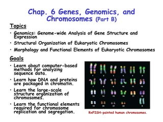
  • 1. Chap. 6 Genes, Genomics, and Chromosomes (Part B) Topics • Genomics: Genome-wide Analysis of Gene Structure and Expression • Structural Organization of Eukaryotic Chromosomes • Morphology and Functional Elements of Eukaryotic Chromosomes Goals • Learn about computer-based methods for analyzing sequence data. • Learn how DNA and proteins are packaged in chromatin. • Learn the large-scale structure organization of chromosomes. • Learn the functional elements required for chromosome replication and segregation. RxFISH-painted human chromosomes.
  • 2. Mining Sequence Data: BLAST Searches An enormous amount of DNA sequence information is available from genome sequencing and sequencing of cloned genes. This data is stored in data banks such as GenBank at the NIH in Bethesda, MD and the EMBL Sequence Data Base at the European Molecular Biology Laboratory in Heidelberg, Germany. Scientists working in the area of bioinformatics use this data to find genes, analyze their properties, and determine phylogenetic relationships between organisms and proteins. A common procedure in which this data is used is the BLAST search (basic local alignment search tool) which is used to compare protein and DNA sequences. An example BLAST search alignment is shown for the human neurofibromatosis 1 (NF1) gene in Fig. 6.25. The alignment shows NF1 is related to the S. cerevisiae Ira GTPase-activating protein (GAP) and suggests the disease is caused by aberrant signal transduction. Computer programs similar to BLAST are used to identify protein sequence motifs (e.g., zinc fingers) in unknown proteins. The identification of structure regions with known function sheds light on overall protein function and helps guide experimental analysis of unknown proteins and genes.
  • 3. BLAST search analysis can identify the members of a protein family originating from gene duplication and speciation mutations. As illustrated for the a- and ß-tubulin protein family in Fig. 6.26, an early gene duplication event created the paralogous a- and ß-tubulin genes. Later speciation mutations lead to evolution of the orthologous members of the a- and ß-tubulin subfamilies. Orthologous proteins are most likely to share the same function. Sequence Comparisons Establish Evolutionary Relationships Among Proteins x
  • 4. Genome Size vs Complexity Genome sequencing has revealed that the morphological complexity of an organism is not strongly correlated with the size of its genome (Fig. 6.27). Alternative splicing of RNAs and post- translational modification of proteins are thought to greatly increase the complexity of the proteins encoded by the genomes of higher organisms. In addition, the relative number of cells formed in a tissue such as the cerebral cortex can be important in increasing complexity (e.g., mice vs humans). Genes can be identified within the sequenced genomes of simple organisms such as yeast and bacteria by searching for open reading frames (ORFS). ORFs are long stretches of triplet codons lacking stop codons. Gene annotation (assignment of likely function) is based on knowledge from biochemical studies and/or alignments with known sequences. In complex organisms such as humans whose genes typically contain introns, more sophisticated algorithms that ID intron splice sites and compare cDNA and other sequence information to genomic DNA sequences must be applied to locate and annotate genes. Using such methods ~25,000 genes have been identified in humans. However, conclusive evidence for synthesis of protein or RNA products is lacking for ~10,000 genes.
  • 5. Extended and Condensed Chromatin Human diploid cells contain about 2 meters of DNA. To fit within nuclei, DNA must be condensed by ~105-fold. DNA exists in cells as a nucleoprotein complex known as chromatin. During interphase when cells are not dividing, chromatin is relatively uncondensed compared to its state in metaphase chromosomes. When released from nuclei with low salt buffer, chromatin displays an extended "beads-on-a-string" morphology, where each bead is a nucleosome (Fig. 6.28). When released in physiological salt concentrations, more condensed fibers of 30 nm diameter are observed. In general, extended chromatin can be transcribed, whereas condensed forms cannot.
  • 6. Structure of Nucleosomes Nucleosomes consist of 147 bp of DNA wrapped in almost two turns around the outside of an octamer of histone proteins (Fig. 6.29). In most nucleosomes, the octamer has a stoichiometry of H2A2H2B2H32H42. Histones are the most abundant DNA-binding proteins in eukaryotic cells. The sequences of the 4 histones that make up the octamer are highly conserved across all organisms, indicating their functions were optimized early in evolution. Histones have a large number of basic amino acids and bind to DNA mostly by salt-bridge interactions to phosphates in the DNA backbone. Another histone, H1, binds to the linker DNA between nucleosomes. Linker DNA is 10-90 bp in length depending upon the organism.
  • 7. Structure of 30-nm Chromatin Fibers In 30-nm fibers, nucleosomes bind to one another in a double helical arrangement (Fig. 6.30). Histone H1 molecules bind to linker DNA between nucleosomes and help stabilize the 30-nm fiber. The stability of 30-nm fibers is modulated by post- translational modification of the tails of histones in the octamers (H4 in particular).
  • 8. Histone Tails and Chromatin Condensation The N- and C-terminal tails of histones project out from the nucleosome core (Fig. 6.31a). They also contain numerous residues that can be modified by acetylation, methylation, etc. (Fig. 6.31b). Acetylation of lysine side-chains by histone acetylases (HATs) neutralizes positive charge and promotes decondensation of 30-nm fibers. Methylation, on the other hand, blocks lysine acetylation, maintains positive charge, and promotes 30-nm fiber condensation. Studies have shown that chromatin condensation is not controlled simply by the net acetylation state of histones. Rather, the sites where acetylation and other modifications occur also are important. The combinations of modifications that specify condensation/decondensation are referred to as the "histone code".
  • 9. Interphase Chromatin Interphase chromatin exists in two different condensation states (Fig. 6.33a). Heterochromatin is a condensed form that has a condensation state similar to chromatin found in metaphase chromosomes. Euchromatin is considerably less condensed. Heterochromatin typically is found at centromere and telomere regions, which remain relatively condensed during interphase. The inactivated copy of the X- chromosome (Barr body) that occurs in cells in females also occurs as heterochromatin. In contrast, most transcribed genes are located in regions of euchromatin. Common modifications occurring in histone H3 in hetero- and euchromatin are illustrated in Fig. 6.33b.
  • 10. Formation of Heterochromatin The trimethylation of histone H3 at lysine 9 (H3K9Me3) plays an important role in promoting chromatin condensation to heterochromatin (Fig. 6.34a). Trimethylated sites are bound by heterochromatin protein 1 (HP1) which self-associates and oligomerizes resulting in heterochromatin. Heterochromatin condensation is thought to spread laterally between “boundary elements” that mark the ends of transcriptionally active euchromatin (Fig. 6.34b). Recruitment of the H3K9 histone methyl transferase (HMT) to HP1 sites promotes heterochromatin spreading by catalyzing H3 methylation.
  • 11. Structure of Interphase Chromosomes FISH analysis performed with fluorescent probes that bind to sequential sequence sites along DNA supports a looped structure for interphase chromosomes (Fig. 6.35). Loops range in size from 1 to 4 million base pairs in mammalian interphase cells. The bases of the loops are located near the center of the chromosome at scaffold-associated regions (SARs), and matrix- attachment regions (MARs). The DNA fibers at the base of the loops are held together by structural maintenance of chromosome (SMC) proteins (Fig. 6.36c) and other non-histone proteins. Transcription units containing expressed genes are located in uncondensed loop regions, away from the more condensed center of the chromosome.
  • 12. Interphase Chromosome Territories In situ hybridization of interphase nuclei with chromosome- specific fluorescently-labeled probes indicates that chromosomes reside within restricted regions of the nucleus rather than appearing throughout the nucleus (Fig. 6.37). Interestingly, the precise positions of chromosomes are not reproducible between cells.
  • 13. Structure of Metaphase Chromosomes In metaphase chromosomes, the number of loops of chromatin is increased and the lengths of the loops are decreased compared to what occurs in interphase chromosomes. In addition, more folded structures called chromonema fibers and higher order structures occur in prophase and metaphase chromatids (Fig. 6.38).
  • 14. Microscopic Structure of Metaphase Chromosomes Because interphase chromosomes are not easily visualized by microscopy techniques, chromosome morphology has been studied mostly using metaphase chromosomes. Metaphase chromosomes are duplicated structures formed after DNA replication is complete. They contain two sister chromatids joined at a structure called the centromere (Fig. 6.39). The ends of chromatids are called telomeres. Centromeres are required for chromatid separation late in mitosis. Telomeres are important in preventing chromosome shortening during replication. The number, sizes, and shapes of metaphase chromosomes constitute the karyotype, which is distinctive for each species.
  • 15. Chromosome Banding Patterns A number of dyes, such as Giemsa reagent, selectively stain different regions of chromosomes forming distinctive bands. For Giemsa reagent, banding is affected by G + C content. Banding patterns are very important in chromosome ID and in looking for chromosomal abnormalities and mapping the locations of genes. The most detailed staining is achieved via multicolor FISH chromosome painting. In this technique, staining is performed using a mixture of DNA probes coupled to several fluorescent dyes (See Slide 1). In Fig. 6.40 below, FISH staining patterns have been converted to false-color images to visualize chromosomes. Standard terminology is used for naming band and gene locations in chromosomes. The short arm is designated "p", and the long arm "q". Arms are further divided into major sections and subsections that are numbered consecutively out from the centromere.
  • 16. Detection of Translocations The analysis of chromosome banding patterns is used to detect anomalies such as truncations and translocations associated with certain genetic disorders and cancers. In chronic myelogenous leukemia, leukemic cells contain a shortened chromosome 22 and a longer chromosome 9 resulting from a translocation event in the q arms of these two chromosomes (Fig. 6.41). The shortened chromosome is distinctive and is referred to as the "Philadelphia chromosome". Multicolor FISH staining (right) is useful in identification of such chromosomes in a chromosome spread.
  • 17. Evolution of Human Chromosomes Through the determination of locations of common chromosomal segments in modern primate chromosomes, investigators have calculated the most likely karyotype of the common ancestor of all primates (Fig. 6.42c). In addition, they have proposed a model for how the human karyotype evolved from that ancestor. Major events in the evolution of the human karyotype include 1) formation of chromosome 2 by fusion of ancestral chromosomes 9 and 11, 2) formation of chromosomes 14 and 15 by breakage of ancestral chromosome 5, and 3) formation of chromosomes 12 and 22 by translocations between ancestral chromosomes 14 and 21. In other cases (e.g., chromosome 1), no significant rearrangements have occurred over time.
  • 18. ID of Functional Chromosomal Elements (I) Studies with yeast have demonstrated that all chromosomes must contain 3 functional elements to replicate and segregate correctly: 1) replication origins, 2) a centromere, and 3) telomeres. Yeast replication origins were identified in plasmid cloning studies. Only yeast plasmids containing a copy of a sequence referred to as the autonomously replicating sequence (ARS) could be transfected into yeast cells (Fig. 6.44a). The haploid S. cerevisiae genome contains many ARSs distributed among its 16 chromosomes.
  • 19. ID of Functional Chromosomal Elements (II) While only ARSs are needed for plasmid replication, an additional sequence identified by cloning procedures was found to be required for efficient segregation of plasmids to yeast daughter cells (Fig. 6.44b). This DNA proved to contain chromosomal centromere sequences (CEN sequences). Yeast CEN sequences are relatively simple (Fig. 6.45, not covered). In humans, they consist of 2-4 x 10⁶ bp of simple sequence DNA composed of a 171 bp repeat unit. The human centromere sequence is bound by specialized nucleosomes containing a centromere-specific histone H3 variant (CENP-A). A large complex of non-histone proteins (the kinetochore) binds to centromeres and attaches them to microtubules of the mitotic spindle apparatus.
  • 20. ID of Functional Chromosomal Elements (III) Yeast transfection studies also showed that linearized plasmids containing ARS and CEN sequences could be maintained in cells only if telomere (TEL) sequences were attached at their ends (Fig. 6.44c). The function of TEL sequences in replication of chromosome ends is illustrated in the next two slides.
  • 21. Function of Telomeres A special mechanism is needed to complete the replication of DNA in DNA strands that have their 3’ ends located at the ends of chromosomes. DNA polymerases cannot complete synthesis of this region of DNA, and without synthesis, chromosomes become shortened with each round of replication (Fig. 6.46). Shortening results in the loss of binding sites for proteins that protect the ends of linear chromosomes from attack by exonucleases. As illustrations of the importance of telomere replication, knockout mice lacking the enzyme that synthesizes DNA at telomeres, telomerase, cannot produce viable offspring after six generations. In addition, telomerase often is switched on in cancer cells.
  • 22. Mechanism of Action of Telomerase Telomere sequences typically consist of tandemly repeating sequence units with a high G content in the strand that has its 3' end at the end of the chromosome. In humans and other vertebrates, the repeating sequence is TTAGGG. This sequence unit repeats over a few thousand base pairs in humans. The mechanism of replication of this DNA is illustrated in Fig. 6.47 for a protozoan species. Replication is carried out by the enzyme known as telomerase. Telomerase is a reverse transcriptase that carries its own internal RNA template which binds to the ssDNA at the chromosome 3’ end and allows this strand to be elongated. Ultimately, DNA Pol a/primase can synthesize a primer on this strand, which is elongated by DNA Pol . Some organisms rely on a different mechanism for replication of telomeric DNA. For example, flies lack telomerase and maintain telomere length by regulated insertion of non-LTR retrotransposons into telomere DNA.