
Recommended
More Related Content
Similar to Myocardial Infarction MI [Autosaved].pptx
Similar to Myocardial Infarction MI [Autosaved].pptx (20)
More from Safoora Qureshi
More from Safoora Qureshi (18)
Recently uploaded
Recently uploaded (20)
Myocardial Infarction MI [Autosaved].pptx
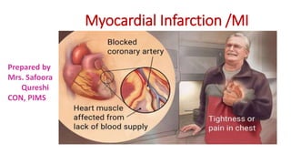
- 1. Myocardial Infarction /MI Prepared by Mrs. Safoora Qureshi CON, PIMS
- 2. Myocardial Infarction/ MI • MI or Heart Attack is the irreversible damage of myocardial tissue cased by prolonged ischemia & hypoxia • MI, occurs as result of sustained ischemia, irreversible myocardial cells death (necrosis) • MI is a disease condition caused by reduced blood flow to the heart muscles due to atherosclerosis & occlusion of a coronary artery by an embolism or thrombosis
- 3. Causes 95% of heart attacks occurs due blockage of one of the major vessels supplying blood to heart • Atherosclerosis and rupture plaque, is the most common cause Other 5% contributing factors are: • Spasm of the artery • Trauma • Rare medical conditions • Electrolyte imbalances such as • hyperkalemia or hypokalemia • Obstruction, from elsewhere in the body • Eating disorders
- 4. Pathophysiology • 80 to 90% of all acute MIs are secondary to thrombus formation • Perfusion to the myocardium distal to the occlusion is halted, result in necrosis • Contractile function of the heart stops in necrotic area(s) • The degree of altered function depend on the area of the heart involved and the size of infarction • The location of MI correlates with the involve coronary artery such as: • Inferior wall MI result from occlusion of right coronary artery • Anterior wall MI, result from occlusions in the left anterior descending artery • Lateral or posterior MIs, result from occlusion of circumflex artery
- 5. Atherosclerosis Spasm Atherosclerosis + Plaque split + thrombosis Gradual obstruction Sudden reversible obstruction Sudden Occlusion, usually irreversible Hypoxia Ischemia Coronary thrombosis Necrosis Angina Unstable angina MI Myocardial infarction Pathophysiology
- 6. Types of MI 1. ST segment Elevation MI (STEMI) 2. Non-ST segment Elevation MI (NSTEMI) 3. Coronary Spasm or unstable Angina
- 7. Clinical Manifestation Pain • Severe, immobilizing chest pain not relieve by position change, or nitrate admin, is the hallmark of an MI • Persistent and unlike other pain • Describe; as a heaviness, pressure, tightness, burning, constriction or crushing • Location; Substernal, retrosternal, or epigastric area • Radiate to: the neck, jaw, arms, or to the back • Occurs; while the patient is active, at rest, or asleep or awake • Time; commonly occurs in morning • No pain / Silent MI: in some pts of DM & cardiac neuropathy Other Symptoms • shortness of breath • sweating • nausea, vomiting, • abnormal heart beating, • anxiety, fatigue, • weakness, • stress, depression
- 8. Complications: Dysrhythmias: • The most common complication, in 80 % of MI pts, is the most common cause of death in prehospital patients Heart Failure • Occurs when pumping power of the heart has diminished. Papillary Muscles dysfunction: • Occurs when infarcted are includes or is adjacent to the papillary muscles that attached to the mitral valve Ventricular aneurysm: infarcted myocardium become thickened and bulged out during contraction Pericarditis: reduce ventricular filling and emptying and cause HF Dressler Syndrome: pericarditis with pericardial effusion and fever as a result of antibody-antigen reaction to necrotic myocardium Cardiogenic Shock: • Occurs when inadequate oxygen and nutrients are supplied to the tissues in sever left ventricular failure
- 9. Diagnosis Unstable Angina and MI • History and physical assessment • ECG: is the primary tool to rule out or confirm, UA or MI • Changes in QRS complex , ST segment and T wave caused by ischemia and infarction can develop quickly in both UA and MI • UA or NSTEMI: tend to have transient thrombosis or partial occlusion • STEMI: tend to have a more extensive MI, associated with prolonged and complete coronary occlusion and development of pathological Q wave on ECG • Serum Cardiac Markers: are released into the blood in large quantities from necrotic heart muscles • Cardio Angiography (PCI): to evaluate the extent of the disease and to determine the most appropriate therapeutic modality
- 10. ST segment & Q Wave Changes In MI
- 12. ENZYMES INITIAL RISE PEAK BACK TO NORMAL Myoglobulin < 2 hrs. 6 – 9 hrs. 1 day CK - MB 3 --- 6 hrs. 12 --- 24 hrs. 2 – 3 days Troponin I < 4 hrs. 14 – 24 hrs. 3 – 5 days CARDIAC MARKERS
- 13. Collaborative Care Emergency Treatment • Strict bed rest • 12- leads, continue ECG monitoring • Maintain IV line • Oxygen therapy via Nasal cath at rate 2 to 4 L/min • Drugs: • Nitroglycerin IV • Morphine sulfate IV • Aspirin • Heparin • Beta-Blockers • AC inhibitors • Monitor oxygen saturation, and vital signs
- 14. stable Angina Non- ST- segment-Elevation MI • Acute intensive drug therapy • Nitroglycerin S/L • Low-molecular-weight Heparin • Clopidogrel / anti-platelets medicine • Glycoprotein IIb/IIIa inhibitors • Coronary Angiography • Percutaneous coronary Intervention PCI
- 15. Unstable Angina Immediate reperfusion therapy • PCI is the first line treatment within 90 /mins of MI • Fibrinolytic therapy to dissolve clots • Streptokinase • urokinase • Recombinant Human tissue- type plasminogen • Anisoylated plasminogen-streptokinase activator complex • Emergency CABG