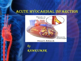
Myocardial infarction
•Download as PPT, PDF•
56 likes•9,830 views

myocardial infarction,symptoms,diagnosis,classification,treatment and comlications

Recommended
More Related Content
What's hot
What's hot (20)
Viewers also liked
Similar to Myocardial infarction
Similar to Myocardial infarction (20)
Recently uploaded
Recently uploaded (20)
Myocardial infarction
- 2. Definition • Otherwise know as heart attack • An MI occurs when there is a diminished blood supply to the heart which leads to myocardial cell damage and ischemia. • Contractile function stops in the necrotic areas of the heart. • Ischemia usually occurs due to blockage of the coronary vessels.
- 3. Definition cont. • This blockage is often the result of thrombus that is superimposed on an ulcerated or unstable atherosclerotic plaque formation in the coronary artery. • MI’s are described by the area of occurrence. • Anterior, Inferior, Lateral or Posterior.
- 5. Coronary artery events • Ischemia – Outer most area, source of arrhythmias, viable if no further infarction. • Injury – Viable tissue found between ischemic and infarcted areas. • Infarction/necrosis – Center area, dead not viable tissue that turn into scar.
- 8. MI Classifications • MI’s can be subcategorized by anatomy and clinical diagnostic information. Anatomic • Transmural and Subendocardial Diagnostic • ST elevations (STEMI) and non ST elevations (NSTEMI).
- 9. Risk Factors • The presence of any risk factor is associated with doubling the risk of an MI. Non Modifiable • Age • Gender • Family history
- 10. Risk Factors Modifiable • Smoking • Diabetes Control • Hypertension • Hyperlipidemia • Obesity • Physical Inactivity
- 11. Pathophysiology • Ischemia develops when there is an increased demand for oxygen or a decreased supply of oxygen. • Ischemia can develop within 10 seconds and if it lasts longer than 20 minutes, irreversible cell and tissue death occurs. • Myocardial cell death begins at the endocardium. The area most distal to the arterial blood supply.
- 12. Pathophysiology • As vessel occlusion continues cell death spreads to the myocardium and eventually to the epicardium. • Severity of the MI depends on three factors. • Level of occlusion • Length of time of occlusion • Presence or absence of collateral circulation
- 13. Symptoms: • Pain is the cardinal symptom of an MI • Anxiety and fear of impending death • Nausea and vomiting • Breathlessness • Collapse/syncope
- 14. Chest Pain • The most common initial manifestation is chest pain or discomfort. • This is not relieved by rest, position change or nitrate administration. • Pain is described by heaviness, pressure, fullness and crushing sensation. • Not everyone experiences this sensation.
- 15. Cardiovascular Changes • Initially the BP and pulse may be elevated. • Later, BP will drop due to decreased cardiac output. • Urine output will decrease • Lung sounds will change to crackles • Jugular veins may become distended and have obvious pulsations.
- 16. DIAGNOSTICS: • Electrocardiogram (ECG) • Blood test (Cardiac enzymes) • Echocardiogram • Nuclear scan • Chest radiographs • Coronary angiography • Exercise stress test. • Cardiac computerized tomography (CT) or magnetic resonance imaging (MRI).
- 17. Diagnostics • After collecting patient health history, a series of EKG’s should be taken to rule out or confirm MI. • 12 lead EKG’s can help to distinguish between ST-elevation MI’s and Non-ST- elevation MI’s.
- 19. Angina: Stable • Chest pain caused by the build up of lactic acid and irritation to the myocardial nerve fibers. • Chest pain caused by the 4 E’s. • Pain is usually relieved with rest, pain meds and nitrates.
- 20. Variable/Prinzmetal/Spasm • Transient ischemia that occurs unpredictably and almost always at rest. • Pain is caused by vasospasm of the arteries. • ST segment elevations will be noted.
- 21. Unstable • Chest pain at rest or with exercise and tends to last greater than 15 minutes. • This results in reversible myocardial ischemia but is a sign that an infarct is soon to come. • EKG will reveal ST segment depression and T wave inversion.
- 22. STEMI • ST segment elevations • T wave changes • Q wave development • Enzyme elevations • Reciprocals
- 23. NSTEMI • ST segment depressions • T wave changes • No Q wave development • Mild enzyme elevations • No reciprocals
- 24. STEMI vs. NSTEMI
- 25. Phases of a STEMI • Hyperacute Phase • Occurs within the first few hours of MI onset. • Leads facing the infarcted surface: ST segment elevation. • Leads facing the uninjured surface: ST segment depression (reciprocals) • T waves become tall, widened and might be taller than the R wave.
- 26. Phases of a STEMI • Fully Evolved Phase • Q wave development • ST elevation • T waves start to become inverted in leads facing the injury.
- 27. Phases of a STEMI • Resolution phase • Weeks after there will be a gradual return of ST segments to baseline. • T waves will gradually return to normal but are the last to change back.
- 28. Serum Cardiac Markers • Myocardial cells produce certain proteins and enzymes associated with cellular functions. • When cell death occurs, these cellular enzymes are released into the blood stream. • CPK and troponin
- 29. CPK • Creatine Phosphokinase • Begin to rise 3 to 12 hours after acute MI. • Peak in 24 hours • Return to normal in 2 to 3 days
- 30. Troponin • Myocardial muscle protein released into circulation after injury. • These are highly specific indicators of MI. • Troponin rises quickly like CK but will continue to stay elevated for 2 weeks. • Myoglobin-lacks cardiac specificity.
- 31. Treatment Options • The immediate goal for any acute MI is to restore normal coronary blood flow to vessels and salvage myocardium. • There are a variety of medical and medicinal therapies to treat an MI.
- 32. General Treatment for the MI patient • Morphine • Oxygen • Nitroglycerin • Aspirin
- 33. Fibrinolytic Therapy • Indicated for patients with STEMI MI’s. • Should be given within 12 hours of symptom onset. • Fibrinolytics will break down clots found within the vessles • Contraindications: post op surgical patients, history of hemorrhagic stroke, ulcer disease, pregnancy, ect.
- 34. Cardiac Catheterization • A diagnostic angiography which includes angioplasty and possible stenting. • Performed by an interventional cardiologist with a cardiac surgeon on stand by. • Percutaneous procedure through the femoral or brachial artery.
- 35. Cardiac Catheterization • Upon arrival to the cath lab all actue MI patients will receive: • A bolus dose of plavix • IV Integrelin • Heparin dose either subcu or IV drip • Angiomax : a DTI may be substituted for heparin and integrelin.
- 36. Coronary artery bypass graft • Surgical treatment where saphenous vein is harvested from the lower leg and used to bypass the occluded vessels.
- 38. Long Term Care • Smoking Cessation and lifestyle modifications. • Aspirin, Beta Blockers and Clopidogrel will be indefinite. • Lipid lowering medication along with diet modifications.
- 39. Complications Vascular Complications • Recurrent ischemia • Recurrent infarction Mechanical Complications • Left ventricular free wall rupture • Ventricular septal rupture • Papillary muscle rupture with acute mitral regurgitation
- 40. COMPLICATION: Myocardial Complications • Diastolic dysfunction • Systolic dysfunction • Congestive heart failure • Hypotension/cardiogenic shock • Right ventricular infarction • Ventricular cavity dilation • Aneurysm formation (true, false)
- 41. References • Bolooki, H.M.& Askari, A. (Published August 8 2010). Acute Myocardial Infarction. Retrieved from http://www.clevelandclinicmeded.com/medicalpubs/disea semanagement/cardiology/acute-myocardial- infarction/#s0050 • Lewis, S., Heitkemper, M., & Dirksen, S. (2004). Medical surgical nursing assessment and management of clinical problems. St. Louis, MO: Mosby. • McCance, K.L., Huether, S.E., Brashers, V.L.& Rote, N.S. (2010). Pathophysiology the biological basis for disease in adults and children. Maryland Heights, MO: Mosby Elsevier.