SlideShare a Scribd company logo
1 of 82
Epithelial to Mesenchymal
Transition
Epithelial vs Mesenchymal cells
(A) Epithelial cells are inter-
connected through tight junctions
(gray), E-cadherin-based junctions
(red), which are connected to the
actin cytoskeleton, gap junctions
(red/blue), and hemidesmosomes
(cyan), which are connected to
the cytokeratin-based intermediate filament cytoskeleton.
Epithelial cells also have specialized cell-ECM interactions
for adhesion to the laminin-rich basement membrane.
CSHL collection
Epithelial vs Mesenchymal cells
(B) Mesenchymal cells show a shift
to a vimentin-based intermediate
filament cytoskeleton and altered
composition of cell-ECM
interactions optimized for adhesion
to the collagen-rich interstitial matrix.
Mesenchymal cells also produce abundant TGFb, growth
factors (GF), and matrix metalloproteinases (MMPs), as
well as components of the extracellular matrix.
CSHL collection
Epithelial vs Mesenchymal cells
Use this slide to practice describing the differences between
these two types of cells
Cell phenotypes in early mouse embryo.
(A) Gastrulating mouse embryo.
(C') Scanning electronic
micrograph of a transverse
section through a mouse embryo
showing the different cell
phenotypes: columnar
epithelium, mesenchyme and
squamous epithelium. (B)
Diagram of the embryonic part
of a mouse embryo. Dashed box
outlines the three germ layers.
(C) Scanning micrograph shown
in C' color-coded for the three
germ layers.
EMT and its reverse MET
.
Diagrammatic representation
of EMT, its reverse MET
(mesenchymal to Epithelial
Transition), egression and
ingression. (A) Epithelial cells
undergo EMT to assume
mesenchymal characteristics.
Conversely, mesenchymal
cells undergo MET to epithelialize. (B) In an ingression, cells
undergo EMT and leave an epithelium, while in an egression
cells undergo MET and join a preexisting epithelium as in
endoderm formation in birds and mammals.
Rear-front to apico-basal polarity
Categories of EMT
Three types of EMT can
be discerned depending
on the physiological tissue
context.
Type1 EMT occurs in
embryogenesis and organ development,
type2 EMT is important for tissue regeneration and organ
fibrosis, and type 3 EMT is associated with cancer
progression and cancer stem cell properties.
EMT and MET
The process of MET
(mesenchymal–epithelial
transition) enables cells that
have undergone EMT to revert
to the epithelial state. MET also
contributes to development and the generation of metastatic
carcinomas. Consecutive rounds of EMT and MET occur
during development. In 1ary EMT, epithelial cells without
prior history of EMT differentiate into mesenchymal cells. In
2ary EMT, cells that have already undergone EMT and MET
initiate a new EMT process, e.g. following dissemination,
cancer cells can revert through MET to an epithelial state.
1ary and 2ary EMT
Steps during EMT and ingression
A conserved series of steps underlie EMT and
ingression process for several developmental
events.
Steps during EMT and ingression
Changes in cell adhesion during EMT
The first cell (left) presents
a cell with epithelial
characteristics and apico-
basal polarity. The two other
cells are transitioning to
mesenchymal status. An
external cue (Growth Factors) starts an STP leading to
cytoskeleton remodeling and loss of apico-basal polarity.
From the review paper EMT-pathways
External cues involved in EMT
Signaling and STP from several different signal
molecules (TGF, Wnt, Shh, and Delta/Notch), as well
as changes in integrin/ECM and injury (inflammation,
hypoxia) can lead to EMT.
Cellular events during EMT
a) The first steps of EMT are
the disassembly of epithelial
cell–cell contacts (tight
junctions, adherens junctions,
desmosomes and gap
junctions) and the loss of cell
polarity through the
disruption of the Crumbs
(Crb), partitioning defective
(PAR) and Scribble (SCRIB)
polarity complexes.
Cellular events during EMT
b| Next, the epithelial actin
architecture reorganizes, and
cells acquire motility and
invasive capacities by
forming lamellipodia,
filopodia and invadopodia,
and by expressing matrix
metalloproteinases (MMPs)
that can degrade extracellular
matrix (ECM) proteins.
Changes in cell adhesion during EMT
The DDR1 complex
activates RhoE, which
weakens actomyosin
contractility at points of
cell-cell contacts (Adherens
junctions, AJ).
TGF receptors located next to tight junctions (TJ) trigger
ubiquitylation and degradation of RhoA and the
destabilization of cortical actin filaments associated with TJ.
From the review paper EMT-pathways
Changes in cell adhesion during EMT
Activation of transcriptional
repressor, such as Snail and
Serpent (Srp/GATA)
downregulates genes
encoding junctional proteins,
including E-cadherins, claudins
and occludin, thus compromising further AJ and TJ. EPB4-
1L5 sequesters p120-catenin weakening AJ attachment to
microfilaments and promotes integrin-ECM adhesion.
Integrin also leads to the activity of Srp, which represses the
apical polarity gene Crumbs (Crb). From the review paper
TFs during EMT
EMT is driven by SNAIL, zinc-finger E-box-binding (ZEB)
and basic helix–loop–helix (bHLH) transcription factors that
repress epithelial marker genes and activate genes
associated with the mesenchymal phenotype. These TFs are
themselves regulated through their cellular localization and
stability.
Snail regulation
Post-translational modifications
(PTM) of the different TFs regulate
their activities, subcellular
localization and stability. For
example phosphorylation (PTM)
of Snail by GSK3β leads to its
export from the nucleus (and
therefore its inactivity as a TF).
Snail phosphorylation by PAK1 on
the other hand promotes the nuclear
retention of SNAIL1 and enhances its activity.
Twist and ZEB regulation
TWIST is phosphorylated by the
MAPK p38, JNK and ERK, which
protects it from degradation, and
thus promotes its nuclear import
and functions. ZEB2 undergoes
another PTM ( sumoylation,
Sumo by Polycomb repressive
complex 2 (PRC2) and is
subsequently exported from the
nucleus, which reduces its activity
as a transcription factor.
EMT during mouse gastrulation
(A) Scheme of the embryonic part of
a early mouse embryo. Dashed box
outlines the primitive streak.
(B') Scanning EM showing a
transverse section through a
mouse primitive streak. (C) Cells
undergo an EMT event at the
primitive streak. (D) Signaling
pathways that regulate the different
EMT steps at the murine primitive
streak.
EMT during mouse gastrulation
(C) Cells undergo an EMT event
at the primitive streak.
(1) First intercellular spaces
appear between cells and
(2) basal lamina breaks down.
(3) Cells acquire a bottle shape,
(4) round up as they travel
through the streak, and
(5) finally acquire a stellate morphology and
migrate away from the streak.
EMT during mouse gastrulation
Signaling in cells located in the
epiblast (left) ensures
maintenance of the epithelial
phenotype via the
expression of Pofut2 and
Sox3. Upon activation by
Wnt, TGF and FGF
proteins, cells in the primitive streak (middle) undergo
apical constriction (indicated by green ring of contracting
actin) to allow invagination before ingression.
EMT during mouse gastrulation
Blue cells in the primitive
streak express Eomes, Mesp1
and Mesp2, which are
responsible for ingression.
RhoA downregulation by
Net1 destabilizes basal
microtubules and
contributes to the breakdown of the basement membrane.
Snail represses Sox3 and E-cadherin, which is also
downregulated post-transcriptionally (PTM). In the
migratory cells (right), Rac1 mediates cell protrusion.
Another EMT example
Signal molecules such as Wnt,
BMP4 retinoic acid (RA) and
FGF lead to EMT. Try to
follow the arrows and
recognize some of the players
from previous slides. The point
here is not to memorize this
pathway, but to understand
common themes and players.
Questions
1) Cite the two basic embryological processes for cell
determination and explain the main difference between them.
2) The experiment below, using sea urchin embryos, illustrate
both processes for cell determination. Identify which one is
represented by each part of the experiment (A and B) and
explain your decision briefly.
A
Abnormal ~ normal
development development
normal normal
development development
B
Paper-based
1) In all the figures where electroporation of DNA in chicken
embryos is performed, the authors show a panel with GFP. Why
is GFP electroporated in these cells, together with the
experimental gene (Snail or Sox)? What kind of control and
information does the GFP signal provide? You may want to read
about electroporation.
(https://en.wikipedia.org/wiki/Electroporation)
2) Based on information in Figure 1E add the appropriate arrow
( or ) between Snail and RhoB as you would find
in a GRN. Justify your choice briefly by describing precisely
the result presented in 1E
Snail RhoB
3) If you were to analyze the promoter sequence of the RhoB
gene, what would you expect to find? Explain briefly
· Sequences for snail to bind as an activator
· Sequences for snail to bind as a repressor
· No binding sequences for Snail
4) The figure to the right is parts 1F and 1G of the paper,
together with a schematic dorsal view of an early chicken
gastrula. The dotted white lines in F and G represent the
position of the primitive streak.
Indicate on the schematic where the electroporation was done.
5) Which cellular structure contains laminin? How is the
absence of laminin on the electroporated side consistent with
the function of snail in EMT? For this question, you want to
also review the steps involved in the epithelial to mesenchymal
transition (PowerPoint on OAKS).
6) This is part of Figure 3.
a)Describe panels B and N, as well as P and Q in your own
word, explaining in particular the effect on Sox3 expression.
b) Predict the expected data for laminin expression. Would it be
up or down in the electroporated area compared to normal?
Justify your answer.
7) Explain what are gene markers
8) Based on the data presented in Figure 6A,B, and F fill, the
GRN below with the correct arrows (. or ). Justify
each placed arrow briefly by describing the appropriate part of
the figure
Developmental Cell
Article
Reciprocal Repression between Sox3 and Snail
Transcription Factors Defines Embryonic
Territories at Gastrulation
Hervé Acloque,1,3 Oscar H. Ocaña,1 Ander Matheu,2 Karine
Rizzoti,2 Clare Wise,2 Robin Lovell-Badge,2
and M. Angela Nieto1,*
1Instituto de Neurociencias, CSIC-UMH, San Juan de Alicante
03550, Spain
2National Institute for Medical Research, London NW7 1AA,
UK
3Present address: UMR 444, INRA-ENVT, Génétique
Cellulaire, Toulouse 31326, France
*Correspondence: [email protected]
DOI 10.1016/j.devcel.2011.07.005
Open access under CC BY license.
SUMMARY
In developing amniote embryos, the first epithelial-
to-mesenchymal transition (EMT) occurs at gastrula-
tion, when a subset of epiblast cells moves to the
primitive streak and undergoes EMT to internalize
and generate the mesoderm and the endoderm. We
show that in the chick embryo this decision to inter-
nalize is mediated by reciprocal transcriptional re-
pression of Snail2 and Sox3 factors. We also show
that the relationship between Sox3 and Snail is
conserved in the mouse embryo and in human
cancer cells. In the embryo, Snail-expressing cells
ingress at the primitive streak, whereas Sox3-posi-
tive cells, which are unable to ingress, ensure the
formation of ectodermal derivatives. Thus, the subdi-
vision of the early embryo into the two main territo-
ries, ectodermal and mesendodermal, is regulated
by changes in cell behavior mediated by the antago-
nistic relationship between Sox3 and Snail transcrip-
tion factors.
INTRODUCTION
The shaping of the early embryo involves the conversion of
a single layer of ectodermal cells (the epiblast) into a
multilayered
structure. This complex biological process starts at gastrulation,
when a subset of the initial epiblast cells moves inside the
embryo to become mesoderm and endoderm (Stern, 2004). In
amniotes, gastrulation occurs where Nodal signaling is strongest
(Brennan et al., 2001; Bertocchini and Stern, 2002). Initially, in
the
chick embryo, cells accumulate at the posterior part through
epithelial cell intercalation (Voiculescu et al., 2007) in a region
now devoid of the hypoblast, a lower layer that inhibits Nodal
signaling (Bertocchini and Stern, 2002). This accumulation
results in the formation of a midline linear structure called the
primitive streak, from which the presumptive mesendodermal
cells ingress upon undergoing an epithelial-to-mesenchymal
transition (EMT). EMT involves a dramatic change in cell
morphology and behavior that allows cells to break the basal
546 Developmental Cell 21, 546–558, September 13, 2011 ª2011
Els
lamina, internalize, and start migrating toward their destinations
(Harrisson et al., 1991; Acloque et al., 2009). Cells that remain
in
the epiblast keep their epithelial character and will contribute to
the ectodermal derivatives, namely the epidermis, the ecto-
dermal placodes, and the anterior central nervous system
(CNS) (Fraser and Stern, 2004). Indeed, much of the CNS will
develop from a subset of the noningressing cells later specified
as neural precursors. Therefore, it is crucial to identify not only
those factors that induce cell ingression at gastrulation but
also those that prevent it, because protection from undergoing
EMT is necessary to ensure the formation of ectodermal deriva-
tives. Indeed, previous studies have shown that committed
neural progenitor cells at the anterior part of the primitive
streak
are protected from signals that induce internalization. The zinc
finger transcription factor Churchill and its target SIP-1 are
required to stop ingression movements through the anterior
primitive streak (Sheng et al., 2003). This safeguard mechanism
operates at stages of neural induction and onset of Churchill
expression (from stage HH4+; Hamburger and Hamilton,
1951), but ingression starts at least as early as stage HH2 (Stern
and Canning, 1990), suggesting that a different mechanism must
exist to protect early ectodermal cells from the EMT inducers.
Among the key factors that induce EMT at gastrulation and
that are conserved during evolution are the members of the
Snail
family (Barrallo-Gimeno and Nieto, 2005). They are
fundamental
for EMT at gastrulation in all species analyzed and for
additional
developmental EMT processes (Acloque et al., 2009). We previ-
ously found that Snail2 (Slug) downregulation prevented migra-
tion from the primitive streak in the chick embryo (Nieto et al.,
1994), and here we show that Snail2 is sufficient to induce
ectopic delamination in otherwise noningressing epiblast cells,
confirming that these cells need to be protected from Snail2
expression and subsequent ingression. Therefore, we set out
to identify factors that might prevent Snail expression at early
gastrulation, as candidates to play an important role in
protecting
epiblast cells from undergoing EMT. We show that Sox3 and
Snail2 are expressed in complementary domains early during
gastrulation; gain- and loss-of-function studies reveal that these
factors antagonize each other to regulate cell ingression. We
show that Sox3-Snail antagonism is implemented through direct
reciprocal transcriptional repression, a relationship that seems
to be conserved in the mouse embryo and in tumor cell lines,
where they also regulate epithelial versus mesenchymal and
evier Inc.
mailto:[email protected]
http://dx.doi.org/10.1016/j.devcel.2011.07.005
http://creativecommons.org/licenses/by/3.0/
Figure 1. Snail2 Expression in the Epiblast Induces
Ectopic EMT and Cell Delamination
(A–C) Chick embryos were coelectroporated with a vector
encoding GFP and another containing the coding region of
Snail2. The embryos were subsequently allowed to
develop for 15 hr. Endogenous Snail2 expression at the
primitive streak and ectopic expression induced by elec-
troporation could be observed in a dorsal view (B) and in
a transverse section through the epiblast (C). Observe the
delamination of cells from the epiblast. Dotted line in (B)
indicates the level of the section shown in (C). Electro-
poration of a control empty vector (pCX) or that containing
GFP failed to induce cell delamination (not shown).
(D–H) Snail2 electroporation was accompanied by the
induction of RhoB expression, a known Snail2 down-
stream target (D and E), cell delamination, and disruption
of the basement membrane as assessed by the absence
of laminin staining (F and G) and by the repression of
E-cadherin expression in the electroporated cells (H).
Developmental Cell
Sox3 and Snail Define Embryonic Territories
invasive properties. Together, our results show that Snail-Sox3
cross-repression regulates cell ingression at gastrulation in
amniotes and suggest that this antagonistic relationship may
also have important implications in cancer.
Developmental Cell 21, 546–5
RESULTS
Snail2 Induces Ectopic Cell Delamination
in the Ectoderm of the Early Chick
Gastrula
We had previously shown that Snail2 knock-
down in the early chick embryo prevents cell
ingression at the primitive streak and neural
crest delamination from the neural tube (Nieto
et al., 1994). To check whether Snail2 is suffi-
cient to trigger EMT and cell delamination, we
ectopically expressed the coding region of
Snail2 by electroporation of the chick blasto-
derm at stage 2 (Hamburger and Hamilton,
1951) in the ectodermal region corresponding
to the prospective neural plate (see Experi-
mental Procedures). Snail2 ectopic expression
induces cell delamination (Figures 1A–1C), sug-
gesting that Snail2 is sufficient to trigger EMT in
a territory that normally keeps its epithelial
integrity at gastrulation. To confirm that this
induced delamination is due to the activation
of a full EMT program, we examined the expres-
sion of previously described Snail2 target genes
known to be necessary for cell delamination,
such as the small GTPase RhoB (del Barrio
and Nieto, 2002) and E-cadherin (Cano et al.,
2000). As shown in Figures 1D and 1E, Snail2
induces ectopic expression of RhoB, together
with the disruption of basal lamina, as assessed
by the loss of laminin expression (Figures 1F and
1G). Figure 1H shows that E-cadherin is down-
regulated in electroporated cells that undergo
EMT, and cells can be seen delaminating from
the epiblast (compare the electroporated and
the control sides). These data indicate that Snail2 is sufficient
to trigger EMT and cell delamination from the chick epiblast,
suggesting that the latter should be protected from Snail ex-
pression in the early embryo.
58, September 13, 2011 ª2011 Elsevier Inc. 547
Figure 2. Snail2 and Sox3 Genes Are Complemen-
tarily Expressed in the Chick Gastrula
(A–C) Snail2 expression starts at the posterior part of the
blastula at stage EGXIV (A), and its expression is associ-
ated with the primitive streak as it progresses in the gas-
trulating embryo (B and C).
(D–F) Sox3 expression is detected in the epiblast and it
gets progressively downregulated at the primitive streak
as it forms.
(G and H) Double labeling for Snail2 (green) and Sox3 (red)
transcripts at the full primitive streak stage (HH4) shows
the complementary expression pattern between Snail2
and Sox3. A transverse vibratome section taken at the
level shown by the dotted line in (G) allows a better
assessment of the mutually exclusive expression pattern
and the continued Snail2 expression in migratory mesen-
dodermal cells emanating from the streak (H).
Developmental Cell
Sox3 and Snail Define Embryonic Territories
Snail2 and Sox3 Expression Domains Delineate
Ingressing versus Noningressing Embryonic Cell
Populations
Because the process of cell ingression has to be tightly
regulated
to maintain the balance between ectodermal (noningressing) and
mesendodermal (ingressing) progenitors, Snail2 is restricted
around the primitive streak, where cells are internalized, from
the first stages of streak formation (Nieto et al., 1994; Figures
2A–2C). To search for genes functionally equivalent to
Churchill
but at the early primitive streak stages, we focused on the Sox3
gene for several reasons: (1) it is highly expressed at epiblast of
early-stage embryos, and its expression disappears in the terri-
tory surrounding the primitive streak (Rex et al., 1997; Figures
2D–2F); (2) Sox3 and Snail2 show mutually exclusive
expression
patterns (Figures 2G and 2H); and (3) Sox2, a closely related
gene, has been shown to prevent the induction of Snail2 by
BMP in the dorsal neural fold (Wakamatsu et al., 2004), but it is
not expressed at the early gastrulation stages in chick. There-
fore, we decided to manipulate either Snail2 or Sox3 expression
548 Developmental Cell 21, 546–558, September 13, 2011 ª2011
Els
in the chick blastoderm by electroporation and assess the
effects on their respective expression. We first induced ectopic
expression of Snail2 by electroporating in regions such as the
anterior epiblast. We found that Sox3 expression was repressed
in 75% of the embryos (Figures 3F–3H; black arrow in H; n =
16),
which is best seen in transverse sections (Figures 3I–3K). GFP
electroporation alone does not have the same effect (Figures
3A–3E; n = 13). We then electroporated a Snail2 dominant-
nega-
tive form lacking the transactivation domain (Morales et al.,
2007;
DN-Snail2). This led to an extension of Sox3 expression to the
primitive streak up to the midline, a region that normally
expresses Snail2 (Figures 3L–3Q; black arrow in N; 53%, n =
17). Conversely, ectopic Sox3 expression at the primitive streak
strongly represses endogenous Snail2 expression (Figures 3R–
3X; 74%; n = 19), indicating that Sox3 and Snail2 act in a
mutually
antagonistic way.
Next, we examined whether the observed changes in the ex-
pression domains of Snail2 and Sox3 correlate with cell
behavior.
With this aim, we followed cell movements near the primitive
Figure 3. Snail2 and Sox3 Behave as Mutual
Transcriptional Repressors in the Chick Embryo
(A–E) Control embryo electroporated with a vector encod-
ing GFP (A) shows the normal pattern of Sox3 expression
in a dorsal view (B) and in a transverse section (C). (D) and
(E) are high power images of the areas demarcated in (C).
(F–K) Similar images taken from an embryo electro-
porated with Snail2 in the anterior epiblast shows the
repression of Sox3 expression in the electroporated
region (arrow in H and sections in I). High power images
comparing the electroporated with the control side
confirm Sox3 downregulation (J and K).
(L–Q) Electroporation of a dominant-negative form of
Snail2 (DN-Snail2) in the primitive streak extended Sox3
expression to the embryonic midline (arrow in N), as better
assessed in the images shown in (O–Q).
(R–X) Ectopic Sox3 expression by electroporation at the
primitive streak inhibited Snail2 expression (black arrow in
T and a section at different magnifications in (U–X).
evier Inc.
D
BF(t=0) GFP (t=0)
A
Cell tracking
(8h)
CB
100
H
G
F
P
ps
2
E
i
GF
Ingressed
0
50
100
non
ingressed
(
%
)
G
F
P
+
S
n
a
i
l
2
Ingressed non
ingressed
0
50
100
(
%
)
L
G
F
P
+
S
o
x
3
KJ
Ingressed non
ingressed
0
50
100
(
%
)
PM ON
Ingressed non
0
50
100
(
%
)
G
F
P
+
D
N
-
S
n
a
i
2
TQ SR
G
F
P
+
D
N
S
o
x
3
ingressed
0
50
100
(
%
)
XU WV
Ingressed non
ingressed
0
50
100
(
%
)
P
+
S
n
a
i
l
2
+
S
o
x
3
Ingressed non
ingressed
0
G
F
P
Figure 4. Sox3 Overexpression or Snail2 Inhibition
Block Ingression at the Primitive Streak
(A–X) Time-lapse confocal analysis of cell movements in
cultured embryos electroporated with GFP alone (A–D),
GFP plus Snail2 (E–H), GFP plus Sox3 (I–L), GFP plus
a dominant-negative form of Snail2 (M–P), GFP plus
a dominant-negative form of Sox3 (Q–T), and GFP, Sox3,
and Snail2 (U–X). BF (t = 0) and GFP (t = 0) show trans-
mitted light and fluorescent images obtained 6 hr after
electroporation, just before the start of time lapse anal-
yses. Colored dots in (B, F, J, N, R, and V) represent the
initial position of cells that were subsequently followed.
Dots in (C, G, K, O, S, and W) represent the final position of
those cells and the lines their tracks. (D), (H), (L), (P), (T),
and (X) show the percentages of ingressing and non-
ingressing cells in each condition. The white dotted lines
indicate the relative position of the midline; black arrows
and ps indicate primitive streak. Control GFP-expressing
embryos showed the normal movements of epiblasts cells
toward the primitive streak and subsequent ingression (C).
Snail2 overexpression increases the percentage of in-
gressing cells (E–H). Ectopic Sox3 expression blocked cell
ingression through the primitive streak without affecting
the convergence movement toward the midline (compare
C and D with K and L). A dominant-negative form of Snail2
also blocked cell ingression (compare C and D with O and
P), whereas a dominant-negative form of Sox3 increased
ingression (compare T with D). Overexpression of both
Snail2 and Sox3 could rescue the ingression behavior of
epiblast cells (compare C and D with W and X).
Developmental Cell
Sox3 and Snail Define Embryonic Territories
streak in embryos electroporated with GFP-containing control
vectors or with GFP plus different Snail2 and Sox3 constructs.
Cells expressing GFP converge at the primitive steak, ingress,
and migrate away in the mesendoderm as expected for a normal
embryo (Figures 4A–4D and Movies S1 and S2 available
online).
Overexpression of wild-type Snail2 increases the proportion of
cells that ingress (Figures 4E–4H and Movies S3 and S4). After
ectopic expression of Sox3, electroporated cells still converge
at the primitive streak but are unable to ingress (Figures 4I–4L
and Movies S5 and S6). This observation confirms that Sox3
needs to be downregulated to allow cell ingression at the primi-
tive streak, but does not impair the convergence of epiblastic
Developmental Cell 21, 546–
cells at the primitive streak. If the defect in cell
ingression is mediated by the observed Snail2
downregulation by Sox3 (Figures 3R–3X), pre-
venting Snail2 function should elicit a similar
defect. Indeed, overexpression of DN-Snail2 in
the primitive streak also inhibits cell ingression
(Figures 4M–4P and Movies S7 and S8), which
is compatible with the observed activation of
Sox3 expression (Figures 3L–3Q). In contrast,
loss of Sox3 function by ectopic expression of
DN-Sox3 close to the primitive streak favors
cell ingression (Figures 4Q–4T and Movies S9
and S10), as also observed after Snail2 overex-
pression (Figures 4E–4H). On the other hand,
overexpression of Snail2 together with Sox3
and GFP in the region of endogenous ingression
rescued the phenotype of Sox3 overexpression,
as the cell movements observed resembled
those in the control embryo (Figures 4U–4X and Movies S11
and S12). Altogether, these results strongly suggest that the
decision to ingress at the primitive streak depends on the inter-
actions between Snail2 and Sox3 transcription factors.
Snail2 and Sox3 Are Reciprocal Direct Transcriptional
Repressors
Next, we investigated whether Snail2 and Sox3 can bind to each
other’s promoter to directly repress expression. We found one
Snail-binding site in sequences located 50 to the Sox3 coding
region, which is conserved in chick, mouse, and human (Fig-
ure S1). First, we used a luciferase reporter driven by the 50
558, September 13, 2011 ª2011 Elsevier Inc. 549
A
B
C
D
E
F
Figure 5. Snail2 and Sox3 Are Direct Mutual Tran-
scriptional Repressors
(A) Conserved Snail-binding site in the Sox3 promoter (see
also Figure S1).
(B) Snail2 overexpression in cultured embryos repressed
the activity of the Sox3 promoter, whereas a deletion that
removes the Snail binding site significantly restores
promoter activity.
(C) Chromatin immunoprecipitation (ChIP) analysis
confirms that Snail2 can directly bind to the Sox3
promoter. ChIP assays were carried out with anti-myc
antibodies on embryo extracts that were previously elec-
troporated with either GFP or myc-Snail2. PCR fragments
were obtained after amplification of the indicated promoter
region from the input (1), histone H3 positive control (2),
IgG negative control (3), and myc immunoprecipitated
fractions (4).
(D) Conserved sequence for Sox transcription factors
binding in the Snail2 promoter (see also Figure S1).
(E) Sox3 overexpression in cultured embryos repressed
Snail2 promoter activity, and the deletion of the Sox-
binding site restored most of the promoter activity.
(F) Chromatin immunoprecipitation (ChIP) analyses con-
firmed that Sox3 can directly bind to the Snail2 promoter.
ChIP assays were carried out as described above.
Developmental Cell
Sox3 and Snail Define Embryonic Territories
Sox3 sequences (Figure 5A), to test whether Snail2 can modu-
late the activity of the Sox3 promoter by coelectroporating
Snail2
with the luciferase construct in the epiblast of preprimitive
streak
stage embryos (see Experimental Procedures). Snail2 reduced
the activity of the Sox3 promoter construct. This repression is
dependent on the presence of the conserved Snail2-binding
box (Figure 5B), because electroporation with a construct
lacking this box significantly restores activity (Figure 5B). To
determine whether Snail2 can bind directly to the Snail box
in vivo, we performed Chromatin immunoprecipitation (ChIP)
experiments after electroporating embryos with GFP and a
myc-tagged version of Snail2. After dissecting the GFP-positive
tissues, we carried out ChIP with an anti-myc antibody. A frag-
ment of genomic DNA corresponding to the Snail-box
containing
region of the Sox3 promoter was amplified in embryos
electropo-
rated with the myc-tagged Snail2 protein (Figure 5C, compare
line 4 for GFP and myc-Snail2 experiments). We then
performed
similar experiments to determine whether Sox3 acts on a similar
way on the Snail2 promoter and found that this is indeed the
case
(Figures 5D–5F). The conserved Sox DNA binding sequence
located in the chick Snail2 promoter is shown in Figure 5D, and
its conservation in other species is shown in Figure S1. Thus,
Snail2 and Sox3 can bind directly to each other’s promoter in
vivo
at conserved elements, providing a molecular mechanism for
their antagonistic role in the cell decision to ingress at gastrula-
tion. This interaction defines two main territories within the
early embryos: the noningressing ectoderm and the ingressing
mesendoderm.
Snail2 Represses Ectodermal Markers without Inducing
Mesodermal Fate
In addition to a strong repression of Sox3 expression in the
epiblast, Snail2 is also able to repress other ectodermal markers,
such as the epidermal marker Dlx5 (Figure 6A; 100%; n = 3)
and
the neuroectodermal marker Otx2, albeit in a limited territory
550 Developmental Cell 21, 546–558, September 13, 2011 ª2011
Els
(Figure 6B; 62%; n = 8). This is consistent with the known role
of Snail as an epithelial repressor (Nieto, 2002). In contrast,
Sox3 induces Dlx5, although it is unable to activate Otx2
expres-
sion (Figure 6A; 80%; n = 5; Figure 6B; 100%; n = 11).
We had observed that, in addition to repressing epithelial
markers, Snail2 induces delamination of epiblast cells by trig-
gering EMT (Figure 1). We wondered whether this
morphological
change is accompanied by the induction of mesendodermal
fate, because primitive streak cells are Snail2 positive and give
rise to mesoderm and endoderm. However, Snail2 electropora-
tion did not induce the expression of the primitive streak and
mesoderm markers Brachyury or Tbx6L in any of the embryos
analyzed (Figures 6C and 6D; n = 5 and n = 8, respectively).
Simi-
larly, Snail2 could not induce the expression of the endodermal
marker Sox17 (Figure 6E; 100%; n = 12). In turn, ectopic
expres-
sion of Sox3 in the primitive streak dramatically reduces the
expression of mesodermal markers (Figures 6C and 6D; 100%;
n = 8 and n = 9, respectively), as expected from the reduction
observed in cell ingression (Figures 4I–4L). Sox3 is unable to
induce Sox17 (Figure 6E; 100%; n = 9), in agreement with the
endodermal fate being determined in cells after ingression (Ta-
katori et al., 2010). Altogether, our data suggest that Snail and
Sox3 do not behave as mesodermal-endodermal or neural
inducers, respectively, but rather as regulators of cell behavior
and movement. This is in agreement with the phenotype of Snail
mutant mouse embryos, which still form mesoderm but are
unable to migrate because of a defective EMT at the primitive
streak (Carver et al., 2001). It is also compatible with Sox3
being
an early neural marker and expressing cells becoming definitive
neural only later when they express Sox2 (Linker and Stern,
2004). Furthermore, as expected from the antagonistic relation-
ship between Snail2 and Sox3, the effects of Snail2 on Dlx5 and
Otx2 expression depend on Sox3 suppression, because overex-
pression of Snail2 together with Sox3 in the presumptive neural
ectoderm rescued their inhibition (Figure 6F; 67%; n = 6).
evier Inc.
Figure 6. Snail2 Represses Neural and Nonneural
Ectodermal Markers without Inducing Meso-
dermal or Endodermal Fate
(A–E) Snail2 or Sox3 were electroporated together with
GFP in the epiblast of stage HH2 embryos. Snail2
repressed the expression of Dlx5, a nonneural ectodermal
marker (A) and Otx2, an anterior neural marker (B),
whereas Sox3 extended Dlx5 expression (A). Snail2 was
unable to induce the expression of mesodermal markers,
as assessed by examining the expression of the T-box
genes Brachyury and Tbx6L (C and D) or the endodermal
marker Sox17 (E), whereas Sox3 strongly repressed
mesodermal (C and D) but not endodermal markers (E).
Overexpression of both Snail2 and Sox3 did not affect the
expression of the ectodermal markers Dlx5 and Otx2 (F),
suggesting that their repression by Snail2 acts through
Sox3 downregulation.
Developmental Cell
Sox3 and Snail Define Embryonic Territories
These results indicate that, even though the antagonistic rela-
tionship between Sox3 and Snail2 divides the early embryo in
two main territories from which either the ectoderm or the mes-
endoderm will form, it is cell adhesion and behavior rather than
the induction of a change of cell fates that drives the
segregation.
Conservation of the Sox3/Snail Antagonistic
Relationship in Mouse Embryos
Next, we wondered whether our observations in the chick could
be extended to other systems, and we compared the expression
of Sox3 and Snail genes in the mouse gastrula. Snail2 is not ex-
pressed in the primitive streak or early mesoderm in mouse
embryos because of a reshuffling of Snail1 and Snail2
expression
domains during evolution (Sefton et al., 1998; Locascio et al.,
2002). Thus, we compared the expression patterns of Snail1
and Sox3. As previously described, Sox3 is strongly expressed
in the epiblast (Wood and Episkopou, 1999; Figure 7A), and
Snail1 is expressed at the primitive streak and in the
mesodermal
cells delaminating from it, but not in the epiblast (Sefton et al.,
1998; Figure 7A). This complementary expression is compatible
with the idea that, in the mouse, Snail1 and Sox3 repress each
other’s transcription. This is also reinforced by the conservation
of the Snail-binding site in the mouse Sox3 promoter and the
Developmental Cell 21, 546–5
presence of two consensus Sox DNA-binding
sequences located in conserved regions of the
mouse Snail1 promoter (Figure S1).
To directly examine the influence of Sox3
on Snail1 expression, we used wild-type cells
(CCE cells; Robertson et al., 1986; Keller et al.,
1993) or Sox3 null mouse ES cells (M. Parsons,
C.W., and R.L.-B., unpublished data) that were
analyzed after 5 or 8 days in the presence or in
the absence of LIF, the latter allowing the
generation of embryoid bodies. Snail1 expres-
sion is activated in Sox3-deficient embryoid
bodies (Figure 7B), suggesting that the absence
of Sox3 leads to a derepression of Snail1
expression. Concomitant with Snail1 activation,
E-cadherin was downregulated (Figure 7B),
indicating the presence of aberrant expression
of Snail1 and downregulation of E-cadherin,
as confirmed by immunohistochemistry (Figure 7C). Further-
more, although embryoid bodies derived from wild-type ES
cells
look round and show a compact morphology, those derived from
Sox3 mutant ES cells are irregular. We excluded both the possi-
bility of these cells dying and the existence of differences in the
rate of cell proliferation (Figure S2). Rather, they are healthy
cells
that appear to disaggregate, resembling a process of EMT (Fig-
ure 7C). These results pointed to a conserved antagonistic rela-
tionship between Sox3 and Snail1 in the mouse.
Further support for the conservation comes from the observa-
tion of gastrulation defects in chimeras obtained after injection
of Sox3 null ES cells in mouse blastocysts (M. Parsons, C.W.,
and R.L.-B., unpublished data). Sox3 is located on the X chro-
mosome; therefore, targeted XY ES cells are null for Sox3.
However, this gastrulation defect was observed in the context
of chimeric embryos, because ubiquitous deletion of a floxed
allele of Sox3 by bactinCre results in generation of live animals
(Rizzoti et al., 2004). This suggests that the presence of Sox3
null cells in a wild-type host embryo is incompatible with
a compensation mechanism that allows normal gastrulation in
an entirely Sox3 null embryo. The latter is likely to involve
Sox2, which, in contrast to the situation in the chick, shows
similar expression at these stages in the mouse (Avilion et al.,
58, September 13, 2011 ª2011 Elsevier Inc. 551
Figure 7. The Antagonistic Relationship between
Sox3 and Snail Factors Is Conserved in the Mouse
(A) Expression of Snail1 and Sox3 in 7,5 d.p.c. mouse
embryos. Snail1 is the family member expressed in
the primitive streak and early mesendodermal cells in the
mouse (Sefton et al., 1998). Note the similarities in the
expression patterns between chick and mouse embryos.
A mutually exclusive expression pattern is observed in the
transverse sections obtained at the level of the primitive
streak.
(B) Snail1 expression is increased in embryoid bodies (EB)
obtained from Sox3 knockout embryonic stem cells
compared to wild-type stem cells (CCE line). Consistent
with the increase in Snail1 expression, E-cadherin tran-
scripts are downregulated in Sox3 null EBs (T-test statis-
tical analysis).
(C) EBs derived from the CCE line are round and smooth,
whereas those derived from Sox3 null ES cells show
irregular edges with budding cells delaminating from the
EBs, leading to abundant isolated cells in the culture
medium (not shown). E-cadherin is strongly down-
regulated in Sox3�/� compared to wild-type (WT) EBs.
Conversely, Snail1 mRNA and protein are detected only in
Sox3�/� EBs, mainly in cells at the edges.
(D) Expression of Snail1 in WT and Sox3�/�; Sox2+/�
mouse embryos. Ectopic Snail1 expression can be
observed in the mutant embryos leading to an extended
region of EMT and cell ingression. The dotted lines in the
whole mounted embryos indicate the level of the sections
shown in the lower panels. Red brackets indicate the
areas of cell delamination in the enlarged pictures and in
the diagrams, which schematically show the defective
phenotype in the gastrulating embryos. Dotted lines in the
diagram indicate the midline.
Developmental Cell
Sox3 and Snail Define Embryonic Territories
2003). Therefore, if the antagonistic relationship between Sox3
and Snail is conserved in the mouse as the EB experiments
indicate, then gastrulation defects similar to those we described
here in the chick should be observed after lowering the Sox2
dose in the Sox3 mutant. Because Sox2 null embryos die at
peri-implantation stages, we analyzed Sox3 null; Sox2 heterozy-
gous embryos and indeed, we found ectopic Snail1 expression
and deformed embryos with an extended area of cell delamina-
tion at the primitive streak (Figure 7D; n = 3). These results
support and extend those found in the Sox3 null EBs, indicative
that the antagonistic relationship with Snail is conserved in the
mouse.
Conservation of the Sox3/Snail Antagonistic
Relationship in Human Cancer Cells
Because Snail factors are also implicated in the repression of
the epithelial phenotype and the induction of EMT during tumor
progression (Thiery et al., 2009), we checked whether human
cancer cell lines also show an antagonistic relationship between
Snail and Sox3 transcription factors. We examined one
epithelial
cell line derived from breast carcinoma (MCF7) and three inde-
pendent mesenchymal and invasive lines, two of them also
derived from breast tumors (MDA231 and MDA435) and one
552 Developmental Cell 21, 546–558, September 13, 2011 ª2011
Elsevier Inc.
from a melanoma (A375P). Sox3 is strongly ex-
pressed in MCF7 cells and it is absent from
A375P, MDA231, and MDA435. Conversely,
Snail1 expression is high in MDA231, MDA435, and A375P and
very low in MCF7 cells (Figure 8A). To address whether the
expression of Snail1 and Sox3 is interdependent, we first inter-
fered with Sox3 expression in MCF7 cells by transfecting
specific
siRNAs (see Experimental Procedures). We found an efficient
Sox3 downregulation concomitant with an increase in Snail1
expression (Figure 8B) accompanied by the decrease in the
epithelial marker Claudin1, and an increase in the mesenchymal
markers Adam12 and Fibronectin (Figure 8B). We did not
observe a reduction in E-cadherin levels in this transient
downre-
gulation of Sox3 expression. Importantly, because MCF7 cells
do not express significant levels of Snail1, we added 2 ng/ml
of its potent inducer TGFb to the cultures. This leads to three-
fold induction of Snail1 expression, which correlates with a
50% decrease of Sox3 transcripts (Figure 8C). Interestingly,
the changes in Sox3 expression were dependent on Snail1,
because Sox3 transcript levels remained unaffected in the
presence of a Snail1-specific siRNA, which prevented Snail1
induction by TGFb (Figure 8C). To examine whether the
interde-
pendent changes in gene expression had an impact on cell
morphology and behavior, we infected the mesenchymal
MDA435 cells with a retrovirus containing the Sox3 coding
sequence to generate MDA435-Sox3 cells stably expressing
A B C
D E F
G
H
Figure 8. The Antagonistic Relationship between Sox3 and
Snail Factors Is Conserved in Human Cancer Cells
(A) Expression of Sox3 and Snail1 in different human cancer
cell lines. Epithelial MCF7 cells express high levels of Sox3
and low levels of Snail1. Conversely,
mesenchymal MDA231, MDA435, and A375P lines are almost
devoid of Sox3 transcripts and show high Snail1 expression.
(B) Efficient Sox3 downregulation by specific siRNA in MCF7
cells is accompanied by an increase in Snail1 and associated
mesenchymal markers Adam12 and
Fibronectin and a decrease in the epithelial marker Claudin1.
(C) TGF-b mediated Snail1 induction in MCF7 cells is
accompanied by a decrease in Sox3 expression. This decrease is
Snail1-dependent, as it is prevented by
transfection with Snail1 siRNA.
(D and E) Sox3 stable overexpression in MDA435 leads to
morphological changes compatible with a partial mesenchymal
to epithelial transition (D). These
morphological changes are associated with a decrease in Snail2
expression and an increase in E-cadherin expression (E).
(F) The migratory behavior of MDA435-Sox3 stable
transfectants was tested in culture using a wound healing assay.
Control MDA435 cells cover the wound in
12 hr, in clear contrast with Sox3-expressing cells.
(G) Invasive behavior of MDA435-Sox3 cells. The nuclei of
cells that invaded the collagen matrix were stained with DAPI
and quantified. MDA435 cells expressing
Sox3 significantly decreased their invasive properties.
(H) Diagram showing the relationship between Snail and Sox3
factors in gastrulating embryos and in cancer cells. In
gastrulating embryos, the mutual repression
between Sox3 and Snail1 regulates the decision to ingress at the
primitive streak. Sox3 expressing cells do not ingress, ensuring
the development of ectodermal
derivatives in developing embryos. In cancer cells, Snail
induces EMT and its expression correlates with invasive
properties. Sox3 represses Snail, thereby
preventing EMT to maintain epithelial integrity.
Developmental Cell
Sox3 and Snail Define Embryonic Territories
Developmental Cell 21, 546–558, September 13, 2011 ª2011
Elsevier Inc. 553
Developmental Cell
Sox3 and Snail Define Embryonic Territories
this transcription factor. We observed morphological changes
compatible with a partial mesenchymal-to-epithelial transition
(Figure 8D), concomitant with the decrease in Snail2 expression
and the reactivation of E-cadherin transcription (Figure 8E). We
next examined cell behavior and found that Sox3 induced
a dramatic decrease in cell motility as assessed in a wound
healing assay in culture (Figure 8F) and a decrease cell invasion
in collagen gels (Figure 8G). In summary, the antagonistic rela-
tionship between Snail1 and Sox3 is maintained in cancer cells,
and their relative expression correlates with their
morphological,
motility and invasive properties.
DISCUSSION
One of the earliest cellular decisions in metazoans is the subdi-
vision of the early embryo into the domains that will give rise
to
the different embryonic layers. In amniote embryos, the first
subdivision occurs at the primitive streak, where ingressing
cells
will later become mesoderm and endoderm and the noningress-
ing cells will become ectoderm. Here we show that the
partition-
ing of these cellular domains at early primitive streak stages is
regulated by interactions between two transcription factors,
Snail and Sox3, which direct ingressing versus noningressing
behaviors, respectively. In the chick embryo, Snail2 and Sox3
act as mutual transcriptional repressors; cells that express
high levels of Snail2 are devoid of Sox3 transcripts and ingress
through the primitive streak. In contrast, cells expressing high
levels of Sox3 lack Snail2 expression and stay in the epiblast.
Both Sox3 ectopic expression and Snail2 inhibition close to the
primitive streak prevent cell ingression while still permitting
the
movement toward the midline. In turn, Snail2 overexpression
or Sox3 inhibition increases cell ingression at the primitive
streak.
This indicates that the interplay between Snail2 and Sox3
controls the delamination from the epiblast, thereby ensuring
the subdivision of the embryo into two populations, ingressing
and noningressing, that will later give rise to the mesendoderm
and to much of the ectoderm, respectively. At later stages, the
posterior neural tube arises from a population of bipotent axial
stem cells set aside within the region of the tail bud (Wilson
et al., 2009). These stem cells can become posterior neural
tissue
or undergo EMT to become paraxial mesoderm, and their
ingres-
sion depends on the repression of Sox2 by Tbx6, as recently
shown in the mouse (Takemoto et al., 2011).
Our data show that Sox3 and Snail behave as direct repres-
sors of each other. Snail is a well-known transcriptional
repressor that controls cell movements both in embryonic devel-
opment and tumor progression (Thiery et al., 2009).
Interestingly,
although the genes of the SoxB1 subgroup (Sox1, Sox2, and
Sox3) are usually described as transcriptional activators (Uchi-
kawa et al., 1999), here we show that like the SoxB2 subgroup
genes (Sox14 and Sox21), Sox3 can also function as a transcrip-
tional repressor depending on the context, as also described for
Sox2 during the differentiation of ES cells (Navarro et al.,
2008).
We demonstrate that Sox3 directly binds to the promoter of its
target gene, Snail2. This is in agreement with recent findings in
the zebrafish embryo showing that Sox3 can repress the expres-
sion of Bozozok, a homeobox gene important for the formation
of
the dorsal organizer and subsequent gastrulation movements
(Shih et al., 2010). We also show that Sox3 represses the
activity
554 Developmental Cell 21, 546–558, September 13, 2011 ª2011
Els
of a Snail2 promoter construct in vivo, now confirming that, in
addition to a transcriptional activator, Sox3 can be considered
as a bona fide transcriptional repressor, depending on context.
Our description of the involvement of Sox3/Snail interactions
in defining embryonic territories in the chick is consistent with
the gastrulation defects observed in chimeras obtained after
injection of Sox3 null ES cells in mouse blastocysts. This
gastru-
lation phenotype observed in the chimeras was difficult to
explain, considering that Sox3 is never expressed in the meso-
derm at gastrulation stages. However, our data on the regulation
of Snail1 and Sox3 expression in embryoid bodies obtained
from
mouse Sox3 null ES cells and our analysis of gastrulating Sox3
null; Sox2 heterozygous embryos provide a simple explanation
for the gastrulation defects first observed in the chimeras con-
taining Sox3 null ES cells and confirm the conservation of the
interplay between Sox3 and Snail in defining ectodermal and
mesendodermal territories.
Our data further clarify recent data showing multiple defects,
including gastrulation defects, in zebrafish embryos after down-
regulation of the full complement of SoxB1 genes (Okuda et al.,
2010). The downregulation of individual SoxB1 genes does not
give rise to severe defects in the fish, reflecting the overlap in
the expression patterns of several family members, particularly
for Sox3 and the fish-specific Sox19a and 19b (Okuda et al.,
2006). In addition, in the fish embryo, the formation and
migration
of mesendoderm does not involve a full EMT, but rather cells
very
quickly re-express the homolog of E-cadherin (Cdh1) and
migrate as a cohesive group (Montero et al., 2005). In the fish,
Cdh1 re-expression is necessary for the proper migration of
the mesendodermal cells (Montero et al., 2005) and indeed it
occurs concomitantly with the loss of snail1a, which is only
transiently expressed in the involuting mesoderm (Blanco
et al., 2007). Furthermore, a dominant-negative form of Sox3 in
the zebrafish induces the formation of multiple organizers (Shih
et al., 2010), indicating that in the fish, as we show here in the
amniote embryo, Sox3 needs to be downregulated for gastrula-
tion to proceed normally. Interestingly, both in fish and
Xenopus,
gastrulation starts concomitantly with the transient activation of
Snail expression (Blanco et al., 2007; Mayor et al., 2000). Thus,
the antagonism between Sox3 and Snail factors shown here in
the amniote embryo, although not directly examined in
zebrafish
or Xenopus, may contribute to the initiation of the gastrulation
process and may therefore be conserved not only in amniotes
but also in anamniote embryos.
A crucial issue that also emerges at gastrulation is the neces-
sary coordination between cell fate and cell behavior. Snail has
been considered as a mesodermal determinant from studies in
Drosophila where it is a repressor of nonmesodermal genes.
However, we would like to argue that the role of Snail is
indepen-
dent from cell fate determination, its main role being the regula-
tion of cell behavior. Our data indicate that Snail2 can trigger
an
ectopic EMT and cell delamination from the epiblast but is
unable to induce the expression of mesodermal or endodermal
markers. This is in agreement with data from Drosophila indi-
cating that low levels of snail do not repress nonmesodermal
genes in the presumptive mesoderm while still able to promote
invagination (Ip et al., 1994) and it is also consistent with data
from the mouse showing that Snail1-deficient embryos can
form mesodermal tissue that expresses Brachyury and Tbx6
evier Inc.
Developmental Cell
Sox3 and Snail Define Embryonic Territories
but these cells are unable to migrate due to defects in EMT and
the continued expression of E-cadherin (Carver et al., 2001;
Mur-
ray and Gridley, 2006). Indeed, fate determination and cell
delamination seem to be two independent processes, both
driven by FGF signaling through FGFR1 in the gastrulating
mouse, exerted on one hand by maintaining the expression of
Snail and on the other by controlling the expression of the
meso-
dermal genes Brachyury and Tbx6 (Ciruna and Rossant, 2001).
Thus, our data show that the definition of the two main embry-
onic territories in the early gastrulating embryo, namely the
ectoderm and the mesendoderm, is governed by the control of
cell behavior driven by the antagonistic role between Snail and
Sox3 factors independently from cell specification, which is
concomitantly coordinated by FGF signaling. Recent data from
ascidian embryos indicate that the subsequent decision for
the mesendoderm to subdivide into mesoderm or endoderm is
determined by the asymmetric partitioning of the Not transcrip-
tion factor in the cells destined to become mesodermal, a mech-
anism that is likely to be conserved in vertebrates because Not
has been already described in Xenopus, fish, and chick embryos
(Takatori et al., 2010).
Pioneer work in the chick embryo showed that when a subset
of ectodermal cells is specified to become the nervous system,
the expression of another transcription factor, Churchill, acts
as an important switch, preventing the ingression of prospective
neural plate cells through the anterior part of the primitive
streak
from late stage HH4 onward (Sheng et al., 2003). The
Sox3/Snail
switch that we describe here to control cell ingression occurs
before neural induction and before the onset of Churchill ex-
pression. The two mechanisms are sequentially implemented in
the embryo, with the Snail/Sox3 axis acting first to ensure that
a subpopulation of ectodermal cells stays in the epiblast. There-
fore, the interplay between Sox3 and Snail controls the first
subdivision of embryonic territories. Subsequently, upon neural
induction, Churchill ensures that the subpopulation of ecto-
dermal cells already specified as neural precursors do not
ingress
through the primitive streak to become mesoderm or endoderm.
Finally, our data obtained in human cancer cell lines suggest
that the mutual repression between Sox3 and Snail is also in
place. Epithelial tumor cells express high levels of Sox3 and
low levels of Snail1, and the opposite is true for mesenchymal
tumor cells. Not only are Snail1 and Sox3 expression levels
associated with the morphological and invasive phenotype, but
also interference with Sox3 or Snail1 expression induces recip-
rocal changes in their expression, compatible with the existence
of a loop of mutual repression, as described in embryos. These
data may have important implications in tumor biology, as Snail
reactivation and EMT contributes to the first steps of the meta-
static cascade in carcinomas and it is considered a target of
anti-invasive drugs (Thiery et al., 2009; Nieto, 2011).
Therefore,
it is important to identify not only how Snail is reactivated in
tumors but also to identify its negative regulators. We propose
Sox3 as a likely candidate.
EXPERIMENTAL PROCEDURES
Chick Embryos and Explant Cultures
Fertilized hen eggs were purchased from Granja Gilbert
(Tarragona, Spain).
The eggs were incubated and opened, and the embryos were
explanted for
Developmen
EC culture as described elsewhere (Flamme, 1987; Chapman et
al., 2001).
Embryos were staged according to Eyal-Giladi and Kochav
(1976) (EG) and
Hamburger and Hamilton (1951) (HH), selecting HH2 embryos
for experiments.
Mouse Embryos, ES Cells, and Human Cell Lines
Mouse embryos were obtained by crossing C57 and CBA mice.
Embryos
dissected at 7.5 dpc were fixed overnight in 4%
paraformaldehyde. CCE
mouse ES cells (Robertson et al., 1986; Keller et al., 1993) were
cultured in
cell culture dishes with DMEM (Invitrogen) supplemented with
10% serum
and LIF (1000 U/ml, Chemicon International). Embryoid bodies
were grown
on bacteriological grade plastic dishes in the same medium in
the absence
of LIF, and total RNA was extracted with Trizol (Life
Technologies) after
different times in culture. MCF7, MDA231, MDA435, and
A375P human tumor
cell lines were purchased from the ATCC (Virginia, USA) and
were cultured in
DMEM supplemented with 10% heat inactivated serum and
0.1% penicillin-
streptomycine (Invitrogen). Cells were transfected with negative
control for
siRNAs or with those directed against Snail1 (3 sequences
tested) or Sox3
(5 sequences tested) using Lipofectamine RNAiMAX following
the manufac-
turer’s instructions (Invitrogen). For RNA/Lipofectamine
complex formation,
siRNAs were used at a working concentration of 100 nM.
Because they
were able to downregulate expression with different
efficiencies, we present
the data obtained with the most efficient oligonucleotide in each
case, whose
sequences are shown in Table S1. RNA was isolated 2 days after
transfection
for the Sox3 interference experiment. When indicated, 2 ng/ml
TGFb was
added to the cells 24 hr after Snail1 siRNA transfection. Total
RNA was ob-
tained at 1 hr, 24 hr, and 48 hr after TGFb administration, and
in all cases
Snail1 induction was impaired (Figure S3). Total RNA was
purified using the
illustra RNAspin Mini kit including DNaseI treatment (GE
Healthcare). Stealth
siRNA (Invitrogen) sequences were as follows: 50-
UCCCAGAUGAGCAUUGG
CAGCGAGG-30 against human Snail1 (SNAI1) and 50-
AGUUCCAGGGUU
AUUCUGUUACAUU-30 against human SOX3.
Viral Production and Generation of Sox3 Stable Expressing
Cells
Retroviral production and infection were carried out as
previously described
(Mani et al., 2008). After infection, MDA435 cells expressing
either pBabe-
PURO or pBabe-PURO-Sox3 were selected with 10 mg/ml
puromycin for
2 weeks.
Migration and Invasion Assays
For migration assays, cells were seeded in six-well culture
dishes at a density
of 1 3 106 cells/well. A wound was made in the center of the
culture 24 hr later,
and phase-contrast pictures were taken at different time
intervals. Invasion
assays on collagen type-IV gels were performed as previously
described
(Cano et al., 2000). Briefly, 6 3 104 cells of each type were
seeded onto the
upper surface of the filters. After 12 hr of incubation, cells
attached in the
lower part of the filters were fixed in methanol, stained with
4,6-diamidinophe-
nylindole (DAPI) and counted.
Electroporation of Chicken Embryos
Explanted embryos at HH2 were placed, vitelline membrane and
filter paper
down, over an electroporation chamber (NEPAGEN) containing
a platinum
electrode connected to the negative pole. A solution containing
expression
plasmids (2 mg/ml in PBS with 0.1% Fastgreen and 6% sucrose)
was injected
between the vitelline membrane and the epiblast. An anodal
electrode was
placed over the hypoblast to cover the injected area. A train of
electric pulses
(5 pulses, 4 Volts, 50 ms, and 0.5 Hz) was applied using an
Intracept TSS10
pulse stimulator (Intracell). In all experiments, the
nonelectroporated right
side of the embryo was used as a control. The embryos were
then cultured
at 38�C (Chapman et al., 2001) to the desired stage. Embryos
were photo-
graphed with a Leica MZFLIII dissecting microscope to record
GFP expression
and fixed overnight in 4% paraformaldehyde (PFA) in PBS at
4�C to be pro-
cessed for in situ hybridization or immunohistochemistry. Cell
death that might
have resulted from the electroporation procedure was excluded
as a factor to
influence cell behavior (Figure S2).
Time-Lapse Confocal Imaging
Six hours after electroporation, chicken embryos at stage HH3+
were washed
in PBS and placed into a glass-bottom culture 35 mm Petri dish
(MatTek)
tal Cell 21, 546–558, September 13, 2011 ª2011 Elsevier Inc.
555
Developmental Cell
Sox3 and Snail Define Embryonic Territories
containing egg albumin. The dish was then located into an
incubation chamber
at 38�C surrounding a Leica inverted confocal microscope for
image acquisi-
tion. One image was captured each 10 min for a total of 8 hr.
Movies were
assembled using the ImageJ software (http://rsbweb.nih.gov/ij/).
Individual
cells were tracked using the ‘‘Manual Tracking’’ plug-in by F.
Cordelières
(http://rsbweb.nih.gov/ij/plugins/track/track.html). Ingression
was quantified
as the percentage of ingressing and noningressing cells after
tracking 20 cells
per field in three fields per movie.
DNA Constructs
pCX-Snail2, pCX-DN-Snail2, and pCX-GFP expression vectors
were previ-
ously described (Morales et al., 2007). Full-length Sox3 or a
truncated domi-
nant negative form of Sox3 similar to that previously described
for the Sox2
Xenopus gene (Kishi et al., 2000) were cloned in pCX at the
EcoRI restriction
site. Snail2 and Sox3 promoters were PCR amplified from chick
genomic
DNA using Phusion High Fidelity DNA polymerase (Finnzyme)
(see Table S1
for primers sequences), sequenced, and inserted in pGL2 basic
using the
KpnI and MluI restriction sites. For viral production, the coding
sequence of
human Sox3 was amplified by PCR (see Table S1 for primers
sequences)
and inserted in the pBabe-PURO vector using the EcoRI
restriction site.
Whole-Mount In Situ Hybridization
Whole-mount in situ hybridization was carried out as described
previously
(Nieto et al., 1996) omitting the proteinase K treatment.
Digoxigenin-labeled
probes were synthesized from the full-length chicken cDNAs of
Brachyury,
Otx2, RhoB (Liu and Jessell, 1998), and Snail2 (Nieto et al.,
1994) and from
Expressed Sequence Tags (EST; Boardman et al., 2002) for
Dlx5
(ChEST808h7), Tbx6L (ChEST90h8), and Sox17
(pgr1n.pk001.g24; Chapman
et al., 2007). Chicken and mouse Sox3 sequences were PCR
amplified from
chicken and mouse genomic DNA (Table S1) and cloned in
pGEMT-easy.
Mouse Snail1 probe was previously described (Sefton et al.,
1998). Hybridized
probes were detected using an alkaline phosphatase-conjugated
anti-digoxi-
genin antibody (Roche, 1:1000) in the presence of NBT/BCIP
substrates
(Roche). For whole-mount fluorescent in situ hybridization,
embryos were
processed as previously described (Acloque et al., 2008).
Briefly, probes
were labeled using digoxigenin- or fluorescein-coupled
nucleotides (Roche,
1:1000) and were sequentially developed with POD-conjugated
anti-fluores-
cein or anti-digoxigenin antibodies (Roche). Peroxidase activity
was succes-
sively detected with the TSA-plus Cy3 and Fluorescein kits
(Perkin Elmer). In
some cases, the embryos were subjected to immunostaining with
anti-GFP
antibody (Invitrogen, 1:1000). After hybridization and/or
immunohistochem-
istry, embryos were fixed in 4% paraformaldehyde in PBS,
washed, and pho-
tographed under a Leica M10 dissecting scope. Some embryos
were subse-
quently embedded in paraffin (Fibrowax) or gelatin, sectioned
at 10 mm or
40 mm, respectively, and photographed using a Leica DMR
microscope under
Nomarski optics and equipped with an Olympus DP70 digital
camera.
Immunohistochemistry
For immunohistochemistry, electroporated embryos were fixed
in PFA 4% in
PBS. For laminin detection, 10 mm cryostat sections were
treated with 0.1%
Triton X-100 (Sigma) in PBS, blocked with 10% FBS in PBS
and incubated
overnight at 4�C with anti-laminin (primary antibody (Sigma)
at 1:1000 dilution.
For E-cadherin detection, chick embryos were embedded in
paraffin and
sectioned at 8 mm. Immunostaining was performed by standard
procedures
using anti-GFP antibody (rabbit polyclonal, Invitrogen; 1:500)
and anti-E-cad-
herin (mouse monoclonal, BD Bioscience; 1:250). After
washing, sections
were incubated for 1 hr with Alexa488 (Invitrogen, 1:1000) and
Cy3 conjugated
(Jackson; 1:1000) secondary antibodies and photographed using
a Leica DMR
microscope.
Embryoid bodies were fixed with ice-cold methanol, rehydrated,
and immu-
nostained by standard procedures using anti-E-cadherin (mouse
monoclonal
ECCD-2, Takara; 1:250) or anti-Snail1 (Abcam). Images were
acquired using
a Leica inverted confocal microscope.
Chromatin Immunoprecipitation
Chicken embryos were electroporated either with GFP and
control myc-Tag,
GFP and myc-Snail2, or GFP and myc-Sox3 expression
plasmids. Eight hours
after electroporation, GFP-positive tissues were dissected from
HH5 embryos.
556 Developmental Cell 21, 546–558, September 13, 2011 ª2011
Els
Tissues were crosslinked with 1% formaldehyde in PBS for 10
min at room
temperature and quenched with 0.125 M glycine for 5 min at
room tempera-
ture. Tissues were then washed three times in PBS and
resuspended in SDS
lysis buffer (1% SDS, 10 mM EDTA, and 50 mM Tris [pH 8.1])
using 100 ml of
buffer for a pool of 15 embryos (corresponding to
approximately 1 3 105 cells).
Lysates were sonicated in an ultrasonic cell disrupter
(Bioruptor, Diagenode
SA, Belgium) for 8 min, with alternating 30 s off and on, frozen
in liquid nitrogen
and stored at �80�C. Each sonicated lysate (about 1 3 105
cells) was diluted
in 900 ml of buffer (0.01% SDS, 1.1% Triton X-100, 1.2 mM
EDTA, 16.7 mM
Tris [pH 8.1], and 167 mM NaCl) in the presence of protease
inhibitors. Ten
microliters was recovered as the input fraction, and the rest was
divided and
incubated overnight at 4�C with anti-myc ChIP grade (ab9132,
Abcam, UK),
anti-H3 ChIP grade (ab1791, Abcam, UK), or rabbit IgG control
(Diagenode,
Belgium) using 1 mg of antibody for tissue lysate.
Immunoprecipitation of
crosslinked Protein/DNA was performed adding 60 ml of a
slurry of Protein
A Agarose beads (Roche) previously saturated with salmon
sperm DNA
(1 mg/ml) and BSA (1 mg/ml). Complexes were washed using
Low Salt
Washing
Solution
(0.1% SDS, 1% Triton X-100, 2 mM EDTA, 20 mM Tris
[pH 8.1], and 150 mM NaCl), High Salt Washing
Epithelial to Mesenchymal Transition Epithelial vs.docx

More Related Content

Similar to Epithelial to Mesenchymal Transition Epithelial vs.docx

Molecular embryology part (2) slideshow
Molecular embryology  part (2) slideshowMolecular embryology  part (2) slideshow
Molecular embryology part (2) slideshowKhaled Elmasry
 
journal dental - gingival
journal  dental  - gingivaljournal  dental  - gingival
journal dental - gingivalyeshenyajk
 
education_11-24
education_11-24education_11-24
education_11-24pharmdude
 
Ldb 145 Geni Mutanti_2014-11-19 Jamora - mercato biomedico 6
Ldb 145 Geni Mutanti_2014-11-19 Jamora - mercato biomedico 6Ldb 145 Geni Mutanti_2014-11-19 Jamora - mercato biomedico 6
Ldb 145 Geni Mutanti_2014-11-19 Jamora - mercato biomedico 6laboratoridalbasso
 
4. 2 Intracellular Binding Partners Of Podocalyxin Lab Study
4. 2 Intracellular Binding Partners Of Podocalyxin Lab Study4. 2 Intracellular Binding Partners Of Podocalyxin Lab Study
4. 2 Intracellular Binding Partners Of Podocalyxin Lab StudyStephanie Roberts
 
Robbins Chapter 1.. Cell as a unit of health and disease
Robbins Chapter 1.. Cell as a unit of health and diseaseRobbins Chapter 1.. Cell as a unit of health and disease
Robbins Chapter 1.. Cell as a unit of health and diseaseAshish Jawarkar
 
s41467-021-21808-x.pdf
s41467-021-21808-x.pdfs41467-021-21808-x.pdf
s41467-021-21808-x.pdfXiaofanLu3
 
Cells & organs ofthe immune system
Cells & organs ofthe immune systemCells & organs ofthe immune system
Cells & organs ofthe immune systemBruno Mmassy
 
Genetics question
Genetics questionGenetics question
Genetics questionsbarkanic
 
Drosophila Melanogaster Genome And its developmental process
Drosophila Melanogaster  Genome And its developmental processDrosophila Melanogaster  Genome And its developmental process
Drosophila Melanogaster Genome And its developmental processSubhradeep sarkar
 
cell surface actin remodeling
cell surface actin remodelingcell surface actin remodeling
cell surface actin remodelingSoM
 
13-miller-chap-5a-lecture.ppt
13-miller-chap-5a-lecture.ppt13-miller-chap-5a-lecture.ppt
13-miller-chap-5a-lecture.pptsoniiKolhi
 
13-miller-chap-5a-lecture.ppt
13-miller-chap-5a-lecture.ppt13-miller-chap-5a-lecture.ppt
13-miller-chap-5a-lecture.pptPedramKashiani
 
13 miller-chap-5a-lecture
13 miller-chap-5a-lecture13 miller-chap-5a-lecture
13 miller-chap-5a-lectureAmit Gupta
 
miller-chap-5a
 miller-chap-5a miller-chap-5a
miller-chap-5aAmit Gupta
 
Phloem diffrentiation
Phloem diffrentiationPhloem diffrentiation
Phloem diffrentiationParul Sharma
 

Similar to Epithelial to Mesenchymal Transition Epithelial vs.docx (20)

Molecular embryology part (2) slideshow
Molecular embryology  part (2) slideshowMolecular embryology  part (2) slideshow
Molecular embryology part (2) slideshow
 
journal dental - gingival
journal  dental  - gingivaljournal  dental  - gingival
journal dental - gingival
 
education_11-24
education_11-24education_11-24
education_11-24
 
Ldb 145 Geni Mutanti_2014-11-19 Jamora - mercato biomedico 6
Ldb 145 Geni Mutanti_2014-11-19 Jamora - mercato biomedico 6Ldb 145 Geni Mutanti_2014-11-19 Jamora - mercato biomedico 6
Ldb 145 Geni Mutanti_2014-11-19 Jamora - mercato biomedico 6
 
4. 2 Intracellular Binding Partners Of Podocalyxin Lab Study
4. 2 Intracellular Binding Partners Of Podocalyxin Lab Study4. 2 Intracellular Binding Partners Of Podocalyxin Lab Study
4. 2 Intracellular Binding Partners Of Podocalyxin Lab Study
 
Robbins Chapter 1.. Cell as a unit of health and disease
Robbins Chapter 1.. Cell as a unit of health and diseaseRobbins Chapter 1.. Cell as a unit of health and disease
Robbins Chapter 1.. Cell as a unit of health and disease
 
Editing example 1
Editing example 1Editing example 1
Editing example 1
 
s41467-021-21808-x.pdf
s41467-021-21808-x.pdfs41467-021-21808-x.pdf
s41467-021-21808-x.pdf
 
Cells & organs ofthe immune system
Cells & organs ofthe immune systemCells & organs ofthe immune system
Cells & organs ofthe immune system
 
Genetics question
Genetics questionGenetics question
Genetics question
 
Drosophila Melanogaster Genome And its developmental process
Drosophila Melanogaster  Genome And its developmental processDrosophila Melanogaster  Genome And its developmental process
Drosophila Melanogaster Genome And its developmental process
 
Summer project poster
Summer project posterSummer project poster
Summer project poster
 
J Cell Sci 2005
J Cell Sci 2005J Cell Sci 2005
J Cell Sci 2005
 
C-Terminal Regions
C-Terminal RegionsC-Terminal Regions
C-Terminal Regions
 
cell surface actin remodeling
cell surface actin remodelingcell surface actin remodeling
cell surface actin remodeling
 
13-miller-chap-5a-lecture.ppt
13-miller-chap-5a-lecture.ppt13-miller-chap-5a-lecture.ppt
13-miller-chap-5a-lecture.ppt
 
13-miller-chap-5a-lecture.ppt
13-miller-chap-5a-lecture.ppt13-miller-chap-5a-lecture.ppt
13-miller-chap-5a-lecture.ppt
 
13 miller-chap-5a-lecture
13 miller-chap-5a-lecture13 miller-chap-5a-lecture
13 miller-chap-5a-lecture
 
miller-chap-5a
 miller-chap-5a miller-chap-5a
miller-chap-5a
 
Phloem diffrentiation
Phloem diffrentiationPhloem diffrentiation
Phloem diffrentiation
 

More from SALU18

AFRICAResearch Paper AssignmentInstructionsOverview.docx
AFRICAResearch Paper AssignmentInstructionsOverview.docxAFRICAResearch Paper AssignmentInstructionsOverview.docx
AFRICAResearch Paper AssignmentInstructionsOverview.docxSALU18
 
Adversarial ProceedingsCritically discuss with your classmates t.docx
Adversarial ProceedingsCritically discuss with your classmates t.docxAdversarial ProceedingsCritically discuss with your classmates t.docx
Adversarial ProceedingsCritically discuss with your classmates t.docxSALU18
 
Advances In Management .docx
Advances In Management                                        .docxAdvances In Management                                        .docx
Advances In Management .docxSALU18
 
African-American Literature An introduction to major African-Americ.docx
African-American Literature An introduction to major African-Americ.docxAfrican-American Literature An introduction to major African-Americ.docx
African-American Literature An introduction to major African-Americ.docxSALU18
 
African American Women and Healthcare I want to explain how heal.docx
African American Women and Healthcare I want to explain how heal.docxAfrican American Women and Healthcare I want to explain how heal.docx
African American Women and Healthcare I want to explain how heal.docxSALU18
 
Advocacy & Legislation in Early Childhood EducationAdvocacy & Le.docx
Advocacy & Legislation in Early Childhood EducationAdvocacy & Le.docxAdvocacy & Legislation in Early Childhood EducationAdvocacy & Le.docx
Advocacy & Legislation in Early Childhood EducationAdvocacy & Le.docxSALU18
 
Advertising is one of the most common forms of visual persuasion we .docx
Advertising is one of the most common forms of visual persuasion we .docxAdvertising is one of the most common forms of visual persuasion we .docx
Advertising is one of the most common forms of visual persuasion we .docxSALU18
 
Adult Health 1 Study GuideSensory Unit Chapters 63 & 64.docx
Adult Health 1 Study GuideSensory Unit Chapters 63 & 64.docxAdult Health 1 Study GuideSensory Unit Chapters 63 & 64.docx
Adult Health 1 Study GuideSensory Unit Chapters 63 & 64.docxSALU18
 
Advertising Campaign Management Part 3Jennifer Sundstrom-F.docx
Advertising Campaign Management Part 3Jennifer Sundstrom-F.docxAdvertising Campaign Management Part 3Jennifer Sundstrom-F.docx
Advertising Campaign Management Part 3Jennifer Sundstrom-F.docxSALU18
 
Adopt-a-Plant Project guidelinesOverviewThe purpose of this.docx
Adopt-a-Plant Project guidelinesOverviewThe purpose of this.docxAdopt-a-Plant Project guidelinesOverviewThe purpose of this.docx
Adopt-a-Plant Project guidelinesOverviewThe purpose of this.docxSALU18
 
ADM2302 M, N, P and Q Assignment # 4 Winter 2020 Page 1 .docx
ADM2302 M, N, P and Q  Assignment # 4 Winter 2020  Page 1 .docxADM2302 M, N, P and Q  Assignment # 4 Winter 2020  Page 1 .docx
ADM2302 M, N, P and Q Assignment # 4 Winter 2020 Page 1 .docxSALU18
 
Adlerian-Based Positive Group Counseling Interventions w ith.docx
Adlerian-Based Positive Group Counseling Interventions w ith.docxAdlerian-Based Positive Group Counseling Interventions w ith.docx
Adlerian-Based Positive Group Counseling Interventions w ith.docxSALU18
 
After completing the assessment, my Signature Theme Report produ.docx
After completing the assessment, my Signature Theme Report produ.docxAfter completing the assessment, my Signature Theme Report produ.docx
After completing the assessment, my Signature Theme Report produ.docxSALU18
 
After careful reading of the case material, consider and fully answe.docx
After careful reading of the case material, consider and fully answe.docxAfter careful reading of the case material, consider and fully answe.docx
After careful reading of the case material, consider and fully answe.docxSALU18
 
AffluentBe unique toConformDebatableDominantEn.docx
AffluentBe unique toConformDebatableDominantEn.docxAffluentBe unique toConformDebatableDominantEn.docx
AffluentBe unique toConformDebatableDominantEn.docxSALU18
 
Advocacy Advoc.docx
Advocacy Advoc.docxAdvocacy Advoc.docx
Advocacy Advoc.docxSALU18
 
Advanced persistent threats (APTs) have been thrust into the spotlig.docx
Advanced persistent threats (APTs) have been thrust into the spotlig.docxAdvanced persistent threats (APTs) have been thrust into the spotlig.docx
Advanced persistent threats (APTs) have been thrust into the spotlig.docxSALU18
 
Advanced persistent threatRecommendations for remediation .docx
Advanced persistent threatRecommendations for remediation .docxAdvanced persistent threatRecommendations for remediation .docx
Advanced persistent threatRecommendations for remediation .docxSALU18
 
Adultism refers to the oppression of young people by adults. The pop.docx
Adultism refers to the oppression of young people by adults. The pop.docxAdultism refers to the oppression of young people by adults. The pop.docx
Adultism refers to the oppression of young people by adults. The pop.docxSALU18
 
ADVANCE v.09212015 •APPLICANT DIVERSITY STATEMENT .docx
ADVANCE v.09212015 •APPLICANT DIVERSITY STATEMENT .docxADVANCE v.09212015 •APPLICANT DIVERSITY STATEMENT .docx
ADVANCE v.09212015 •APPLICANT DIVERSITY STATEMENT .docxSALU18
 

More from SALU18 (20)

AFRICAResearch Paper AssignmentInstructionsOverview.docx
AFRICAResearch Paper AssignmentInstructionsOverview.docxAFRICAResearch Paper AssignmentInstructionsOverview.docx
AFRICAResearch Paper AssignmentInstructionsOverview.docx
 
Adversarial ProceedingsCritically discuss with your classmates t.docx
Adversarial ProceedingsCritically discuss with your classmates t.docxAdversarial ProceedingsCritically discuss with your classmates t.docx
Adversarial ProceedingsCritically discuss with your classmates t.docx
 
Advances In Management .docx
Advances In Management                                        .docxAdvances In Management                                        .docx
Advances In Management .docx
 
African-American Literature An introduction to major African-Americ.docx
African-American Literature An introduction to major African-Americ.docxAfrican-American Literature An introduction to major African-Americ.docx
African-American Literature An introduction to major African-Americ.docx
 
African American Women and Healthcare I want to explain how heal.docx
African American Women and Healthcare I want to explain how heal.docxAfrican American Women and Healthcare I want to explain how heal.docx
African American Women and Healthcare I want to explain how heal.docx
 
Advocacy & Legislation in Early Childhood EducationAdvocacy & Le.docx
Advocacy & Legislation in Early Childhood EducationAdvocacy & Le.docxAdvocacy & Legislation in Early Childhood EducationAdvocacy & Le.docx
Advocacy & Legislation in Early Childhood EducationAdvocacy & Le.docx
 
Advertising is one of the most common forms of visual persuasion we .docx
Advertising is one of the most common forms of visual persuasion we .docxAdvertising is one of the most common forms of visual persuasion we .docx
Advertising is one of the most common forms of visual persuasion we .docx
 
Adult Health 1 Study GuideSensory Unit Chapters 63 & 64.docx
Adult Health 1 Study GuideSensory Unit Chapters 63 & 64.docxAdult Health 1 Study GuideSensory Unit Chapters 63 & 64.docx
Adult Health 1 Study GuideSensory Unit Chapters 63 & 64.docx
 
Advertising Campaign Management Part 3Jennifer Sundstrom-F.docx
Advertising Campaign Management Part 3Jennifer Sundstrom-F.docxAdvertising Campaign Management Part 3Jennifer Sundstrom-F.docx
Advertising Campaign Management Part 3Jennifer Sundstrom-F.docx
 
Adopt-a-Plant Project guidelinesOverviewThe purpose of this.docx
Adopt-a-Plant Project guidelinesOverviewThe purpose of this.docxAdopt-a-Plant Project guidelinesOverviewThe purpose of this.docx
Adopt-a-Plant Project guidelinesOverviewThe purpose of this.docx
 
ADM2302 M, N, P and Q Assignment # 4 Winter 2020 Page 1 .docx
ADM2302 M, N, P and Q  Assignment # 4 Winter 2020  Page 1 .docxADM2302 M, N, P and Q  Assignment # 4 Winter 2020  Page 1 .docx
ADM2302 M, N, P and Q Assignment # 4 Winter 2020 Page 1 .docx
 
Adlerian-Based Positive Group Counseling Interventions w ith.docx
Adlerian-Based Positive Group Counseling Interventions w ith.docxAdlerian-Based Positive Group Counseling Interventions w ith.docx
Adlerian-Based Positive Group Counseling Interventions w ith.docx
 
After completing the assessment, my Signature Theme Report produ.docx
After completing the assessment, my Signature Theme Report produ.docxAfter completing the assessment, my Signature Theme Report produ.docx
After completing the assessment, my Signature Theme Report produ.docx
 
After careful reading of the case material, consider and fully answe.docx
After careful reading of the case material, consider and fully answe.docxAfter careful reading of the case material, consider and fully answe.docx
After careful reading of the case material, consider and fully answe.docx
 
AffluentBe unique toConformDebatableDominantEn.docx
AffluentBe unique toConformDebatableDominantEn.docxAffluentBe unique toConformDebatableDominantEn.docx
AffluentBe unique toConformDebatableDominantEn.docx
 
Advocacy Advoc.docx
Advocacy Advoc.docxAdvocacy Advoc.docx
Advocacy Advoc.docx
 
Advanced persistent threats (APTs) have been thrust into the spotlig.docx
Advanced persistent threats (APTs) have been thrust into the spotlig.docxAdvanced persistent threats (APTs) have been thrust into the spotlig.docx
Advanced persistent threats (APTs) have been thrust into the spotlig.docx
 
Advanced persistent threatRecommendations for remediation .docx
Advanced persistent threatRecommendations for remediation .docxAdvanced persistent threatRecommendations for remediation .docx
Advanced persistent threatRecommendations for remediation .docx
 
Adultism refers to the oppression of young people by adults. The pop.docx
Adultism refers to the oppression of young people by adults. The pop.docxAdultism refers to the oppression of young people by adults. The pop.docx
Adultism refers to the oppression of young people by adults. The pop.docx
 
ADVANCE v.09212015 •APPLICANT DIVERSITY STATEMENT .docx
ADVANCE v.09212015 •APPLICANT DIVERSITY STATEMENT .docxADVANCE v.09212015 •APPLICANT DIVERSITY STATEMENT .docx
ADVANCE v.09212015 •APPLICANT DIVERSITY STATEMENT .docx
 

Recently uploaded

Contemporary philippine arts from the regions_PPT_Module_12 [Autosaved] (1).pptx
Contemporary philippine arts from the regions_PPT_Module_12 [Autosaved] (1).pptxContemporary philippine arts from the regions_PPT_Module_12 [Autosaved] (1).pptx
Contemporary philippine arts from the regions_PPT_Module_12 [Autosaved] (1).pptxRoyAbrique
 
A Critique of the Proposed National Education Policy Reform
A Critique of the Proposed National Education Policy ReformA Critique of the Proposed National Education Policy Reform
A Critique of the Proposed National Education Policy ReformChameera Dedduwage
 
Beyond the EU: DORA and NIS 2 Directive's Global Impact
Beyond the EU: DORA and NIS 2 Directive's Global ImpactBeyond the EU: DORA and NIS 2 Directive's Global Impact
Beyond the EU: DORA and NIS 2 Directive's Global ImpactPECB
 
Separation of Lanthanides/ Lanthanides and Actinides
Separation of Lanthanides/ Lanthanides and ActinidesSeparation of Lanthanides/ Lanthanides and Actinides
Separation of Lanthanides/ Lanthanides and ActinidesFatimaKhan178732
 
Accessible design: Minimum effort, maximum impact
Accessible design: Minimum effort, maximum impactAccessible design: Minimum effort, maximum impact
Accessible design: Minimum effort, maximum impactdawncurless
 
POINT- BIOCHEMISTRY SEM 2 ENZYMES UNIT 5.pptx
POINT- BIOCHEMISTRY SEM 2 ENZYMES UNIT 5.pptxPOINT- BIOCHEMISTRY SEM 2 ENZYMES UNIT 5.pptx
POINT- BIOCHEMISTRY SEM 2 ENZYMES UNIT 5.pptxSayali Powar
 
CARE OF CHILD IN INCUBATOR..........pptx
CARE OF CHILD IN INCUBATOR..........pptxCARE OF CHILD IN INCUBATOR..........pptx
CARE OF CHILD IN INCUBATOR..........pptxGaneshChakor2
 
Introduction to ArtificiaI Intelligence in Higher Education
Introduction to ArtificiaI Intelligence in Higher EducationIntroduction to ArtificiaI Intelligence in Higher Education
Introduction to ArtificiaI Intelligence in Higher Educationpboyjonauth
 
Nutritional Needs Presentation - HLTH 104
Nutritional Needs Presentation - HLTH 104Nutritional Needs Presentation - HLTH 104
Nutritional Needs Presentation - HLTH 104misteraugie
 
Web & Social Media Analytics Previous Year Question Paper.pdf
Web & Social Media Analytics Previous Year Question Paper.pdfWeb & Social Media Analytics Previous Year Question Paper.pdf
Web & Social Media Analytics Previous Year Question Paper.pdfJayanti Pande
 
Sanyam Choudhary Chemistry practical.pdf
Sanyam Choudhary Chemistry practical.pdfSanyam Choudhary Chemistry practical.pdf
Sanyam Choudhary Chemistry practical.pdfsanyamsingh5019
 
BASLIQ CURRENT LOOKBOOK LOOKBOOK(1) (1).pdf
BASLIQ CURRENT LOOKBOOK  LOOKBOOK(1) (1).pdfBASLIQ CURRENT LOOKBOOK  LOOKBOOK(1) (1).pdf
BASLIQ CURRENT LOOKBOOK LOOKBOOK(1) (1).pdfSoniaTolstoy
 
Student login on Anyboli platform.helpin
Student login on Anyboli platform.helpinStudent login on Anyboli platform.helpin
Student login on Anyboli platform.helpinRaunakKeshri1
 
The basics of sentences session 2pptx copy.pptx
The basics of sentences session 2pptx copy.pptxThe basics of sentences session 2pptx copy.pptx
The basics of sentences session 2pptx copy.pptxheathfieldcps1
 
Employee wellbeing at the workplace.pptx
Employee wellbeing at the workplace.pptxEmployee wellbeing at the workplace.pptx
Employee wellbeing at the workplace.pptxNirmalaLoungPoorunde1
 
Grant Readiness 101 TechSoup and Remy Consulting
Grant Readiness 101 TechSoup and Remy ConsultingGrant Readiness 101 TechSoup and Remy Consulting
Grant Readiness 101 TechSoup and Remy ConsultingTechSoup
 
“Oh GOSH! Reflecting on Hackteria's Collaborative Practices in a Global Do-It...
“Oh GOSH! Reflecting on Hackteria's Collaborative Practices in a Global Do-It...“Oh GOSH! Reflecting on Hackteria's Collaborative Practices in a Global Do-It...
“Oh GOSH! Reflecting on Hackteria's Collaborative Practices in a Global Do-It...Marc Dusseiller Dusjagr
 
Organic Name Reactions for the students and aspirants of Chemistry12th.pptx
Organic Name Reactions  for the students and aspirants of Chemistry12th.pptxOrganic Name Reactions  for the students and aspirants of Chemistry12th.pptx
Organic Name Reactions for the students and aspirants of Chemistry12th.pptxVS Mahajan Coaching Centre
 
microwave assisted reaction. General introduction
microwave assisted reaction. General introductionmicrowave assisted reaction. General introduction
microwave assisted reaction. General introductionMaksud Ahmed
 
Mastering the Unannounced Regulatory Inspection
Mastering the Unannounced Regulatory InspectionMastering the Unannounced Regulatory Inspection
Mastering the Unannounced Regulatory InspectionSafetyChain Software
 

Recently uploaded (20)

Contemporary philippine arts from the regions_PPT_Module_12 [Autosaved] (1).pptx
Contemporary philippine arts from the regions_PPT_Module_12 [Autosaved] (1).pptxContemporary philippine arts from the regions_PPT_Module_12 [Autosaved] (1).pptx
Contemporary philippine arts from the regions_PPT_Module_12 [Autosaved] (1).pptx
 
A Critique of the Proposed National Education Policy Reform
A Critique of the Proposed National Education Policy ReformA Critique of the Proposed National Education Policy Reform
A Critique of the Proposed National Education Policy Reform
 
Beyond the EU: DORA and NIS 2 Directive's Global Impact
Beyond the EU: DORA and NIS 2 Directive's Global ImpactBeyond the EU: DORA and NIS 2 Directive's Global Impact
Beyond the EU: DORA and NIS 2 Directive's Global Impact
 
Separation of Lanthanides/ Lanthanides and Actinides
Separation of Lanthanides/ Lanthanides and ActinidesSeparation of Lanthanides/ Lanthanides and Actinides
Separation of Lanthanides/ Lanthanides and Actinides
 
Accessible design: Minimum effort, maximum impact
Accessible design: Minimum effort, maximum impactAccessible design: Minimum effort, maximum impact
Accessible design: Minimum effort, maximum impact
 
POINT- BIOCHEMISTRY SEM 2 ENZYMES UNIT 5.pptx
POINT- BIOCHEMISTRY SEM 2 ENZYMES UNIT 5.pptxPOINT- BIOCHEMISTRY SEM 2 ENZYMES UNIT 5.pptx
POINT- BIOCHEMISTRY SEM 2 ENZYMES UNIT 5.pptx
 
CARE OF CHILD IN INCUBATOR..........pptx
CARE OF CHILD IN INCUBATOR..........pptxCARE OF CHILD IN INCUBATOR..........pptx
CARE OF CHILD IN INCUBATOR..........pptx
 
Introduction to ArtificiaI Intelligence in Higher Education
Introduction to ArtificiaI Intelligence in Higher EducationIntroduction to ArtificiaI Intelligence in Higher Education
Introduction to ArtificiaI Intelligence in Higher Education
 
Nutritional Needs Presentation - HLTH 104
Nutritional Needs Presentation - HLTH 104Nutritional Needs Presentation - HLTH 104
Nutritional Needs Presentation - HLTH 104
 
Web & Social Media Analytics Previous Year Question Paper.pdf
Web & Social Media Analytics Previous Year Question Paper.pdfWeb & Social Media Analytics Previous Year Question Paper.pdf
Web & Social Media Analytics Previous Year Question Paper.pdf
 
Sanyam Choudhary Chemistry practical.pdf
Sanyam Choudhary Chemistry practical.pdfSanyam Choudhary Chemistry practical.pdf
Sanyam Choudhary Chemistry practical.pdf
 
BASLIQ CURRENT LOOKBOOK LOOKBOOK(1) (1).pdf
BASLIQ CURRENT LOOKBOOK  LOOKBOOK(1) (1).pdfBASLIQ CURRENT LOOKBOOK  LOOKBOOK(1) (1).pdf
BASLIQ CURRENT LOOKBOOK LOOKBOOK(1) (1).pdf
 
Student login on Anyboli platform.helpin
Student login on Anyboli platform.helpinStudent login on Anyboli platform.helpin
Student login on Anyboli platform.helpin
 
The basics of sentences session 2pptx copy.pptx
The basics of sentences session 2pptx copy.pptxThe basics of sentences session 2pptx copy.pptx
The basics of sentences session 2pptx copy.pptx
 
Employee wellbeing at the workplace.pptx
Employee wellbeing at the workplace.pptxEmployee wellbeing at the workplace.pptx
Employee wellbeing at the workplace.pptx
 
Grant Readiness 101 TechSoup and Remy Consulting
Grant Readiness 101 TechSoup and Remy ConsultingGrant Readiness 101 TechSoup and Remy Consulting
Grant Readiness 101 TechSoup and Remy Consulting
 
“Oh GOSH! Reflecting on Hackteria's Collaborative Practices in a Global Do-It...
“Oh GOSH! Reflecting on Hackteria's Collaborative Practices in a Global Do-It...“Oh GOSH! Reflecting on Hackteria's Collaborative Practices in a Global Do-It...
“Oh GOSH! Reflecting on Hackteria's Collaborative Practices in a Global Do-It...
 
Organic Name Reactions for the students and aspirants of Chemistry12th.pptx
Organic Name Reactions  for the students and aspirants of Chemistry12th.pptxOrganic Name Reactions  for the students and aspirants of Chemistry12th.pptx
Organic Name Reactions for the students and aspirants of Chemistry12th.pptx
 
microwave assisted reaction. General introduction
microwave assisted reaction. General introductionmicrowave assisted reaction. General introduction
microwave assisted reaction. General introduction
 
Mastering the Unannounced Regulatory Inspection
Mastering the Unannounced Regulatory InspectionMastering the Unannounced Regulatory Inspection
Mastering the Unannounced Regulatory Inspection
 

Epithelial to Mesenchymal Transition Epithelial vs.docx

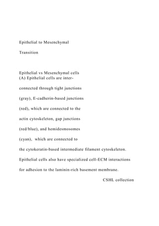
  • 1. Epithelial to Mesenchymal Transition Epithelial vs Mesenchymal cells (A) Epithelial cells are inter- connected through tight junctions (gray), E-cadherin-based junctions (red), which are connected to the actin cytoskeleton, gap junctions (red/blue), and hemidesmosomes (cyan), which are connected to the cytokeratin-based intermediate filament cytoskeleton. Epithelial cells also have specialized cell-ECM interactions for adhesion to the laminin-rich basement membrane. CSHL collection
  • 2. Epithelial vs Mesenchymal cells (B) Mesenchymal cells show a shift to a vimentin-based intermediate filament cytoskeleton and altered composition of cell-ECM interactions optimized for adhesion to the collagen-rich interstitial matrix. Mesenchymal cells also produce abundant TGFb, growth factors (GF), and matrix metalloproteinases (MMPs), as well as components of the extracellular matrix. CSHL collection Epithelial vs Mesenchymal cells Use this slide to practice describing the differences between these two types of cells Cell phenotypes in early mouse embryo.
  • 3. (A) Gastrulating mouse embryo. (C') Scanning electronic micrograph of a transverse section through a mouse embryo showing the different cell phenotypes: columnar epithelium, mesenchyme and squamous epithelium. (B) Diagram of the embryonic part of a mouse embryo. Dashed box outlines the three germ layers. (C) Scanning micrograph shown in C' color-coded for the three germ layers. EMT and its reverse MET . Diagrammatic representation of EMT, its reverse MET (mesenchymal to Epithelial Transition), egression and ingression. (A) Epithelial cells undergo EMT to assume mesenchymal characteristics. Conversely, mesenchymal
  • 4. cells undergo MET to epithelialize. (B) In an ingression, cells undergo EMT and leave an epithelium, while in an egression cells undergo MET and join a preexisting epithelium as in endoderm formation in birds and mammals. Rear-front to apico-basal polarity Categories of EMT Three types of EMT can be discerned depending on the physiological tissue context. Type1 EMT occurs in embryogenesis and organ development, type2 EMT is important for tissue regeneration and organ fibrosis, and type 3 EMT is associated with cancer progression and cancer stem cell properties.
  • 5. EMT and MET The process of MET (mesenchymal–epithelial transition) enables cells that have undergone EMT to revert to the epithelial state. MET also contributes to development and the generation of metastatic carcinomas. Consecutive rounds of EMT and MET occur during development. In 1ary EMT, epithelial cells without prior history of EMT differentiate into mesenchymal cells. In 2ary EMT, cells that have already undergone EMT and MET initiate a new EMT process, e.g. following dissemination, cancer cells can revert through MET to an epithelial state. 1ary and 2ary EMT Steps during EMT and ingression A conserved series of steps underlie EMT and
  • 6. ingression process for several developmental events. Steps during EMT and ingression Changes in cell adhesion during EMT The first cell (left) presents a cell with epithelial characteristics and apico- basal polarity. The two other cells are transitioning to mesenchymal status. An external cue (Growth Factors) starts an STP leading to cytoskeleton remodeling and loss of apico-basal polarity. From the review paper EMT-pathways External cues involved in EMT Signaling and STP from several different signal
  • 7. molecules (TGF, Wnt, Shh, and Delta/Notch), as well as changes in integrin/ECM and injury (inflammation, hypoxia) can lead to EMT. Cellular events during EMT a) The first steps of EMT are the disassembly of epithelial cell–cell contacts (tight junctions, adherens junctions, desmosomes and gap junctions) and the loss of cell polarity through the disruption of the Crumbs (Crb), partitioning defective (PAR) and Scribble (SCRIB) polarity complexes. Cellular events during EMT b| Next, the epithelial actin
  • 8. architecture reorganizes, and cells acquire motility and invasive capacities by forming lamellipodia, filopodia and invadopodia, and by expressing matrix metalloproteinases (MMPs) that can degrade extracellular matrix (ECM) proteins. Changes in cell adhesion during EMT The DDR1 complex activates RhoE, which weakens actomyosin contractility at points of cell-cell contacts (Adherens junctions, AJ). TGF receptors located next to tight junctions (TJ) trigger
  • 9. ubiquitylation and degradation of RhoA and the destabilization of cortical actin filaments associated with TJ. From the review paper EMT-pathways Changes in cell adhesion during EMT Activation of transcriptional repressor, such as Snail and Serpent (Srp/GATA) downregulates genes encoding junctional proteins, including E-cadherins, claudins and occludin, thus compromising further AJ and TJ. EPB4- 1L5 sequesters p120-catenin weakening AJ attachment to microfilaments and promotes integrin-ECM adhesion. Integrin also leads to the activity of Srp, which represses the apical polarity gene Crumbs (Crb). From the review paper
  • 10. TFs during EMT EMT is driven by SNAIL, zinc-finger E-box-binding (ZEB) and basic helix–loop–helix (bHLH) transcription factors that repress epithelial marker genes and activate genes associated with the mesenchymal phenotype. These TFs are themselves regulated through their cellular localization and stability. Snail regulation Post-translational modifications (PTM) of the different TFs regulate their activities, subcellular localization and stability. For example phosphorylation (PTM) of Snail by GSK3β leads to its export from the nucleus (and therefore its inactivity as a TF). Snail phosphorylation by PAK1 on
  • 11. the other hand promotes the nuclear retention of SNAIL1 and enhances its activity. Twist and ZEB regulation TWIST is phosphorylated by the MAPK p38, JNK and ERK, which protects it from degradation, and thus promotes its nuclear import and functions. ZEB2 undergoes another PTM ( sumoylation, Sumo by Polycomb repressive complex 2 (PRC2) and is subsequently exported from the nucleus, which reduces its activity as a transcription factor. EMT during mouse gastrulation (A) Scheme of the embryonic part of a early mouse embryo. Dashed box
  • 12. outlines the primitive streak. (B') Scanning EM showing a transverse section through a mouse primitive streak. (C) Cells undergo an EMT event at the primitive streak. (D) Signaling pathways that regulate the different EMT steps at the murine primitive streak. EMT during mouse gastrulation (C) Cells undergo an EMT event at the primitive streak. (1) First intercellular spaces appear between cells and (2) basal lamina breaks down. (3) Cells acquire a bottle shape, (4) round up as they travel through the streak, and
  • 13. (5) finally acquire a stellate morphology and migrate away from the streak. EMT during mouse gastrulation EMT during mouse gastrulation Signaling in cells located in the epiblast (left) ensures maintenance of the epithelial phenotype via the expression of Pofut2 and Sox3. Upon activation by Wnt, TGF and FGF proteins, cells in the primitive streak (middle) undergo apical constriction (indicated by green ring of contracting actin) to allow invagination before ingression. EMT during mouse gastrulation Blue cells in the primitive
  • 14. streak express Eomes, Mesp1 and Mesp2, which are responsible for ingression. RhoA downregulation by Net1 destabilizes basal microtubules and contributes to the breakdown of the basement membrane. Snail represses Sox3 and E-cadherin, which is also downregulated post-transcriptionally (PTM). In the migratory cells (right), Rac1 mediates cell protrusion. Another EMT example Signal molecules such as Wnt, BMP4 retinoic acid (RA) and FGF lead to EMT. Try to follow the arrows and recognize some of the players from previous slides. The point
  • 15. here is not to memorize this pathway, but to understand common themes and players. Questions 1) Cite the two basic embryological processes for cell determination and explain the main difference between them. 2) The experiment below, using sea urchin embryos, illustrate both processes for cell determination. Identify which one is represented by each part of the experiment (A and B) and explain your decision briefly. A
  • 16. Abnormal ~ normal development development normal normal development development B Paper-based 1) In all the figures where electroporation of DNA in chicken embryos is performed, the authors show a panel with GFP. Why is GFP electroporated in these cells, together with the experimental gene (Snail or Sox)? What kind of control and information does the GFP signal provide? You may want to read about electroporation. (https://en.wikipedia.org/wiki/Electroporation) 2) Based on information in Figure 1E add the appropriate arrow ( or ) between Snail and RhoB as you would find in a GRN. Justify your choice briefly by describing precisely the result presented in 1E Snail RhoB
  • 17. 3) If you were to analyze the promoter sequence of the RhoB gene, what would you expect to find? Explain briefly · Sequences for snail to bind as an activator · Sequences for snail to bind as a repressor · No binding sequences for Snail 4) The figure to the right is parts 1F and 1G of the paper, together with a schematic dorsal view of an early chicken gastrula. The dotted white lines in F and G represent the position of the primitive streak. Indicate on the schematic where the electroporation was done. 5) Which cellular structure contains laminin? How is the absence of laminin on the electroporated side consistent with the function of snail in EMT? For this question, you want to also review the steps involved in the epithelial to mesenchymal transition (PowerPoint on OAKS). 6) This is part of Figure 3. a)Describe panels B and N, as well as P and Q in your own word, explaining in particular the effect on Sox3 expression. b) Predict the expected data for laminin expression. Would it be up or down in the electroporated area compared to normal?
  • 18. Justify your answer. 7) Explain what are gene markers 8) Based on the data presented in Figure 6A,B, and F fill, the GRN below with the correct arrows (. or ). Justify each placed arrow briefly by describing the appropriate part of
  • 19. the figure Developmental Cell Article Reciprocal Repression between Sox3 and Snail Transcription Factors Defines Embryonic Territories at Gastrulation Hervé Acloque,1,3 Oscar H. Ocaña,1 Ander Matheu,2 Karine Rizzoti,2 Clare Wise,2 Robin Lovell-Badge,2 and M. Angela Nieto1,* 1Instituto de Neurociencias, CSIC-UMH, San Juan de Alicante 03550, Spain 2National Institute for Medical Research, London NW7 1AA, UK 3Present address: UMR 444, INRA-ENVT, Génétique Cellulaire, Toulouse 31326, France *Correspondence: [email protected] DOI 10.1016/j.devcel.2011.07.005 Open access under CC BY license. SUMMARY In developing amniote embryos, the first epithelial- to-mesenchymal transition (EMT) occurs at gastrula- tion, when a subset of epiblast cells moves to the primitive streak and undergoes EMT to internalize and generate the mesoderm and the endoderm. We show that in the chick embryo this decision to inter- nalize is mediated by reciprocal transcriptional re-
  • 20. pression of Snail2 and Sox3 factors. We also show that the relationship between Sox3 and Snail is conserved in the mouse embryo and in human cancer cells. In the embryo, Snail-expressing cells ingress at the primitive streak, whereas Sox3-posi- tive cells, which are unable to ingress, ensure the formation of ectodermal derivatives. Thus, the subdi- vision of the early embryo into the two main territo- ries, ectodermal and mesendodermal, is regulated by changes in cell behavior mediated by the antago- nistic relationship between Sox3 and Snail transcrip- tion factors. INTRODUCTION The shaping of the early embryo involves the conversion of a single layer of ectodermal cells (the epiblast) into a multilayered structure. This complex biological process starts at gastrulation, when a subset of the initial epiblast cells moves inside the embryo to become mesoderm and endoderm (Stern, 2004). In amniotes, gastrulation occurs where Nodal signaling is strongest (Brennan et al., 2001; Bertocchini and Stern, 2002). Initially, in the chick embryo, cells accumulate at the posterior part through epithelial cell intercalation (Voiculescu et al., 2007) in a region now devoid of the hypoblast, a lower layer that inhibits Nodal
  • 21. signaling (Bertocchini and Stern, 2002). This accumulation results in the formation of a midline linear structure called the primitive streak, from which the presumptive mesendodermal cells ingress upon undergoing an epithelial-to-mesenchymal transition (EMT). EMT involves a dramatic change in cell morphology and behavior that allows cells to break the basal 546 Developmental Cell 21, 546–558, September 13, 2011 ª2011 Els lamina, internalize, and start migrating toward their destinations (Harrisson et al., 1991; Acloque et al., 2009). Cells that remain in the epiblast keep their epithelial character and will contribute to the ectodermal derivatives, namely the epidermis, the ecto- dermal placodes, and the anterior central nervous system (CNS) (Fraser and Stern, 2004). Indeed, much of the CNS will develop from a subset of the noningressing cells later specified as neural precursors. Therefore, it is crucial to identify not only those factors that induce cell ingression at gastrulation but also those that prevent it, because protection from undergoing EMT is necessary to ensure the formation of ectodermal deriva-
  • 22. tives. Indeed, previous studies have shown that committed neural progenitor cells at the anterior part of the primitive streak are protected from signals that induce internalization. The zinc finger transcription factor Churchill and its target SIP-1 are required to stop ingression movements through the anterior primitive streak (Sheng et al., 2003). This safeguard mechanism operates at stages of neural induction and onset of Churchill expression (from stage HH4+; Hamburger and Hamilton, 1951), but ingression starts at least as early as stage HH2 (Stern and Canning, 1990), suggesting that a different mechanism must exist to protect early ectodermal cells from the EMT inducers. Among the key factors that induce EMT at gastrulation and that are conserved during evolution are the members of the Snail family (Barrallo-Gimeno and Nieto, 2005). They are fundamental for EMT at gastrulation in all species analyzed and for additional developmental EMT processes (Acloque et al., 2009). We previ-
  • 23. ously found that Snail2 (Slug) downregulation prevented migra- tion from the primitive streak in the chick embryo (Nieto et al., 1994), and here we show that Snail2 is sufficient to induce ectopic delamination in otherwise noningressing epiblast cells, confirming that these cells need to be protected from Snail2 expression and subsequent ingression. Therefore, we set out to identify factors that might prevent Snail expression at early gastrulation, as candidates to play an important role in protecting epiblast cells from undergoing EMT. We show that Sox3 and Snail2 are expressed in complementary domains early during gastrulation; gain- and loss-of-function studies reveal that these factors antagonize each other to regulate cell ingression. We show that Sox3-Snail antagonism is implemented through direct reciprocal transcriptional repression, a relationship that seems to be conserved in the mouse embryo and in tumor cell lines, where they also regulate epithelial versus mesenchymal and evier Inc. mailto:[email protected]
  • 24. http://dx.doi.org/10.1016/j.devcel.2011.07.005 http://creativecommons.org/licenses/by/3.0/ Figure 1. Snail2 Expression in the Epiblast Induces Ectopic EMT and Cell Delamination (A–C) Chick embryos were coelectroporated with a vector encoding GFP and another containing the coding region of Snail2. The embryos were subsequently allowed to develop for 15 hr. Endogenous Snail2 expression at the primitive streak and ectopic expression induced by elec- troporation could be observed in a dorsal view (B) and in a transverse section through the epiblast (C). Observe the delamination of cells from the epiblast. Dotted line in (B) indicates the level of the section shown in (C). Electro- poration of a control empty vector (pCX) or that containing GFP failed to induce cell delamination (not shown). (D–H) Snail2 electroporation was accompanied by the induction of RhoB expression, a known Snail2 down- stream target (D and E), cell delamination, and disruption
  • 25. of the basement membrane as assessed by the absence of laminin staining (F and G) and by the repression of E-cadherin expression in the electroporated cells (H). Developmental Cell Sox3 and Snail Define Embryonic Territories invasive properties. Together, our results show that Snail-Sox3 cross-repression regulates cell ingression at gastrulation in amniotes and suggest that this antagonistic relationship may also have important implications in cancer. Developmental Cell 21, 546–5 RESULTS Snail2 Induces Ectopic Cell Delamination in the Ectoderm of the Early Chick Gastrula We had previously shown that Snail2 knock- down in the early chick embryo prevents cell ingression at the primitive streak and neural crest delamination from the neural tube (Nieto et al., 1994). To check whether Snail2 is suffi- cient to trigger EMT and cell delamination, we ectopically expressed the coding region of
  • 26. Snail2 by electroporation of the chick blasto- derm at stage 2 (Hamburger and Hamilton, 1951) in the ectodermal region corresponding to the prospective neural plate (see Experi- mental Procedures). Snail2 ectopic expression induces cell delamination (Figures 1A–1C), sug- gesting that Snail2 is sufficient to trigger EMT in a territory that normally keeps its epithelial integrity at gastrulation. To confirm that this induced delamination is due to the activation of a full EMT program, we examined the expres- sion of previously described Snail2 target genes known to be necessary for cell delamination, such as the small GTPase RhoB (del Barrio and Nieto, 2002) and E-cadherin (Cano et al., 2000). As shown in Figures 1D and 1E, Snail2 induces ectopic expression of RhoB, together with the disruption of basal lamina, as assessed
  • 27. by the loss of laminin expression (Figures 1F and 1G). Figure 1H shows that E-cadherin is down- regulated in electroporated cells that undergo EMT, and cells can be seen delaminating from the epiblast (compare the electroporated and the control sides). These data indicate that Snail2 is sufficient to trigger EMT and cell delamination from the chick epiblast, suggesting that the latter should be protected from Snail ex- pression in the early embryo. 58, September 13, 2011 ª2011 Elsevier Inc. 547 Figure 2. Snail2 and Sox3 Genes Are Complemen- tarily Expressed in the Chick Gastrula (A–C) Snail2 expression starts at the posterior part of the blastula at stage EGXIV (A), and its expression is associ- ated with the primitive streak as it progresses in the gas- trulating embryo (B and C). (D–F) Sox3 expression is detected in the epiblast and it gets progressively downregulated at the primitive streak
  • 28. as it forms. (G and H) Double labeling for Snail2 (green) and Sox3 (red) transcripts at the full primitive streak stage (HH4) shows the complementary expression pattern between Snail2 and Sox3. A transverse vibratome section taken at the level shown by the dotted line in (G) allows a better assessment of the mutually exclusive expression pattern and the continued Snail2 expression in migratory mesen- dodermal cells emanating from the streak (H). Developmental Cell Sox3 and Snail Define Embryonic Territories Snail2 and Sox3 Expression Domains Delineate Ingressing versus Noningressing Embryonic Cell Populations Because the process of cell ingression has to be tightly regulated to maintain the balance between ectodermal (noningressing) and mesendodermal (ingressing) progenitors, Snail2 is restricted around the primitive streak, where cells are internalized, from the first stages of streak formation (Nieto et al., 1994; Figures 2A–2C). To search for genes functionally equivalent to
  • 29. Churchill but at the early primitive streak stages, we focused on the Sox3 gene for several reasons: (1) it is highly expressed at epiblast of early-stage embryos, and its expression disappears in the terri- tory surrounding the primitive streak (Rex et al., 1997; Figures 2D–2F); (2) Sox3 and Snail2 show mutually exclusive expression patterns (Figures 2G and 2H); and (3) Sox2, a closely related gene, has been shown to prevent the induction of Snail2 by BMP in the dorsal neural fold (Wakamatsu et al., 2004), but it is not expressed at the early gastrulation stages in chick. There- fore, we decided to manipulate either Snail2 or Sox3 expression 548 Developmental Cell 21, 546–558, September 13, 2011 ª2011 Els in the chick blastoderm by electroporation and assess the effects on their respective expression. We first induced ectopic expression of Snail2 by electroporating in regions such as the anterior epiblast. We found that Sox3 expression was repressed in 75% of the embryos (Figures 3F–3H; black arrow in H; n = 16), which is best seen in transverse sections (Figures 3I–3K). GFP
  • 30. electroporation alone does not have the same effect (Figures 3A–3E; n = 13). We then electroporated a Snail2 dominant- nega- tive form lacking the transactivation domain (Morales et al., 2007; DN-Snail2). This led to an extension of Sox3 expression to the primitive streak up to the midline, a region that normally expresses Snail2 (Figures 3L–3Q; black arrow in N; 53%, n = 17). Conversely, ectopic Sox3 expression at the primitive streak strongly represses endogenous Snail2 expression (Figures 3R– 3X; 74%; n = 19), indicating that Sox3 and Snail2 act in a mutually antagonistic way. Next, we examined whether the observed changes in the ex- pression domains of Snail2 and Sox3 correlate with cell behavior. With this aim, we followed cell movements near the primitive Figure 3. Snail2 and Sox3 Behave as Mutual Transcriptional Repressors in the Chick Embryo (A–E) Control embryo electroporated with a vector encod- ing GFP (A) shows the normal pattern of Sox3 expression
  • 31. in a dorsal view (B) and in a transverse section (C). (D) and (E) are high power images of the areas demarcated in (C). (F–K) Similar images taken from an embryo electro- porated with Snail2 in the anterior epiblast shows the repression of Sox3 expression in the electroporated region (arrow in H and sections in I). High power images comparing the electroporated with the control side confirm Sox3 downregulation (J and K). (L–Q) Electroporation of a dominant-negative form of Snail2 (DN-Snail2) in the primitive streak extended Sox3 expression to the embryonic midline (arrow in N), as better assessed in the images shown in (O–Q). (R–X) Ectopic Sox3 expression by electroporation at the primitive streak inhibited Snail2 expression (black arrow in T and a section at different magnifications in (U–X). evier Inc. D
  • 32. BF(t=0) GFP (t=0) A Cell tracking (8h) CB 100 H G F P ps 2 E i GF Ingressed 0 50 100
  • 38. P Figure 4. Sox3 Overexpression or Snail2 Inhibition Block Ingression at the Primitive Streak (A–X) Time-lapse confocal analysis of cell movements in cultured embryos electroporated with GFP alone (A–D), GFP plus Snail2 (E–H), GFP plus Sox3 (I–L), GFP plus a dominant-negative form of Snail2 (M–P), GFP plus a dominant-negative form of Sox3 (Q–T), and GFP, Sox3, and Snail2 (U–X). BF (t = 0) and GFP (t = 0) show trans- mitted light and fluorescent images obtained 6 hr after electroporation, just before the start of time lapse anal- yses. Colored dots in (B, F, J, N, R, and V) represent the initial position of cells that were subsequently followed. Dots in (C, G, K, O, S, and W) represent the final position of those cells and the lines their tracks. (D), (H), (L), (P), (T), and (X) show the percentages of ingressing and non- ingressing cells in each condition. The white dotted lines indicate the relative position of the midline; black arrows
  • 39. and ps indicate primitive streak. Control GFP-expressing embryos showed the normal movements of epiblasts cells toward the primitive streak and subsequent ingression (C). Snail2 overexpression increases the percentage of in- gressing cells (E–H). Ectopic Sox3 expression blocked cell ingression through the primitive streak without affecting the convergence movement toward the midline (compare C and D with K and L). A dominant-negative form of Snail2 also blocked cell ingression (compare C and D with O and P), whereas a dominant-negative form of Sox3 increased ingression (compare T with D). Overexpression of both Snail2 and Sox3 could rescue the ingression behavior of epiblast cells (compare C and D with W and X). Developmental Cell Sox3 and Snail Define Embryonic Territories streak in embryos electroporated with GFP-containing control vectors or with GFP plus different Snail2 and Sox3 constructs. Cells expressing GFP converge at the primitive steak, ingress, and migrate away in the mesendoderm as expected for a normal
  • 40. embryo (Figures 4A–4D and Movies S1 and S2 available online). Overexpression of wild-type Snail2 increases the proportion of cells that ingress (Figures 4E–4H and Movies S3 and S4). After ectopic expression of Sox3, electroporated cells still converge at the primitive streak but are unable to ingress (Figures 4I–4L and Movies S5 and S6). This observation confirms that Sox3 needs to be downregulated to allow cell ingression at the primi- tive streak, but does not impair the convergence of epiblastic Developmental Cell 21, 546– cells at the primitive streak. If the defect in cell ingression is mediated by the observed Snail2 downregulation by Sox3 (Figures 3R–3X), pre- venting Snail2 function should elicit a similar defect. Indeed, overexpression of DN-Snail2 in the primitive streak also inhibits cell ingression (Figures 4M–4P and Movies S7 and S8), which is compatible with the observed activation of Sox3 expression (Figures 3L–3Q). In contrast,
  • 41. loss of Sox3 function by ectopic expression of DN-Sox3 close to the primitive streak favors cell ingression (Figures 4Q–4T and Movies S9 and S10), as also observed after Snail2 overex- pression (Figures 4E–4H). On the other hand, overexpression of Snail2 together with Sox3 and GFP in the region of endogenous ingression rescued the phenotype of Sox3 overexpression, as the cell movements observed resembled those in the control embryo (Figures 4U–4X and Movies S11 and S12). Altogether, these results strongly suggest that the decision to ingress at the primitive streak depends on the inter- actions between Snail2 and Sox3 transcription factors. Snail2 and Sox3 Are Reciprocal Direct Transcriptional Repressors Next, we investigated whether Snail2 and Sox3 can bind to each other’s promoter to directly repress expression. We found one Snail-binding site in sequences located 50 to the Sox3 coding region, which is conserved in chick, mouse, and human (Fig- ure S1). First, we used a luciferase reporter driven by the 50 558, September 13, 2011 ª2011 Elsevier Inc. 549
  • 42. A B C D E F Figure 5. Snail2 and Sox3 Are Direct Mutual Tran- scriptional Repressors (A) Conserved Snail-binding site in the Sox3 promoter (see also Figure S1). (B) Snail2 overexpression in cultured embryos repressed the activity of the Sox3 promoter, whereas a deletion that removes the Snail binding site significantly restores promoter activity. (C) Chromatin immunoprecipitation (ChIP) analysis confirms that Snail2 can directly bind to the Sox3 promoter. ChIP assays were carried out with anti-myc
  • 43. antibodies on embryo extracts that were previously elec- troporated with either GFP or myc-Snail2. PCR fragments were obtained after amplification of the indicated promoter region from the input (1), histone H3 positive control (2), IgG negative control (3), and myc immunoprecipitated fractions (4). (D) Conserved sequence for Sox transcription factors binding in the Snail2 promoter (see also Figure S1). (E) Sox3 overexpression in cultured embryos repressed Snail2 promoter activity, and the deletion of the Sox- binding site restored most of the promoter activity. (F) Chromatin immunoprecipitation (ChIP) analyses con- firmed that Sox3 can directly bind to the Snail2 promoter. ChIP assays were carried out as described above. Developmental Cell Sox3 and Snail Define Embryonic Territories Sox3 sequences (Figure 5A), to test whether Snail2 can modu- late the activity of the Sox3 promoter by coelectroporating Snail2
  • 44. with the luciferase construct in the epiblast of preprimitive streak stage embryos (see Experimental Procedures). Snail2 reduced the activity of the Sox3 promoter construct. This repression is dependent on the presence of the conserved Snail2-binding box (Figure 5B), because electroporation with a construct lacking this box significantly restores activity (Figure 5B). To determine whether Snail2 can bind directly to the Snail box in vivo, we performed Chromatin immunoprecipitation (ChIP) experiments after electroporating embryos with GFP and a myc-tagged version of Snail2. After dissecting the GFP-positive tissues, we carried out ChIP with an anti-myc antibody. A frag- ment of genomic DNA corresponding to the Snail-box containing region of the Sox3 promoter was amplified in embryos electropo- rated with the myc-tagged Snail2 protein (Figure 5C, compare line 4 for GFP and myc-Snail2 experiments). We then performed similar experiments to determine whether Sox3 acts on a similar
  • 45. way on the Snail2 promoter and found that this is indeed the case (Figures 5D–5F). The conserved Sox DNA binding sequence located in the chick Snail2 promoter is shown in Figure 5D, and its conservation in other species is shown in Figure S1. Thus, Snail2 and Sox3 can bind directly to each other’s promoter in vivo at conserved elements, providing a molecular mechanism for their antagonistic role in the cell decision to ingress at gastrula- tion. This interaction defines two main territories within the early embryos: the noningressing ectoderm and the ingressing mesendoderm. Snail2 Represses Ectodermal Markers without Inducing Mesodermal Fate In addition to a strong repression of Sox3 expression in the epiblast, Snail2 is also able to repress other ectodermal markers, such as the epidermal marker Dlx5 (Figure 6A; 100%; n = 3) and the neuroectodermal marker Otx2, albeit in a limited territory 550 Developmental Cell 21, 546–558, September 13, 2011 ª2011 Els (Figure 6B; 62%; n = 8). This is consistent with the known role
  • 46. of Snail as an epithelial repressor (Nieto, 2002). In contrast, Sox3 induces Dlx5, although it is unable to activate Otx2 expres- sion (Figure 6A; 80%; n = 5; Figure 6B; 100%; n = 11). We had observed that, in addition to repressing epithelial markers, Snail2 induces delamination of epiblast cells by trig- gering EMT (Figure 1). We wondered whether this morphological change is accompanied by the induction of mesendodermal fate, because primitive streak cells are Snail2 positive and give rise to mesoderm and endoderm. However, Snail2 electropora- tion did not induce the expression of the primitive streak and mesoderm markers Brachyury or Tbx6L in any of the embryos analyzed (Figures 6C and 6D; n = 5 and n = 8, respectively). Simi- larly, Snail2 could not induce the expression of the endodermal marker Sox17 (Figure 6E; 100%; n = 12). In turn, ectopic expres- sion of Sox3 in the primitive streak dramatically reduces the expression of mesodermal markers (Figures 6C and 6D; 100%;
  • 47. n = 8 and n = 9, respectively), as expected from the reduction observed in cell ingression (Figures 4I–4L). Sox3 is unable to induce Sox17 (Figure 6E; 100%; n = 9), in agreement with the endodermal fate being determined in cells after ingression (Ta- katori et al., 2010). Altogether, our data suggest that Snail and Sox3 do not behave as mesodermal-endodermal or neural inducers, respectively, but rather as regulators of cell behavior and movement. This is in agreement with the phenotype of Snail mutant mouse embryos, which still form mesoderm but are unable to migrate because of a defective EMT at the primitive streak (Carver et al., 2001). It is also compatible with Sox3 being an early neural marker and expressing cells becoming definitive neural only later when they express Sox2 (Linker and Stern, 2004). Furthermore, as expected from the antagonistic relation- ship between Snail2 and Sox3, the effects of Snail2 on Dlx5 and Otx2 expression depend on Sox3 suppression, because overex- pression of Snail2 together with Sox3 in the presumptive neural
  • 48. ectoderm rescued their inhibition (Figure 6F; 67%; n = 6). evier Inc. Figure 6. Snail2 Represses Neural and Nonneural Ectodermal Markers without Inducing Meso- dermal or Endodermal Fate (A–E) Snail2 or Sox3 were electroporated together with GFP in the epiblast of stage HH2 embryos. Snail2 repressed the expression of Dlx5, a nonneural ectodermal marker (A) and Otx2, an anterior neural marker (B), whereas Sox3 extended Dlx5 expression (A). Snail2 was unable to induce the expression of mesodermal markers, as assessed by examining the expression of the T-box genes Brachyury and Tbx6L (C and D) or the endodermal marker Sox17 (E), whereas Sox3 strongly repressed mesodermal (C and D) but not endodermal markers (E). Overexpression of both Snail2 and Sox3 did not affect the expression of the ectodermal markers Dlx5 and Otx2 (F), suggesting that their repression by Snail2 acts through
  • 49. Sox3 downregulation. Developmental Cell Sox3 and Snail Define Embryonic Territories These results indicate that, even though the antagonistic rela- tionship between Sox3 and Snail2 divides the early embryo in two main territories from which either the ectoderm or the mes- endoderm will form, it is cell adhesion and behavior rather than the induction of a change of cell fates that drives the segregation. Conservation of the Sox3/Snail Antagonistic Relationship in Mouse Embryos Next, we wondered whether our observations in the chick could be extended to other systems, and we compared the expression of Sox3 and Snail genes in the mouse gastrula. Snail2 is not ex- pressed in the primitive streak or early mesoderm in mouse embryos because of a reshuffling of Snail1 and Snail2 expression domains during evolution (Sefton et al., 1998; Locascio et al., 2002). Thus, we compared the expression patterns of Snail1 and Sox3. As previously described, Sox3 is strongly expressed
  • 50. in the epiblast (Wood and Episkopou, 1999; Figure 7A), and Snail1 is expressed at the primitive streak and in the mesodermal cells delaminating from it, but not in the epiblast (Sefton et al., 1998; Figure 7A). This complementary expression is compatible with the idea that, in the mouse, Snail1 and Sox3 repress each other’s transcription. This is also reinforced by the conservation of the Snail-binding site in the mouse Sox3 promoter and the Developmental Cell 21, 546–5 presence of two consensus Sox DNA-binding sequences located in conserved regions of the mouse Snail1 promoter (Figure S1). To directly examine the influence of Sox3 on Snail1 expression, we used wild-type cells (CCE cells; Robertson et al., 1986; Keller et al., 1993) or Sox3 null mouse ES cells (M. Parsons, C.W., and R.L.-B., unpublished data) that were analyzed after 5 or 8 days in the presence or in the absence of LIF, the latter allowing the generation of embryoid bodies. Snail1 expres-
  • 51. sion is activated in Sox3-deficient embryoid bodies (Figure 7B), suggesting that the absence of Sox3 leads to a derepression of Snail1 expression. Concomitant with Snail1 activation, E-cadherin was downregulated (Figure 7B), indicating the presence of aberrant expression of Snail1 and downregulation of E-cadherin, as confirmed by immunohistochemistry (Figure 7C). Further- more, although embryoid bodies derived from wild-type ES cells look round and show a compact morphology, those derived from Sox3 mutant ES cells are irregular. We excluded both the possi- bility of these cells dying and the existence of differences in the rate of cell proliferation (Figure S2). Rather, they are healthy cells that appear to disaggregate, resembling a process of EMT (Fig- ure 7C). These results pointed to a conserved antagonistic rela- tionship between Sox3 and Snail1 in the mouse. Further support for the conservation comes from the observa-
  • 52. tion of gastrulation defects in chimeras obtained after injection of Sox3 null ES cells in mouse blastocysts (M. Parsons, C.W., and R.L.-B., unpublished data). Sox3 is located on the X chro- mosome; therefore, targeted XY ES cells are null for Sox3. However, this gastrulation defect was observed in the context of chimeric embryos, because ubiquitous deletion of a floxed allele of Sox3 by bactinCre results in generation of live animals (Rizzoti et al., 2004). This suggests that the presence of Sox3 null cells in a wild-type host embryo is incompatible with a compensation mechanism that allows normal gastrulation in an entirely Sox3 null embryo. The latter is likely to involve Sox2, which, in contrast to the situation in the chick, shows similar expression at these stages in the mouse (Avilion et al., 58, September 13, 2011 ª2011 Elsevier Inc. 551 Figure 7. The Antagonistic Relationship between Sox3 and Snail Factors Is Conserved in the Mouse (A) Expression of Snail1 and Sox3 in 7,5 d.p.c. mouse embryos. Snail1 is the family member expressed in
  • 53. the primitive streak and early mesendodermal cells in the mouse (Sefton et al., 1998). Note the similarities in the expression patterns between chick and mouse embryos. A mutually exclusive expression pattern is observed in the transverse sections obtained at the level of the primitive streak. (B) Snail1 expression is increased in embryoid bodies (EB) obtained from Sox3 knockout embryonic stem cells compared to wild-type stem cells (CCE line). Consistent with the increase in Snail1 expression, E-cadherin tran- scripts are downregulated in Sox3 null EBs (T-test statis- tical analysis). (C) EBs derived from the CCE line are round and smooth, whereas those derived from Sox3 null ES cells show irregular edges with budding cells delaminating from the EBs, leading to abundant isolated cells in the culture medium (not shown). E-cadherin is strongly down- regulated in Sox3�/� compared to wild-type (WT) EBs.
  • 54. Conversely, Snail1 mRNA and protein are detected only in Sox3�/� EBs, mainly in cells at the edges. (D) Expression of Snail1 in WT and Sox3�/�; Sox2+/� mouse embryos. Ectopic Snail1 expression can be observed in the mutant embryos leading to an extended region of EMT and cell ingression. The dotted lines in the whole mounted embryos indicate the level of the sections shown in the lower panels. Red brackets indicate the areas of cell delamination in the enlarged pictures and in the diagrams, which schematically show the defective phenotype in the gastrulating embryos. Dotted lines in the diagram indicate the midline. Developmental Cell Sox3 and Snail Define Embryonic Territories 2003). Therefore, if the antagonistic relationship between Sox3 and Snail is conserved in the mouse as the EB experiments indicate, then gastrulation defects similar to those we described here in the chick should be observed after lowering the Sox2 dose in the Sox3 mutant. Because Sox2 null embryos die at
  • 55. peri-implantation stages, we analyzed Sox3 null; Sox2 heterozy- gous embryos and indeed, we found ectopic Snail1 expression and deformed embryos with an extended area of cell delamina- tion at the primitive streak (Figure 7D; n = 3). These results support and extend those found in the Sox3 null EBs, indicative that the antagonistic relationship with Snail is conserved in the mouse. Conservation of the Sox3/Snail Antagonistic Relationship in Human Cancer Cells Because Snail factors are also implicated in the repression of the epithelial phenotype and the induction of EMT during tumor progression (Thiery et al., 2009), we checked whether human cancer cell lines also show an antagonistic relationship between Snail and Sox3 transcription factors. We examined one epithelial cell line derived from breast carcinoma (MCF7) and three inde- pendent mesenchymal and invasive lines, two of them also derived from breast tumors (MDA231 and MDA435) and one 552 Developmental Cell 21, 546–558, September 13, 2011 ª2011 Elsevier Inc. from a melanoma (A375P). Sox3 is strongly ex-
  • 56. pressed in MCF7 cells and it is absent from A375P, MDA231, and MDA435. Conversely, Snail1 expression is high in MDA231, MDA435, and A375P and very low in MCF7 cells (Figure 8A). To address whether the expression of Snail1 and Sox3 is interdependent, we first inter- fered with Sox3 expression in MCF7 cells by transfecting specific siRNAs (see Experimental Procedures). We found an efficient Sox3 downregulation concomitant with an increase in Snail1 expression (Figure 8B) accompanied by the decrease in the epithelial marker Claudin1, and an increase in the mesenchymal markers Adam12 and Fibronectin (Figure 8B). We did not observe a reduction in E-cadherin levels in this transient downre- gulation of Sox3 expression. Importantly, because MCF7 cells do not express significant levels of Snail1, we added 2 ng/ml of its potent inducer TGFb to the cultures. This leads to three- fold induction of Snail1 expression, which correlates with a 50% decrease of Sox3 transcripts (Figure 8C). Interestingly, the changes in Sox3 expression were dependent on Snail1,
  • 57. because Sox3 transcript levels remained unaffected in the presence of a Snail1-specific siRNA, which prevented Snail1 induction by TGFb (Figure 8C). To examine whether the interde- pendent changes in gene expression had an impact on cell morphology and behavior, we infected the mesenchymal MDA435 cells with a retrovirus containing the Sox3 coding sequence to generate MDA435-Sox3 cells stably expressing A B C D E F G H Figure 8. The Antagonistic Relationship between Sox3 and Snail Factors Is Conserved in Human Cancer Cells (A) Expression of Sox3 and Snail1 in different human cancer cell lines. Epithelial MCF7 cells express high levels of Sox3 and low levels of Snail1. Conversely, mesenchymal MDA231, MDA435, and A375P lines are almost devoid of Sox3 transcripts and show high Snail1 expression. (B) Efficient Sox3 downregulation by specific siRNA in MCF7
  • 58. cells is accompanied by an increase in Snail1 and associated mesenchymal markers Adam12 and Fibronectin and a decrease in the epithelial marker Claudin1. (C) TGF-b mediated Snail1 induction in MCF7 cells is accompanied by a decrease in Sox3 expression. This decrease is Snail1-dependent, as it is prevented by transfection with Snail1 siRNA. (D and E) Sox3 stable overexpression in MDA435 leads to morphological changes compatible with a partial mesenchymal to epithelial transition (D). These morphological changes are associated with a decrease in Snail2 expression and an increase in E-cadherin expression (E). (F) The migratory behavior of MDA435-Sox3 stable transfectants was tested in culture using a wound healing assay. Control MDA435 cells cover the wound in 12 hr, in clear contrast with Sox3-expressing cells. (G) Invasive behavior of MDA435-Sox3 cells. The nuclei of cells that invaded the collagen matrix were stained with DAPI and quantified. MDA435 cells expressing Sox3 significantly decreased their invasive properties. (H) Diagram showing the relationship between Snail and Sox3 factors in gastrulating embryos and in cancer cells. In gastrulating embryos, the mutual repression between Sox3 and Snail1 regulates the decision to ingress at the primitive streak. Sox3 expressing cells do not ingress, ensuring
  • 59. the development of ectodermal derivatives in developing embryos. In cancer cells, Snail induces EMT and its expression correlates with invasive properties. Sox3 represses Snail, thereby preventing EMT to maintain epithelial integrity. Developmental Cell Sox3 and Snail Define Embryonic Territories Developmental Cell 21, 546–558, September 13, 2011 ª2011 Elsevier Inc. 553 Developmental Cell Sox3 and Snail Define Embryonic Territories this transcription factor. We observed morphological changes compatible with a partial mesenchymal-to-epithelial transition (Figure 8D), concomitant with the decrease in Snail2 expression and the reactivation of E-cadherin transcription (Figure 8E). We next examined cell behavior and found that Sox3 induced a dramatic decrease in cell motility as assessed in a wound healing assay in culture (Figure 8F) and a decrease cell invasion in collagen gels (Figure 8G). In summary, the antagonistic rela-
  • 60. tionship between Snail1 and Sox3 is maintained in cancer cells, and their relative expression correlates with their morphological, motility and invasive properties. DISCUSSION One of the earliest cellular decisions in metazoans is the subdi- vision of the early embryo into the domains that will give rise to the different embryonic layers. In amniote embryos, the first subdivision occurs at the primitive streak, where ingressing cells will later become mesoderm and endoderm and the noningress- ing cells will become ectoderm. Here we show that the partition- ing of these cellular domains at early primitive streak stages is regulated by interactions between two transcription factors, Snail and Sox3, which direct ingressing versus noningressing behaviors, respectively. In the chick embryo, Snail2 and Sox3 act as mutual transcriptional repressors; cells that express high levels of Snail2 are devoid of Sox3 transcripts and ingress
  • 61. through the primitive streak. In contrast, cells expressing high levels of Sox3 lack Snail2 expression and stay in the epiblast. Both Sox3 ectopic expression and Snail2 inhibition close to the primitive streak prevent cell ingression while still permitting the movement toward the midline. In turn, Snail2 overexpression or Sox3 inhibition increases cell ingression at the primitive streak. This indicates that the interplay between Snail2 and Sox3 controls the delamination from the epiblast, thereby ensuring the subdivision of the embryo into two populations, ingressing and noningressing, that will later give rise to the mesendoderm and to much of the ectoderm, respectively. At later stages, the posterior neural tube arises from a population of bipotent axial stem cells set aside within the region of the tail bud (Wilson et al., 2009). These stem cells can become posterior neural tissue or undergo EMT to become paraxial mesoderm, and their ingres- sion depends on the repression of Sox2 by Tbx6, as recently
  • 62. shown in the mouse (Takemoto et al., 2011). Our data show that Sox3 and Snail behave as direct repres- sors of each other. Snail is a well-known transcriptional repressor that controls cell movements both in embryonic devel- opment and tumor progression (Thiery et al., 2009). Interestingly, although the genes of the SoxB1 subgroup (Sox1, Sox2, and Sox3) are usually described as transcriptional activators (Uchi- kawa et al., 1999), here we show that like the SoxB2 subgroup genes (Sox14 and Sox21), Sox3 can also function as a transcrip- tional repressor depending on the context, as also described for Sox2 during the differentiation of ES cells (Navarro et al., 2008). We demonstrate that Sox3 directly binds to the promoter of its target gene, Snail2. This is in agreement with recent findings in the zebrafish embryo showing that Sox3 can repress the expres- sion of Bozozok, a homeobox gene important for the formation of the dorsal organizer and subsequent gastrulation movements (Shih et al., 2010). We also show that Sox3 represses the
  • 63. activity 554 Developmental Cell 21, 546–558, September 13, 2011 ª2011 Els of a Snail2 promoter construct in vivo, now confirming that, in addition to a transcriptional activator, Sox3 can be considered as a bona fide transcriptional repressor, depending on context. Our description of the involvement of Sox3/Snail interactions in defining embryonic territories in the chick is consistent with the gastrulation defects observed in chimeras obtained after injection of Sox3 null ES cells in mouse blastocysts. This gastru- lation phenotype observed in the chimeras was difficult to explain, considering that Sox3 is never expressed in the meso- derm at gastrulation stages. However, our data on the regulation of Snail1 and Sox3 expression in embryoid bodies obtained from mouse Sox3 null ES cells and our analysis of gastrulating Sox3 null; Sox2 heterozygous embryos provide a simple explanation for the gastrulation defects first observed in the chimeras con- taining Sox3 null ES cells and confirm the conservation of the interplay between Sox3 and Snail in defining ectodermal and
  • 64. mesendodermal territories. Our data further clarify recent data showing multiple defects, including gastrulation defects, in zebrafish embryos after down- regulation of the full complement of SoxB1 genes (Okuda et al., 2010). The downregulation of individual SoxB1 genes does not give rise to severe defects in the fish, reflecting the overlap in the expression patterns of several family members, particularly for Sox3 and the fish-specific Sox19a and 19b (Okuda et al., 2006). In addition, in the fish embryo, the formation and migration of mesendoderm does not involve a full EMT, but rather cells very quickly re-express the homolog of E-cadherin (Cdh1) and migrate as a cohesive group (Montero et al., 2005). In the fish, Cdh1 re-expression is necessary for the proper migration of the mesendodermal cells (Montero et al., 2005) and indeed it occurs concomitantly with the loss of snail1a, which is only transiently expressed in the involuting mesoderm (Blanco et al., 2007). Furthermore, a dominant-negative form of Sox3 in
  • 65. the zebrafish induces the formation of multiple organizers (Shih et al., 2010), indicating that in the fish, as we show here in the amniote embryo, Sox3 needs to be downregulated for gastrula- tion to proceed normally. Interestingly, both in fish and Xenopus, gastrulation starts concomitantly with the transient activation of Snail expression (Blanco et al., 2007; Mayor et al., 2000). Thus, the antagonism between Sox3 and Snail factors shown here in the amniote embryo, although not directly examined in zebrafish or Xenopus, may contribute to the initiation of the gastrulation process and may therefore be conserved not only in amniotes but also in anamniote embryos. A crucial issue that also emerges at gastrulation is the neces- sary coordination between cell fate and cell behavior. Snail has been considered as a mesodermal determinant from studies in Drosophila where it is a repressor of nonmesodermal genes. However, we would like to argue that the role of Snail is indepen-
  • 66. dent from cell fate determination, its main role being the regula- tion of cell behavior. Our data indicate that Snail2 can trigger an ectopic EMT and cell delamination from the epiblast but is unable to induce the expression of mesodermal or endodermal markers. This is in agreement with data from Drosophila indi- cating that low levels of snail do not repress nonmesodermal genes in the presumptive mesoderm while still able to promote invagination (Ip et al., 1994) and it is also consistent with data from the mouse showing that Snail1-deficient embryos can form mesodermal tissue that expresses Brachyury and Tbx6 evier Inc. Developmental Cell Sox3 and Snail Define Embryonic Territories but these cells are unable to migrate due to defects in EMT and the continued expression of E-cadherin (Carver et al., 2001; Mur- ray and Gridley, 2006). Indeed, fate determination and cell delamination seem to be two independent processes, both
  • 67. driven by FGF signaling through FGFR1 in the gastrulating mouse, exerted on one hand by maintaining the expression of Snail and on the other by controlling the expression of the meso- dermal genes Brachyury and Tbx6 (Ciruna and Rossant, 2001). Thus, our data show that the definition of the two main embry- onic territories in the early gastrulating embryo, namely the ectoderm and the mesendoderm, is governed by the control of cell behavior driven by the antagonistic role between Snail and Sox3 factors independently from cell specification, which is concomitantly coordinated by FGF signaling. Recent data from ascidian embryos indicate that the subsequent decision for the mesendoderm to subdivide into mesoderm or endoderm is determined by the asymmetric partitioning of the Not transcrip- tion factor in the cells destined to become mesodermal, a mech- anism that is likely to be conserved in vertebrates because Not has been already described in Xenopus, fish, and chick embryos (Takatori et al., 2010). Pioneer work in the chick embryo showed that when a subset
  • 68. of ectodermal cells is specified to become the nervous system, the expression of another transcription factor, Churchill, acts as an important switch, preventing the ingression of prospective neural plate cells through the anterior part of the primitive streak from late stage HH4 onward (Sheng et al., 2003). The Sox3/Snail switch that we describe here to control cell ingression occurs before neural induction and before the onset of Churchill ex- pression. The two mechanisms are sequentially implemented in the embryo, with the Snail/Sox3 axis acting first to ensure that a subpopulation of ectodermal cells stays in the epiblast. There- fore, the interplay between Sox3 and Snail controls the first subdivision of embryonic territories. Subsequently, upon neural induction, Churchill ensures that the subpopulation of ecto- dermal cells already specified as neural precursors do not ingress through the primitive streak to become mesoderm or endoderm. Finally, our data obtained in human cancer cell lines suggest
  • 69. that the mutual repression between Sox3 and Snail is also in place. Epithelial tumor cells express high levels of Sox3 and low levels of Snail1, and the opposite is true for mesenchymal tumor cells. Not only are Snail1 and Sox3 expression levels associated with the morphological and invasive phenotype, but also interference with Sox3 or Snail1 expression induces recip- rocal changes in their expression, compatible with the existence of a loop of mutual repression, as described in embryos. These data may have important implications in tumor biology, as Snail reactivation and EMT contributes to the first steps of the meta- static cascade in carcinomas and it is considered a target of anti-invasive drugs (Thiery et al., 2009; Nieto, 2011). Therefore, it is important to identify not only how Snail is reactivated in tumors but also to identify its negative regulators. We propose Sox3 as a likely candidate. EXPERIMENTAL PROCEDURES Chick Embryos and Explant Cultures Fertilized hen eggs were purchased from Granja Gilbert (Tarragona, Spain).
  • 70. The eggs were incubated and opened, and the embryos were explanted for Developmen EC culture as described elsewhere (Flamme, 1987; Chapman et al., 2001). Embryos were staged according to Eyal-Giladi and Kochav (1976) (EG) and Hamburger and Hamilton (1951) (HH), selecting HH2 embryos for experiments. Mouse Embryos, ES Cells, and Human Cell Lines Mouse embryos were obtained by crossing C57 and CBA mice. Embryos dissected at 7.5 dpc were fixed overnight in 4% paraformaldehyde. CCE mouse ES cells (Robertson et al., 1986; Keller et al., 1993) were cultured in cell culture dishes with DMEM (Invitrogen) supplemented with 10% serum and LIF (1000 U/ml, Chemicon International). Embryoid bodies were grown on bacteriological grade plastic dishes in the same medium in the absence of LIF, and total RNA was extracted with Trizol (Life Technologies) after
  • 71. different times in culture. MCF7, MDA231, MDA435, and A375P human tumor cell lines were purchased from the ATCC (Virginia, USA) and were cultured in DMEM supplemented with 10% heat inactivated serum and 0.1% penicillin- streptomycine (Invitrogen). Cells were transfected with negative control for siRNAs or with those directed against Snail1 (3 sequences tested) or Sox3 (5 sequences tested) using Lipofectamine RNAiMAX following the manufac- turer’s instructions (Invitrogen). For RNA/Lipofectamine complex formation, siRNAs were used at a working concentration of 100 nM. Because they were able to downregulate expression with different efficiencies, we present the data obtained with the most efficient oligonucleotide in each case, whose sequences are shown in Table S1. RNA was isolated 2 days after transfection for the Sox3 interference experiment. When indicated, 2 ng/ml TGFb was
  • 72. added to the cells 24 hr after Snail1 siRNA transfection. Total RNA was ob- tained at 1 hr, 24 hr, and 48 hr after TGFb administration, and in all cases Snail1 induction was impaired (Figure S3). Total RNA was purified using the illustra RNAspin Mini kit including DNaseI treatment (GE Healthcare). Stealth siRNA (Invitrogen) sequences were as follows: 50- UCCCAGAUGAGCAUUGG CAGCGAGG-30 against human Snail1 (SNAI1) and 50- AGUUCCAGGGUU AUUCUGUUACAUU-30 against human SOX3. Viral Production and Generation of Sox3 Stable Expressing Cells Retroviral production and infection were carried out as previously described (Mani et al., 2008). After infection, MDA435 cells expressing either pBabe- PURO or pBabe-PURO-Sox3 were selected with 10 mg/ml puromycin for 2 weeks. Migration and Invasion Assays For migration assays, cells were seeded in six-well culture dishes at a density
  • 73. of 1 3 106 cells/well. A wound was made in the center of the culture 24 hr later, and phase-contrast pictures were taken at different time intervals. Invasion assays on collagen type-IV gels were performed as previously described (Cano et al., 2000). Briefly, 6 3 104 cells of each type were seeded onto the upper surface of the filters. After 12 hr of incubation, cells attached in the lower part of the filters were fixed in methanol, stained with 4,6-diamidinophe- nylindole (DAPI) and counted. Electroporation of Chicken Embryos Explanted embryos at HH2 were placed, vitelline membrane and filter paper down, over an electroporation chamber (NEPAGEN) containing a platinum electrode connected to the negative pole. A solution containing expression plasmids (2 mg/ml in PBS with 0.1% Fastgreen and 6% sucrose) was injected between the vitelline membrane and the epiblast. An anodal
  • 74. electrode was placed over the hypoblast to cover the injected area. A train of electric pulses (5 pulses, 4 Volts, 50 ms, and 0.5 Hz) was applied using an Intracept TSS10 pulse stimulator (Intracell). In all experiments, the nonelectroporated right side of the embryo was used as a control. The embryos were then cultured at 38�C (Chapman et al., 2001) to the desired stage. Embryos were photo- graphed with a Leica MZFLIII dissecting microscope to record GFP expression and fixed overnight in 4% paraformaldehyde (PFA) in PBS at 4�C to be pro- cessed for in situ hybridization or immunohistochemistry. Cell death that might have resulted from the electroporation procedure was excluded as a factor to influence cell behavior (Figure S2). Time-Lapse Confocal Imaging Six hours after electroporation, chicken embryos at stage HH3+ were washed in PBS and placed into a glass-bottom culture 35 mm Petri dish (MatTek)
  • 75. tal Cell 21, 546–558, September 13, 2011 ª2011 Elsevier Inc. 555 Developmental Cell Sox3 and Snail Define Embryonic Territories containing egg albumin. The dish was then located into an incubation chamber at 38�C surrounding a Leica inverted confocal microscope for image acquisi- tion. One image was captured each 10 min for a total of 8 hr. Movies were assembled using the ImageJ software (http://rsbweb.nih.gov/ij/). Individual cells were tracked using the ‘‘Manual Tracking’’ plug-in by F. Cordelières (http://rsbweb.nih.gov/ij/plugins/track/track.html). Ingression was quantified as the percentage of ingressing and noningressing cells after tracking 20 cells per field in three fields per movie. DNA Constructs pCX-Snail2, pCX-DN-Snail2, and pCX-GFP expression vectors were previ- ously described (Morales et al., 2007). Full-length Sox3 or a
  • 76. truncated domi- nant negative form of Sox3 similar to that previously described for the Sox2 Xenopus gene (Kishi et al., 2000) were cloned in pCX at the EcoRI restriction site. Snail2 and Sox3 promoters were PCR amplified from chick genomic DNA using Phusion High Fidelity DNA polymerase (Finnzyme) (see Table S1 for primers sequences), sequenced, and inserted in pGL2 basic using the KpnI and MluI restriction sites. For viral production, the coding sequence of human Sox3 was amplified by PCR (see Table S1 for primers sequences) and inserted in the pBabe-PURO vector using the EcoRI restriction site. Whole-Mount In Situ Hybridization Whole-mount in situ hybridization was carried out as described previously (Nieto et al., 1996) omitting the proteinase K treatment. Digoxigenin-labeled probes were synthesized from the full-length chicken cDNAs of Brachyury,
  • 77. Otx2, RhoB (Liu and Jessell, 1998), and Snail2 (Nieto et al., 1994) and from Expressed Sequence Tags (EST; Boardman et al., 2002) for Dlx5 (ChEST808h7), Tbx6L (ChEST90h8), and Sox17 (pgr1n.pk001.g24; Chapman et al., 2007). Chicken and mouse Sox3 sequences were PCR amplified from chicken and mouse genomic DNA (Table S1) and cloned in pGEMT-easy. Mouse Snail1 probe was previously described (Sefton et al., 1998). Hybridized probes were detected using an alkaline phosphatase-conjugated anti-digoxi- genin antibody (Roche, 1:1000) in the presence of NBT/BCIP substrates (Roche). For whole-mount fluorescent in situ hybridization, embryos were processed as previously described (Acloque et al., 2008). Briefly, probes were labeled using digoxigenin- or fluorescein-coupled nucleotides (Roche, 1:1000) and were sequentially developed with POD-conjugated anti-fluores-
  • 78. cein or anti-digoxigenin antibodies (Roche). Peroxidase activity was succes- sively detected with the TSA-plus Cy3 and Fluorescein kits (Perkin Elmer). In some cases, the embryos were subjected to immunostaining with anti-GFP antibody (Invitrogen, 1:1000). After hybridization and/or immunohistochem- istry, embryos were fixed in 4% paraformaldehyde in PBS, washed, and pho- tographed under a Leica M10 dissecting scope. Some embryos were subse- quently embedded in paraffin (Fibrowax) or gelatin, sectioned at 10 mm or 40 mm, respectively, and photographed using a Leica DMR microscope under Nomarski optics and equipped with an Olympus DP70 digital camera. Immunohistochemistry For immunohistochemistry, electroporated embryos were fixed in PFA 4% in PBS. For laminin detection, 10 mm cryostat sections were treated with 0.1%
  • 79. Triton X-100 (Sigma) in PBS, blocked with 10% FBS in PBS and incubated overnight at 4�C with anti-laminin (primary antibody (Sigma) at 1:1000 dilution. For E-cadherin detection, chick embryos were embedded in paraffin and sectioned at 8 mm. Immunostaining was performed by standard procedures using anti-GFP antibody (rabbit polyclonal, Invitrogen; 1:500) and anti-E-cad- herin (mouse monoclonal, BD Bioscience; 1:250). After washing, sections were incubated for 1 hr with Alexa488 (Invitrogen, 1:1000) and Cy3 conjugated (Jackson; 1:1000) secondary antibodies and photographed using a Leica DMR microscope. Embryoid bodies were fixed with ice-cold methanol, rehydrated, and immu- nostained by standard procedures using anti-E-cadherin (mouse monoclonal ECCD-2, Takara; 1:250) or anti-Snail1 (Abcam). Images were acquired using a Leica inverted confocal microscope.
  • 80. Chromatin Immunoprecipitation Chicken embryos were electroporated either with GFP and control myc-Tag, GFP and myc-Snail2, or GFP and myc-Sox3 expression plasmids. Eight hours after electroporation, GFP-positive tissues were dissected from HH5 embryos. 556 Developmental Cell 21, 546–558, September 13, 2011 ª2011 Els Tissues were crosslinked with 1% formaldehyde in PBS for 10 min at room temperature and quenched with 0.125 M glycine for 5 min at room tempera- ture. Tissues were then washed three times in PBS and resuspended in SDS lysis buffer (1% SDS, 10 mM EDTA, and 50 mM Tris [pH 8.1]) using 100 ml of buffer for a pool of 15 embryos (corresponding to approximately 1 3 105 cells). Lysates were sonicated in an ultrasonic cell disrupter (Bioruptor, Diagenode SA, Belgium) for 8 min, with alternating 30 s off and on, frozen in liquid nitrogen and stored at �80�C. Each sonicated lysate (about 1 3 105 cells) was diluted in 900 ml of buffer (0.01% SDS, 1.1% Triton X-100, 1.2 mM
  • 81. EDTA, 16.7 mM Tris [pH 8.1], and 167 mM NaCl) in the presence of protease inhibitors. Ten microliters was recovered as the input fraction, and the rest was divided and incubated overnight at 4�C with anti-myc ChIP grade (ab9132, Abcam, UK), anti-H3 ChIP grade (ab1791, Abcam, UK), or rabbit IgG control (Diagenode, Belgium) using 1 mg of antibody for tissue lysate. Immunoprecipitation of crosslinked Protein/DNA was performed adding 60 ml of a slurry of Protein A Agarose beads (Roche) previously saturated with salmon sperm DNA (1 mg/ml) and BSA (1 mg/ml). Complexes were washed using Low Salt Washing Solution (0.1% SDS, 1% Triton X-100, 2 mM EDTA, 20 mM Tris [pH 8.1], and 150 mM NaCl), High Salt Washing