1. Higher-order nuclear organization in growth arrest of human
mammary epithelial cells: a novel role for telomere-associated
protein TIN2
Patrick Kaminker1, Cedric Plachot2, Sahn-Ho Kim3, Peter Chung3, Danielle Crippen1, Ole
W. Petersen4, Mina J. Bissell3, Judith Campisi1,3, and Sophie A. Lelièvre2,*
1 Buck Institute for Age Research, 8001 Redwood Boulevard, Novato, California 94945, USA
2 Department of Basic Medical Sciences and Cancer Center, Purdue University, 625 Harrison Street,
West Lafayette, Indiana 47907-2026, USA
3 Life Sciences Division, Lawrence Berkeley National Laboratory, 1 Cyclotron Road, Berkeley,
California, 94720, USA
4 Structural Cell Biology Unit, The Panum Institute, University of Copenhagen, Blegdamsvej 3,
DK-2200, Copenhagen, Denmark
Summary
Nuclear organization, such as the formation of specific nuclear subdomains, is generally thought to
be involved in the control of cellular phenotype; however, there are relatively few specific examples
of how mammalian nuclei organize during radical changes in phenotype, such as those occurring
during differentiation and growth arrest. Using human mammary epithelial cells in which growth
arrest is essential for morphological differentiation, we show that the arrest of cell proliferation is
accompanied by a reorganization of the telomere-associated protein, TIN2, into one to three large
nuclear subdomains. The large TIN2 domains do not contain telomeres and occur concomitant with
the continued presence of TIN2 at telomeres. The TIN2 domains were sensitive to DNase, but not
RNase, occurred frequently, but not exclusively near nucleoli, and overlapped often with dense
domains containing heterochromatin protein 1γ. Expression of truncated forms of TIN2
simultaneously prevented the formation of TIN2 domains and relaxed the stringent morphogenesis-
induced growth arrest in human mammary epithelial cells. Here we show that a novel extra-telomeric
organization of TIN2 is associated with the control of cell proliferation and identify TIN2 as an
important regulator of mammary epithelial differentiation.
Keywords
Nuclear structure; Three-dimensional culture; Breast; Morphogenesis; Quiescence; Heterochromatin
protein 1
Introduction
Changes in higher-order nuclear organization may be a key event in the control of cellular
phenotypes, particularly the changes in phenotype that occur during development and
differentiation (reviewed by Lelièvre et al., 2000; Müller and Leutz, 2001). In lower eukaryotes,
telomeres are among the nuclear structures that have been shown to undergo higher-order
*
Author for correspondence (lelievre@purdue.edu).
NIH Public Access
Author Manuscript
J Cell Sci. Author manuscript; available in PMC 2010 September 3.
Published in final edited form as:
J Cell Sci. 2005 March 15; 118(Pt 6): 1321–1330. doi:10.1242/jcs.01709.
NIH-PAAuthorManuscriptNIH-PAAuthorManuscriptNIH-PAAuthorManuscript
2. organization, which is important for cell phenotype. Telomeres are the repetitive DNA
sequence and specialized proteins that cap the ends of linear chromosomes, and prevent their
recombination or degradation by DNA repair processes. Telomeres have long been recognized
as important nuclear organizers and regulators of cell phenotype in yeast (Gotta and Gasser,
1996). Specifically, yeast telomeres and their associated proteins organize into clusters at the
nuclear periphery, and this clustering is associated with the formation of chromatin domains
that determine the pattern of gene expression (Maillet et al., 1996; Gotta et al., 1996). In the
somatic cells of higher eukaryotes, however, telomeres are generally randomly distributed
throughout the nucleus, and telomeric functions other than their crucial role in chromosome
end protection have not been reported.
The structure and function of telomeres depend on the activities of telomere-associated
proteins. In mammalian cells, the telomeric end structure is controlled by several telomere-
associated proteins, including TRF1, TRF2 and TIN2 (van Steensel and de Lange, 1997; Kim
et al., 1999; Kim et al., 2003a). TRF1 and TRF2 bind exclusively to the double-stranded
telomeric repeat sequence (Chong et al., 1995; Bilaud et al., 1997), and as such constitute
primary telomere-associated proteins. These proteins are thought to function by promoting a
closed or capped end structure that protects the chromosome ends from being recognized as
‘broken’ DNA; these proteins are also thought to negatively regulate telomere length by
limiting the access of telomerase, the reverse transcriptase that can add telomeric DNA repeats
to chromosome ends de novo. TIN2 also participates in chromosome end protection (Kim et
al., 2004) and negatively regulates telomere length, although it does not bind telomeric DNA
directly (Kim et al., 1999). Rather, TIN2 binds TRF1 (Kim et al., 1999) and indirectly
influences telomere structure, possibly by altering the conformation of TRF1 (Kim et al.,
2003a). In addition, TIN2 binds the telomeric proteins TRF2 and PTOP, also known as PIP1
(Kim et al., 2004; Liu et al., 2004; Ye et al., 2004). Thus, TIN2 is a secondary telomere-
associated protein. To date, yeast homologues of TIN2 have not been identified (Kim et al.,
1999; Kim et al., 2003b), and the full range of TIN2 functions in mammalian cells is not yet
known.
The functional differentiation of the mammary epithelium depends on the growth arrest and
proper arrangement of the epithelial cells into glandular structures termed acini. Among the
intracellular alterations that are crucial for mammary epithelial cell differentiation, the role of
nuclear reorganization is the least well understood and has been only sporadically investigated.
We have shown that acinar differentiation entails the redistribution of nuclear proteins such as
heterochromatin-associated protein H3K9m, splicing factor SRm160, and the nuclear mitotic
apparatus protein NuMA (Lelièvre et al., 1998; Plachot and Lelièvre, 2004). Conversely, we
have demonstrated that altering the distribution of NuMA in acinar cells perturbs their
differentiation (Lelièvre et al., 1998). These findings suggest that the spatial organization of
nuclear components may play an important role in controlling the phenotype of mammalian
cells.
Given the importance of telomere organization in controlling gene expression in yeast, and the
importance of nuclear organization in the differentiation of human mammary epithelial cells
(HMECs), we asked whether the organization of telomeres and/or their associated proteins was
important for the control of mammary epithelial phenotypes. To do so, we used three-
dimensional (3D) cell culture models that recapitulate many aspects of HMEC differentiation.
We show that TIN2 undergoes a striking reorganization into large nuclear domains when
HMECs arrest proliferation, a prerequisite for acinar differentiation. The formation of large
TIN2 domains is not accompanied by clustering of telomeres or TIN2 binding partners, the
telomeric proteins TRF1 and TRF2. In addition, both formation of large TIN2 domains and
mammary-cell growth arrest are impaired upon expression of truncated forms of TIN2. Our
findings reveal a higher-order nuclear organization associated with growth arrest and define a
Kaminker et al. Page 2
J Cell Sci. Author manuscript; available in PMC 2010 September 3.
NIH-PAAuthorManuscriptNIH-PAAuthorManuscriptNIH-PAAuthorManuscript
3. novel nuclear organizing principle in mammalian cells based on the distribution of telomere-
associated proteins.
Materials and Methods
Cell culture and differentiation
HMT-3522 non-neoplastic (S1) HMECs (Briand et al., 1987) were cultured in serum-free H14
medium (GIBCO BRL, St Louis, MO) as described (Petersen et al., 1992). Cells of the HMEC
cell line 184 (strain 184) cultured in MCDB 170 medium (Cambrex Biosciences, Walkersville,
MD) as described (Hammond et al., 1984), are termed post-selection HMECs because they
spontaneously overcame the p16-mediated cell-cycle arrest of primary HMECs (Yaswen and
Stampfer, 2002). To induce differentiation, cells were cultured for 10 days on tissue culture
surfaces coated with 40 μl/cm2 Matrigel™ (BD Biosciences, Bedford, MA), a laminin-rich
extracellular matrix (ECM); and in culture medium containing 5% Matrigel™ (Plachot and
Lelièvre, 2004). Culture in collagen I was performed as described (Weaver et al., 2002). Briefly,
cells were embedded in a collagen mixture consisting of DMEM-F12, 0.1 M HEPES, 0.04 M
NaHCO3, cellagen solution AC-5 (ICN Biomedicals, Aurora, OH) diluted 1:4, pH 7.4.
Multicellular structures were removed from the collagen gel by incubating with collagenase
D (Roche, Indianapolis, IN) for 30 minutes at 37°C.
Synchronization of cells of the HMEC strain 184
184 HMECs were synchronized as described (Stampfer et al., 1993). Briefly cells were cultured
in MEGM medium (Clonetics, La Jolla, CA) supplemented with transferrin (5 μg/ml) and
isoproterenol (10−5M) (Sigma, St Louis, MO) and seeded at 50% confluence. Cells were
allowed to recover from plating for 24 hours, rinsed twice in PBS, and then given MEGM
lacking EGF and supplemented with 8 μg/ml EGF blocking antibody (MAb 225, American
Type Culture Collection hybridoma clone HB-8508). After 48 hours, cells were either
processed for immunostaining (G0 phase) or released from growth arrest by washing twice
with PBS and replacing the medium with MEGM supplemented with 25 ng/ml EGF and then
immunostained at 16 and 20 hours (S and G2-M phases).
Retroviral infections
Production of retroviruses that express wild-type or truncated TIN2 proteins has been described
(Kim et al., 1999). We added a C-terminal V5 epitope tag by PCR to create TIN2-V5 and
cloned the fragment into the same retroviral vector (pLXSN). Proliferating S1 or 184 HMECs
(25–30% confluent) were infected for 6 hours each on three consecutive days with viruses
expressing either TIN2-V5, TIN2-13, myc-TIN2-15 (Kim et al., 1999), hTERT (Counter et
al., 1998) or GFP, and selected in 200 μg/ml G418 (Invitrogen Life Technologies, Carlsbad,
CA). hTERT expression was verified by TRAP assay (Chemicon, Temecula, CA), GFP
expression was verified by fluorescence microscopy, and mutant and wild-type TIN2
expression was verified by western analysis, immunofluorescence and analysis of telomere
length.
Western blot analysis
Total protein extracts were prepared in Laemmli buffer containing 2% sodium dodecyl sulfate.
For TIN2 and TIN2-13 expression analysis, 30 μg protein were separated on 4–12%
polyacrylamide gradient gels (Invitrogen), and transferred to a nitrocellulose membrane. The
membrane was blocked and incubated with rabbit polyclonal anti-TIN2 antibody that
recognizes both full-length TIN2 and N-terminally truncated TIN2-13, followed by secondary
antibody, as described (Kim et al., 1999). Horseradish peroxidase-conjugated secondary
antibody was detected by enhanced chemiluminescence (Amersham, Piscataway, NJ).
Kaminker et al. Page 3
J Cell Sci. Author manuscript; available in PMC 2010 September 3.
NIH-PAAuthorManuscriptNIH-PAAuthorManuscriptNIH-PAAuthorManuscript
4. Analysis of telomere length
Genomic DNA was isolated from HMECs S1 cells infected with insertless vector, TIN2-13,
or TIN2-15 constructs, digested with HinfI and RsaI, and analyzed by Southern blotting using
a telomere (TTAGGG)3 probe as described (Harley et al., 1990; Kim et al., 1999). Mean
terminal restriction fragment lengths were determined using a phosphorimager (Amersham)
and Imagequant software (Harley et al., 1990).
Immunofluorescence analysis
Monolayer or 3D cultures in four-well chamber slides (Nalge Nunc International, Naperville,
IL) were either permeabilized with 0.5% peroxide and carbonyl-free Triton X-100 (Sigma) in
cytoskeleton buffer (100 mM NaCl, 300 mM sucrose, 10 mM PIPES, pH 6.8, 5 mM MgCl2,
1 mM pefabloc, 10 μg/ml aprotinin, 250 μM NaF) prior to fixation in 4% paraformaldehyde
(Sigma), or fixed with 4% paraformaldehyde prior to immunostaining (Lelièvre et al., 1998).
In some experiments, 3D cultures were embedded in sucrose, frozen in Tissue-Tek OCT
(Sakura Finetek, Torrance, CA) and 5–20 μm frozen sections were cut and used for
immunostaining. Primary antibodies were mouse monoclonal anti-PML (Santa Cruz
Biotechnology, Santa Cruz, CA), anti-TRF1 (Oncogene Research Products, San Diego, CA),
anti-TRF2 (Imgenex, San Diego, CA), anti-nucleolin (Santa Cruz Biotechnology), anti-HP
1γ (Chemicon), anti-c-myc (clone 9E10, Roche) and rabbit polyclonal anti-TIN2 (Kim et al.,
1999). Nuclei were counterstained with 4′,6-diamidino-2-phenylindole (DAPI) or propidium
iodide.
Normal human breast biopsies were obtained from women undergoing reduction
mammoplasty for cosmetic reasons. The use of human material has been reviewed by the
Regional Scientific-Ethical Committees for Copenhagen and Frederiksberg, Denmark and
approved with reference (KF) 01–161/98. Tissue cryosections were dried for 15 minutes at
room temperature, incubated for 5 minutes in 0.5% Triton X-100 in PBS and fixed in 2%
paraformaldehyde. After blocking with 10% goat serum, sections were incubated overnight
with TIN2 antibody or pre-immune serum from the same rabbit (1:100 in PBS with 10% goat
serum) and then for 60 minutes with FITC-conjugated anti-rabbit IgG. Sections were
counterstained with 1 μg/ml propidium iodide.
Immunocytochemistry and fluorescence in situ hybridization (FISH)
We used a modification of a FISH assay established to preserve initial immunocytochemical
staining (Lansdorp et al., 1996). Immunocytochemistry was performed as described above.
Following secondary antibody incubation and washing, cells were post-fixed with 4%
paraformaldehyde in PBS, washed three times for 5 minutes each with PBS, dehydrated in
ethanol and air-dried. An 18-mer biotinylated-(C3TA2)3 peptide-nucleic acid (PNA) probe
(Applied Biosystems, Framingham, MA) was hybridized as described (Lansdorp et al.,
1996). Following hybridization, samples were incubated with 0.5 μg/ml fluorescein-
conjugated streptavidin, counterstained with DAPI, and mounted in anti-fade medium (Vector
Laboratories, Burlingame, CA).
Growth analysis
Cells cultured in 3D for 6 or 10 days were assayed for 5-bromo-2-deoxyuridine (BrdU)
incorporation using a commercial labeling and detection kit (Roche). The BrdU-labeling index
was determined by scoring 200–400 DAPI-stained cells for BrdU positivity in four independent
experiments. In parallel experiments, acini diameters were measured using a scaled eye-piece.
Kaminker et al. Page 4
J Cell Sci. Author manuscript; available in PMC 2010 September 3.
NIH-PAAuthorManuscriptNIH-PAAuthorManuscriptNIH-PAAuthorManuscript
5. DNase I and RNase A treatments
Cells were permeabilized using 0.5% peroxide and carbonyl-free Triton X-100 (Sigma) in the
presence of protease and phosphatase inhibitors in cytoskeleton buffer (100 mM NaCl, 300
mM sucrose, 10 mM PIPES, pH 6.8, 5 mM MgCl2), then incubated with 130 μg/ml DNase I
(Worthington Biochemical Corporation, Lakewood, NJ) or 100 μg/ml DNase-free RNase A
(Roche) in the presence of protease and phosphatase inhibitors in cytoskeleton buffer for 30
minutes at 37 °C. Cells were then fixed as described for immunostaining.
Results
TIN2 organizes into large nuclear domains during HMEC acinar differentiation
To understand whether and how telomeres and/or their associated proteins might influence
mammalian cell phenotypes, we followed the localization of telomeres and primary and
secondary telomere-associated proteins during the morphological differentiation of HMECs
in 3D culture. The differentiation of HMECs under these conditions is accompanied by an
arrest of cell proliferation (Petersen et al., 1992; Lelièvre et al., 1998), chromatin remodeling
and changes in gene expression (Bissell et al., 2003; Plachot and Lelièvre, 2004). We initiated
our studies using the non-neoplastic human breast epithelial cell line, HMT-3522 (S1), which
forms tissue-like acini when cultured in 3D in the presence of Matrigel™. During the 10-day
morphogenesis process, S1 cells proliferate for 5–6 days, then arrest growth, deposit an
endogenous basement membrane, and polarize around a central lumen (Petersen et al., 1992).
We immunostained proliferating and differentiated S1 cells for telomere-associated proteins
including TIN2, Ku, ATM, TRF2 and TRF1. Among these proteins, only TIN2 showed a
dramatic redistribution upon completion of acinar differentiation (Fig. 1A).
In human fibroblasts, TIN2 localizes exclusively to telomeres, which appear randomly
distributed as small foci throughout the nucleus (Kim et al., 1999). TIN2 showed a similar
random punctate pattern in the nuclei of proliferating S1 cells cultured as monolayers.
However, when S1 cells underwent acinar differentiation in 3D culture, TIN2 reorganized into
large domains (Fig. 1A). Each nucleus contained one to three large TIN2 domains that coexisted
with the small foci seen in monolayer cultures. The small TIN2 foci probably corresponded to
telomeres, which remained dispersed throughout the nuclei after differentiation, as determined
by separate staining for telomeres using a PNA telomeric probe or immunostaining for primary
telomere-associated proteins TRF1 or TRF2. It was not possible to co-stain 3D cultures for
telomeres and TIN2 owing to high non-specific signals from the Matrigel™ after fixation.
However, sectioning of 3D culture of acini followed by FISH using the telomeric PNA probe
showed that the telomeres did not cluster after differentiation (Fig. 1A). Thus, TIN2 remained
at telomeres upon differentiation, but additionally clustered into large nuclear domains. These
domains did not result from the clustering of either telomeres or the TIN2 telomeric binding
partners TRF1 and TRF2.
We detected large TIN2 domains in >80% of the nuclei present in S1 acini. We detected TIN2
only as small foci that overlapped with telomere-binding protein TRF2 in proliferating finite
life span HMECs, strain 184 (Hammond et al., 1984; Yaswen and Stampfer, 2002) (see Fig.
1C). However, similarly to S1 cells, large TIN2 domains were observed in the majority of 184
cells that arrested proliferation and underwent morphological differentiation in 3D culture (see
Fig. 1D, discussed below). Moreover, we detected large TIN2 domains in biopsies from normal
human breast tissue, where many of the epithelial cells in the acini showed clustered TIN2
immunostaining (Fig. 1B). Thus, the formation of large TIN2 domains was not unique to S1
cells, and was not restricted to cultured cells.
Kaminker et al. Page 5
J Cell Sci. Author manuscript; available in PMC 2010 September 3.
NIH-PAAuthorManuscriptNIH-PAAuthorManuscriptNIH-PAAuthorManuscript
6. To confirm that the large domains recognized by our affinity-purified antibody indeed
correspond to TIN2, we expressed a C-terminally V5-epitope-tagged TIN2 protein in 184
HMECs using retroviral transduction. We allowed the cells to form acini in 3D culture, then
dually stained with V5 and TIN2 antibodies. The antibodies showed >95% colocalization,
identifying TIN2 in both small foci and large domains (Fig. 1D). We conclude that TIN2
organizes into large domains when HMECs undergo morphological differentiation.
TIN2 domains are frequently perinucleolar and associated with HP 1γ
To determine whether large TIN2 domains overlap with other nuclear structures, we performed
dual immunostaining for TIN2 and the nucleolar protein nucleolin. Whereas TIN2 domains
did not colocalize with nucleoli, they frequently (>55%) were perinucleolar (Fig. 2A). As
nucleoli are sites of intense RNA metabolism and are also linked to gene silencing (Olson et
al., 2002), we asked whether integrity of the large TIN2 domains depended on intact RNA- or
DNA-rich structures. We used PML as a control because its distribution into distinct domains
is not dramatically altered upon RNase A or DNase I treatment (Szekely et al., 1999). TIN2
domains did not substantially overlap with PML domains, which contain proteins that
participate in a variety of cellular processes including transcription (Borden, 2002). Moreover,
RNase A treatment left both the PML and TIN2 domains intact (Fig. 2B), but DNase I treatment
markedly and selectively eliminated the large TIN2 structures (Fig. 2B). This finding raised
the possibility that the large TIN2 domains associate with chromatin, as disappearance of
protein domains following DNase I treatment is considered a good indicator that such domains
are part of DNA-rich regions (Szekely et al., 1999). This possibility was supported by staining
acini for both TIN2 and heterochromatin protein 1γ (HP 1γ), which is known to participate in
chromatin packaging and gene silencing (Li et al., 2002) (Fig. 2C). HP 1γ was widely but
unevenly distributed throughout the nucleus, showing areas of relatively light staining, as well
as regions of dense focal staining. The majority (80%) of large TIN2 domains colocalized with
dense focal HP 1γ staining. These findings suggest that TIN2 may participate in organizing
chromatin during breast acinar differentiation, a possibility we are currently investigating in
greater detail.
Formation of large TIN2 domains coincides with arrest of cell proliferation
Morphological differentiation into acini entails an arrest of cell proliferation, in addition to the
formation of a polarity axis (Petersen et al., 1992; Weaver et al., 1997; Lelièvre et al., 1998).
To determine whether the formation of large TIN2 domains was associated with polarity or
growth arrest, we cultured S1 cells in collagen I, rather than the laminin-rich Matrigel™. Under
these conditions, the cells arrest proliferation, form multicellular structures of sizes similar to
the acini formed in Matrigel™, but the cells inversely polarize (Gudjonsson et al., 2002; Weaver
et al., 2002). Large TIN2 domains were present in the majority of nuclei when S1 cells were
cultured for 10 days in collagen I (Fig. 3A). Thus, formation of large TIN2 domains did not
depend on acinar polarity.
We next asked whether growth arrest was necessary for the formation of large TIN2 domains.
We cultured S1 and 184 HMECs as subconfluent monolayers in a defined medium, then
arrested proliferation by providing medium lacking EGF. After 3 days, most of the cells
withdrew from the cell cycle (Lelièvre et al., 1998) (not shown), and TIN2 formed large
domains in about 80% of the nuclei (Fig. 3A,B). S1 cells also arrested proliferation upon
reaching confluence, even in the presence of EGF, and TIN2 formed large domains in the
majority of confluent S1 cells (not shown). Thus, the formation of large TIN2 domains was
associated with growth arrest, rather than an absence of EGF or acinus formation per se.
Consistent with our observations in 3D cultures, the TIN2 domains that formed upon growth
arrest in monolayer cultures frequently localized adjacent to nucleoli (Fig. 3B) and partially
Kaminker et al. Page 6
J Cell Sci. Author manuscript; available in PMC 2010 September 3.
NIH-PAAuthorManuscriptNIH-PAAuthorManuscriptNIH-PAAuthorManuscript
7. or totally overlapped with HP 1γ domains (Fig. 3C). To more definitively determine the
relationship between large TIN2 domains and telomeres, we co-stained growth-arrested S1
monolayer for telomeres and TIN2. Although occasional telomeric foci could be observed
within a large TIN2 domain, most of the large TIN2 domains were devoid of telomeres and
most of the telomeres were outside the large TIN2 domains (Fig. 4A). Dual staining for TIN2
and TRF1 or TRF2 likewise showed that only TIN2 formed large domains in growth-arrested
cells (Fig. 4A), and that the majority of TRF1 and TRF2 focal staining was excluded from
these domains. The foci-like distribution of TIN2 outside large domains overlapped with TRF2,
a marker of telomeres, indicating that TIN2 remains at telomeres in growth-arrested cells but
additionally forms large domains outside the telomeres (Fig. 4B).
To more accurately define the cell-cycle dependence of TIN2 domain formation, we co-stained
S1 cells for TIN2 and Ki-67, a cell proliferation marker that displays distinct nuclear
distributions depending on the phase of the cell cycle (Braun et al., 1988). In proliferating cells
(day 3 of 3D culture or monolayers in the presence of EGF), ~30% of cycling (Ki-67 positive)
cells displayed large TIN2 domains. By contrast, ~80% of non-cycling (Ki-67 negative) cells
displayed large TIN2 domains (Fig. 5A). Examination of the Ki-67 staining pattern and TIN2
distribution showed that large TIN2 domains were present primarily during the G0 and G1
phases of the cell cycle, whereas only a few cells in S phase, and virtually no cells in the G2
and M phases, had these large domains (Fig. 5B). To confirm that large TIN2 domains form
primarily in G0 and G1, we synchronized 184 HMECs in monolayer culture by incubating in
medium lacking EGF and supplemented with EGF blocking antibody. We immunostained the
cells for Ki-67 and TIN2 while growth-arrested, as well as 16 and 20 hours after release from
growth arrest, which corresponded to the mid-S and G2-M phases of the cell cycle, respectively,
as confirmed by Ki-67 staining. The majority of 184 cells displaying large TIN2 domains were
negative for Ki-67 (Fig. 5C). Thus, TIN2 formed large domains primarily when cells were
quiescent.
Truncated forms of TIN2 prevent formation of large TIN2 domains and growth arrest
To determine whether the change in TIN2 organization is related to the status of growth in
HMECs, we infected S1 cells with retroviruses expressing either N-terminally (TIN2-13)- or
C-terminally (TIN2-15) truncated forms of TIN2 (Fig. 6A). These mutants interfere with the
telomere length control function of wild-type TIN2 in a dominant-negative fashion (Kim et
al., 1999). Viruses lacking an insert (vector control), expressing green fluorescent protein
(GFP) or expressing hTERT (catalytic subunit of human telomerase) served as controls for the
effects of infection, expression of an ectopic protein, and expression of a telomeric protein that
does not interact with TIN2, respectively. Western analysis confirmed expression of TIN2-13
(Fig. 6B). However, because our polyclonal antibody was raised against an N-terminally
truncated protein, TIN2-15 was undetectable by western analysis. In addition, detection of
TIN2-15 in protein extracts using an antibody against the myc-epitope tag was marginal,
indicating that TIN2-15 is unstable or the anti-myc antibody does not detect this protein readily
on western blots. However, TIN2-15 was detectable in cell nuclei in monolayer and 3D cultures
by anti-myc immunostaining (Fig. 6C). To confirm the expression of both truncated proteins,
we performed Southern blot analysis using a telomeric probe to assess the effect of TIN2-13
and TIN2-15 on telomere length (Kim et al., 1999). Cells infected with either TIN2-13 or
TIN2-15 constructs showed a 27% and 46% increase in mean telomere length, respectively,
compared with vector control after seven population doublings, indicating that both TIN2
mutants were expressed and biologically active.
In contrast to control cells, cells that expressed TIN2-13 or TIN2-15 formed heterogeneous
and disorganized acini. TIN2-15 was more potent than TIN2-13 in this regard (Fig. 6D).
TIN2-15-expressing cells formed acini that were up to fourfold larger than control acini,
Kaminker et al. Page 7
J Cell Sci. Author manuscript; available in PMC 2010 September 3.
NIH-PAAuthorManuscriptNIH-PAAuthorManuscriptNIH-PAAuthorManuscript
8. indicating a loss of growth control. In addition, very few of these acini showed basal
localization of collagen IV, indicating loss of acinar polarity (Fig. 6E,F). GFP and hTERT-
expressing S1 cells did not display any detectable alteration in acinar morphogenesis (Fig. 6G).
To further investigate the effects of TIN2 mutants on proliferation, we assessed cell cycle
activity by Ki-67 immunostaining and BrdU incorporation. After 10 days in 3D culture, both
TIN2-13- and TIN2-15-expressing S1 cells showed substantially more Ki-67 staining (Fig. 6F)
than control cells. Moreover, the sharp drop in BrdU-positive cells usually observed between
days 6 and 10 of acinus formation was less pronounced in TIN2-13-expressing cells, and
essentially eliminated in TIN2-15-expressing cells (decrease in BrdU positive index: 64.6
±8.8% in control, 52.98±4.4% in TIN2-13 and 16.6±9.8% in TIN2-15). Similarly, TIN2-15-
expressing cells cultured as monolayers were resistant to EGF withdrawal-induced growth
arrest, as indicated by the high number of BrdU-positive cells compared with controls after 3
days in medium lacking EGF (not shown). Thus, cells expressing mutant forms of TIN2 had
a diminished capacity to respond to signals for growth arrest.
To determine whether the TIN2 mutants acted in a dominant-negative fashion to prevent the
formation of large TIN2 domains, we examined the distribution of TIN2 in TIN2-13- and
TIN2-15-expressing cells cultured in 3D for 10 days. Formation of large TIN2 domains was
reduced sharply, approximately 3- and 4.5-fold respectively, in the aberrant acini formed by
TIN2-13- and TIN2-15-expressing cells. Thus, nuclei with large TIN2 domains were observed
in 80% of control cells, 27% of TIN2-13-expressing cells and 18% of TIN2-15-expressing
cells. Within the disorganized acini, nuclei with prominent TIN2 domains could be seen
alongside nuclei devoid of large TIN2 domains or showing only fragmented TIN2 domains
(Fig. 6H). Expression of TIN2-15 was not accompanied by the formation of large domains
containing TIN2-15 (see Fig. 6C). Thus, the TIN2 mutants greatly diminished formation of
large TIN2 domains and prevented efficient growth arrest.
Discussion
Our findings provide the first evidence that a mammalian telomere-associated protein forms a
novel nuclear structure, which is not associated with telomeres, and that formation of this
structure is important for the growth arrest of HMECs under monolayer and 3D culture
conditions. This protein, TIN2, associates with telomeres indirectly by binding to TRF1 (Kim
et al., 1999) and TRF2 (Kim et al., 2004). We show here that TIN2 also forms large non-
telomeric domains in a non-neoplastic human mammary epithelial cell line and a finite life
span human mammary-cell strain. In addition, large TIN2 domains were detectable in normal
human breast tissue, indicating that these structures do form in vivo.
The large TIN2 domains observed in mammary epithelial cells were not accompanied by
clustering of telomeric DNA or the TIN2 binding partners TRF1 and TRF2 proteins. The
clustering or aggregation of telomeric components has been described during spermatogenesis
in mammalian cells (Zalensky et al., 1997). In male germ cells, clustering of telomeric DNA
is mediated by a telomere-binding protein complex (hSTBP) that includes a variant of histone
H2B but does not contain TRF1 or TRF2 (Gineitis et al., 2000). This higher organization of
telomeric DNA is established during early meiosis and is proposed to be important for
fertilization and early development (Zalensky et al., 1997). By contrast, we have found no
evidence for higher-order telomere clusters (i.e. large domains that include telomeric DNA) in
somatic mammalian cells, in agreement with previous studies (Ludérus et al., 1996) that used
tumor-derived and non-transformed mammalian cells. However, these studies did not explore
the effects of growth arrest or tissue differentiation on telomere organization. Our results show
that although the striking phenotypic changes that accompany acinar morphogenesis do not
alter telomere organization, acinar morphogenesis is accompanied by higher-order nuclear
Kaminker et al. Page 8
J Cell Sci. Author manuscript; available in PMC 2010 September 3.
NIH-PAAuthorManuscriptNIH-PAAuthorManuscriptNIH-PAAuthorManuscript
9. organization of the secondary telomere-associated protein, TIN2. Thus, in contrast to the
clustering of telomeres in germ cells, our findings suggest that telomere components, but not
telomeres, may cluster in somatic cells.
Using a variety of cell culture manipulations, we determined that the important step for the
formation of large TIN2 domains was exit from the cell cycle. The importance of the large
TIN2 domains for the growth arrest of HMECs was evident from the behavior of cells
expressing mutant forms of TIN2. Truncated forms of TIN2 prevented the formation of large
TIN2 domains, and simultaneously interfered with the ability of cells to arrest proliferation
both in 3D and monolayer culture. These data suggest that TIN2 reorganization is probably
crucial for proper HMEC growth control and hence subsequent acinar differentiation. We do
not yet know whether large TIN2 domain formation and its influence on growth arrest occur
in multiple cell types, or are restricted to HMECs. Preliminary data suggest that large TIN2
domains do not form naturally in growth-arrested fibroblasts (unpublished data), raising the
possibility that their formation is restricted to all or certain types of epithelial cells.
Although dual staining for TIN2 and telomeric DNA could not be achieved in 3D culture, dual
staining on growth-arrested cells in monolayer culture showed that telomeres were not
constituents of the large TIN2 domains. These data suggest that there is an extra-telomeric
function for TIN2 upon growth arrest of HMECs. It is important to emphasize that the
reorganization of TIN2 observed upon growth arrest in HMECs was not associated with loss
of TIN2 from telomeres. Rather, the reorganization entailed a gain of TIN2 at mostly
perinucleolar sites. The mechanisms that trigger recruitment of TIN2 to extra-telomeric
domains in growth-arrested HMECs are not yet understood but may involve binding to as yet
unknown partners. Our finding that the large TIN2 domains frequently associate with HP 1γ
suggests that TIN2 may be recruited to extra-telomeric sites by chromatin-associated proteins.
In support of this possibility, other HP 1 variants have been shown to interact with TRF1-PIN2
in mice and Ku70 in humans (Netzer et al., 2001; Song et al., 2001).
The role of TIN2 within HP 1-TIN2 domains remains to be deciphered. TIN2 domains were
DNase sensitive, suggesting an association with chromatin, and they frequently associated with
HP 1γ, a known heterochromatin-associated protein. Therefore, our current hypothesis is that
large TIN2 domains may promote chromatin compaction in association with other chromatin
components. Indeed the highly conserved HP 1 proteins are key transcriptional regulators and
are critical for the functional and structural organization of the nucleus (Kellum, 2003).
Notably, the binding of HP 1 variants (including HP 1γ) to many different proteins indicates
a central role for this protein family in nuclear function. Thus, TIN2 may have the capability
to organize chromatin, possibly in conjunction with HP 1, although so far TIN2 has only been
shown to promote the compaction of telomeric chromatin in association with TRF1 (Kim et
al., 2003a). The participation of large TIN2 domains in chromatin compaction is suggested
further by the frequent localization of these domains to perinucleolar regions, where
heterochromatin and/or regions of gene silencing have been located (Olson et al., 2002).
Chromatin remodeling is a key event in differentiation processes in both non-mammalian and
mammalian cells (Müller and Leutz, 2001; Plachot and Lelièvre, 2004). Most interestingly, it
was recently demonstrated that alterations in chromatin structure also precede exit from the
cell cycle (Barbie et al., 2004): perinucleolar replication foci were shown to persist throughout
S phase prior to exit from the cell cycle. In addition, pRB and histone deacetylase complexes
localized to perinucleolar replication sites and thus, might be poised to establish repressive
chromatin structures in the vicinity of the nucleolus. One possibility is that HP 1 intervenes at
this step via its ability to propagate heterochromatin and hence help silence genes that promote
cell proliferation. TIN2 may facilitate this process in HMECs by promoting chromatin
compaction owing to its influence on a binding partner, similar to its effect on the compaction
Kaminker et al. Page 9
J Cell Sci. Author manuscript; available in PMC 2010 September 3.
NIH-PAAuthorManuscriptNIH-PAAuthorManuscriptNIH-PAAuthorManuscript
10. of telomeric DNA (Kim et al., 2003a). This could result in the formation of a highly dense
chromatin structure capable of repressing gene activity. The expression of truncated forms of
TIN2, one of which was shown to be defective in telomeric DNA compaction (Kim et al.,
2003a), was sufficient to prevent the formation of large TIN2 domains and growth arrest. These
results suggest that these TIN2 domains may have a repressive effect on genes that promote
proliferation.
Whether telomere organization influences the formation of large extra-telomeric TIN2
domains as well as the function of TIN2 in these large domains is an exciting question. TIN2
is a critical regulator of telomere length, and TIN2-induced changes in telomere length may,
in turn, affect telomere organization and the formation of large TIN2 domains. Indeed
expression of truncated forms of TIN2 in S1 cells affected both telomere length and the
formation of large TIN2 domains. The molecular tools currently available do not allow us to
specifically target TIN2 located in the large perinucleolar domains and thus to assess whether
the organization of large TIN2 domains is sufficient to control growth arrest. The identification
of TIN2 binding partners within the large domains will be critical to address this question.
In yeast, clustering of telomeric DNA and associated proteins influences cellular phenotype;
our findings indicate that mammary epithelial cell phenotypes are influenced by the
organization of a secondary telomere-associated protein. It will be of interest to examine other
differentiation systems for TIN2 organization and also for the organization of other telomere-
associated proteins. Although telomeric sequences tend to be highly conserved across species,
mammalian telomeric proteins show significantly greater divergence (Li et al., 2000; de Lange,
2004), suggesting that the telomeric proteins of higher organisms may have functions in
addition to telomere protection and regulation of telomere length.
Acknowledgments
We thank Martha Stampfer and Paul Yaswen for 184 cells, Robert Weinberg for hTERT cDNA, Fritz Rank for breast
biopsy samples and Tove Marianne Lund for technical assistance. We also thank Carol Prives, Sybille Galosy and
Derek Radisky and in particular Zena Werb for critical reading of the manuscript and insightful comments. This work
was supported by the Department of Energy, Office of Health and Environmental Research (DE-AC03 SF0098 to
M.J.B. and J.C.), Walther Cancer Institute (WCI-110-114 to S.A.L.), Jim and Diane Robbers Foundation at the Purdue
Cancer Center (S.A.L.), California Breast Cancer Research Program (88AV01 to S-H.K.), Danish Research Council
and Novo Nordisk Foundation to O.W.P., National Institutes of Health (CA64786 to M.J.B. and AG09909 to J.C.),
Department of Defense Breast Cancer Research Program (DAMD17-02-1-0438 to M.J.B.), California Breast Cancer
Research Program (7FB0018 to P.K.), Joyce Fox Jordan Cancer Research Program at the Purdue Cancer Center (C.P.),
Department of Energy, Undergraduate Laboratory Fellowship program (P.C.) and National Institutes of Health training
grant (AG00266).
References
Barbie DA, Kudlow BA, Frock R, Zhao J, Johnson BR, Dyson N, Harlow E, Kennedy BK. Nuclear
reorganization of mammalian DNA synthesis prior to cell cycle exit. Mol Cell Biol 2004;24:595–607.
[PubMed: 14701733]
Bilaud T, Brun C, Ancelin K, Koering CE, Laroche T, Gilson E. Telomeric localization of TRF2, a novel
human telobox protein. Nat Genet 1997;17:236–239. [PubMed: 9326951]
Bissell MJ, Rizki A, Mian IS. Tissue architecture: the ultimate regulator of breast epithelial function.
Curr Opin Cell Biol 2003;6:753–762. [PubMed: 14644202]
Borden KL. Pondering the promyelocytic leukemia protein (PML) puzzle: possible functions for PML
nuclear bodies. Mol Cell Biol 2002;22:5259–5269. [PubMed: 12101223]
Braun N, Papadopoulos T, Muller-Hermelink HK. Cell cycle dependent distribution of the proliferation-
associated Ki-67 antigen in human embryonic lung cells. Virchows Arch B Cell Pathol Incl Mol Pathol
1988;56:25–33. [PubMed: 2907198]
Kaminker et al. Page 10
J Cell Sci. Author manuscript; available in PMC 2010 September 3.
NIH-PAAuthorManuscriptNIH-PAAuthorManuscriptNIH-PAAuthorManuscript
11. Briand P, Petersen OW, van Deurs B. A new diploid nontumorigenic human breast epithelial cell line
isolated and propagated in chemically defined medium. In Vitro Cell Dev Biol 1987;23:181–188.
[PubMed: 3558253]
Chong L, van Steensel B, Broccoli D, Erdjument-Bromage H, Hanish J, Tempst P, de Lange T. A human
telomeric protein. Science 1995;270:1663–1667. [PubMed: 7502076]
Counter CM, Meyerson M, Eaton EN, Ellisen LW, Caddle SD, Haber DA, Weinberg RA. Telomerase
activity is restored in human cells by ectopic expression of hTERT (hEST2), the catalytic subunit of
telomerase. Oncogene 1998;16:1217–1222. [PubMed: 9528864]
de Lange T. T-loops and the origin of telomeres. Nat Rev Mol Cell Biol 2004;5:323–329. [PubMed:
15071557]
Gineitis AA, Zalenskaya IA, Yau PM, Bradbury EM, Zalensky AO. Human sperm telomere-binding
complex involves histone H2B and secures telomere membrane attachment. J Cell Biol
2000;151:1591–1598. [PubMed: 11134086]
Gotta M, Gasser SM. Nuclear organization and transcriptional silencing in yeast. Experientia
1996;52:1136–1147. [PubMed: 8988257]
Gotta M, Laroche T, Formenton A, Maillet L, Scherthan H, Gasser SM. The clustering of telomeres and
colocalization with Rap1, Sir3, and Sir4 proteins in wild type Saccharomyces cerevisiae. J Cell Biol
1996;134:1349–1363. [PubMed: 8830766]
Gudjonsson T, Rønnov-Jessen L, Villadsen R, Rank F, Bissell MJ, Petersen OW. Normal and tumor-
derived myoepithelial cells differ in their ability to signal to luminal breast epithelial cells for polarity
and basement membrane deposition. J Cell Sci 2002;115:39–50. [PubMed: 11801722]
Hammond SL, Ham RG, Stampfer MR. Serum-free growth of human mammary epithelial cells: rapid
clonal growth in defined medium and extended serial passage with pituitary extract. Proc Natl Acad
Sci USA 1984;81:5435–5439. [PubMed: 6591199]
Harley CB, Futcher C, Greider CW. Telomeres shorten during ageing of human fibroblasts. Nature
1990;345:458–460. [PubMed: 2342578]
Kellum R. HP1 complexes and heterochromatin assembly. Curr Top Microbiol Immunol 2003;274:53–
77. [PubMed: 12596904]
Kim SH, Beausejour C, Davalos AR, Kaminker P, Heo SJ, Campisi J. TIN2 mediates functions of TRF2
at human telomeres. J Biol Chem 2004;279:43799–43804. [PubMed: 15292264]
Kim SH, Han S, You YH, Chen DJ, Campisi J. The human telomere-associated protein TIN2 stimulates
interactions between telomeric DNA tracts in vitro. EMBO Rep 2003a;4:685–691. [PubMed:
12835755]
Kim SH, Parrinello S, Kim J, Campisi J. Mus musculus and Mus spretus homologues of the human
telomere-associated protein TIN2. Genomics 2003b;81:422–32. [PubMed: 12676566]
Kim SH, Kaminker P, Campisi J. TIN2, a new regulator of telomere length in human cells. Nat Genet
1999;23:405–412. [PubMed: 10581025]
Lansdorp PM, Verwoerd NP, van de Rijke FM, Dragowska V, Little MT, Dirks RW, Raap AK, Tanke
HJ. Heterogeneity in telomere length of human chromosomes. Hum Mol Genet 1996;5:685–691.
[PubMed: 8733138]
Lelièvre SA, Weaver VM, Nickerson JA, Larabell CA, Bhaumik A, Petersen OW, Bissell MJ. Tissue
phenotype depends on reciprocal interactions between the extracellular matrix and the structural
organization of the nucleus. Proc Natl Acad Sci USA 1998;95:14711–14716. [PubMed: 9843954]
Lelièvre SA, Bissell MJ, Pujuguet P. Cell nucleus in context. Crit Rev Eukaryot Gene Expr 2000;10:13–
20. [PubMed: 10813390]
Li B, Oestreich S, de Lange T. Identification of human Rap1: implications for telomere evolution. Cell
2000;101:471–483. [PubMed: 10850490]
Li Y, Kirschmann DA, Wallrath LL. Does heterochromatin protein 1 always follow code? Proc Natl Acad
Sci USA 2002;99:16462–16469. [PubMed: 12151603]
Liu D, Safari A, O’Connor MS, Chan DW, Laegeler A, Qin J, Songyang Z. PTOP interacts with POT1
and regulates its localization to telomeres. Nat Cell Biol 2004;6:673–680. [PubMed: 15181449]
Ludérus ME, van Steensel B, Chong L, Sibon OC, Cremers FF, de Lange T. Structure, subnuclear
distribution, and nuclear matrix association of the mammalian telomeric complex. J Cell Biol
1996;135:867–881. [PubMed: 8922373]
Kaminker et al. Page 11
J Cell Sci. Author manuscript; available in PMC 2010 September 3.
NIH-PAAuthorManuscriptNIH-PAAuthorManuscriptNIH-PAAuthorManuscript
12. Maillet L, Boscheron C, Gotta M, Marcand S, Gilson E, Gasser SM. Evidence for silencing compartments
within the yeast nucleus: a role for telomere proximity and Sir protein concentration in silencer-
mediated repression. Genes Dev 1996;10:1796–1811. [PubMed: 8698239]
Müller C, Leutz A. Chromatin remodeling in development and differentiation. Curr Opin Genet Dev
2001;11:167–174. [PubMed: 11250140]
Netzer C, Rieger L, Brero A, Zhang CD, Hinzke M, Kohlhase J, Bohlander SK. SALL1, the gene mutated
in Townes-Brocks syndrome, encodes a transcriptional repressor which interacts with TRF1/PIN2
and localizes to pericentromeric heterochromatin. Hum Mol Genet 2001;10:3017–3024. [PubMed:
11751684]
Olson MO, Hingorani K, Szebeni A. Conventional and nonconventional roles of the nucleolus. Int Rev
Cytol 2002;219:199–266. [PubMed: 12211630]
Petersen OW, Ronnov-Jessen L, Howlett AR, Bissell MJ. Interaction with basement membrane serves
to rapidly distinguish growth and differentiation pattern of normal and malignant human breast
epithelial cells [published erratum appears in Proc Natl Acad Sci USA (1993) 90, 2556]. Proc Natl
Acad Sci USA 1992;89:9064–9068. [PubMed: 1384042]
Plachot C, Lelièvre SA. DNA methylation control of tissue polarity and cellular differentiation in the
mammary epithelium. Exp Cell Res 2004;298:122–132. [PubMed: 15242767]
Szekely L, Kiss C, Mattsson K, Kashuba E, Pokrovskaja K, Juhasz A, Holmvall P, Klein G. Human
herpesvirus-8-encoded LNA-1 accumulates in heterochromatin- associated nuclear bodies. J Gen
Virol 1999;80:2889–2900. [PubMed: 10580050]
Song K, Jung Y, Jung D, Lee I. Human Ku70 interacts with heterochromatin protein 1alpha. J Biol Chem
2001;276:8321–8327. [PubMed: 11112778]
Stampfer MR, Pan CH, Hosoda J, Bartholomew J, Mendelsohn J, Yaswen P. Blockage of EGF receptor
signal transduction causes reversible arrest of normal and immortal human mammary epithelial cells
with synchronous reentry into the cell cycle. Exp Cell Res 1993;208:175–188. [PubMed: 7689475]
van Steensel B, de Lange T. Control of telomere length by the human telomeric protein TRF1. Nature
1997;385:740–743. [PubMed: 9034193]
Weaver VM, Petersen OW, Wang F, Larabell CA, Briand P, Damsky C, Bissell MJ. Reversion of the
malignant phenotype of human breast cells in three- dimensional culture and in vivo by integrin
blocking antibodies. J Cell Biol 1997;137:231–245. [PubMed: 9105051]
Weaver VM, Lelièvre S, Lakins JN, Chrenek MA, Jones JC, Giancotti F, Werb Z, Bissell MJ. beta4
integrin-dependent formation of polarized three-dimensional architecture confers resistance to
apoptosis in normal and malignant mammary epithelium. Cancer Cell 2002;2:205–216. [PubMed:
12242153]
Yaswen P, Stampfer MR. Molecular Changes accompanying senescence and immortalization of cultured
human mammary epithelial cells. Int J Biochem Cell Biol 2002;34:1382–1394. [PubMed: 12200033]
Ye JZ, Hockemeyer D, Krutchinsky AN, Loayza D, Hooper SM, Chait BT, de Lange T. POT1-interacting
protein PIP1: a telomere length regulator that recruits POT1 to the TIN2/TRF1 complex. Genes Dev
2004;18:1649–1654. [PubMed: 15231715]
Zalensky AO, Tomilin NV, Zalenskaya IA, Teplitz RL, Bradbury EM. Telomere-telomere interactions
and candidate telomere binding protein(s) in mammalian sperm cells. Exp Cell Res 1997;232:29–
41. [PubMed: 9141618]
Kaminker et al. Page 12
J Cell Sci. Author manuscript; available in PMC 2010 September 3.
NIH-PAAuthorManuscriptNIH-PAAuthorManuscriptNIH-PAAuthorManuscript
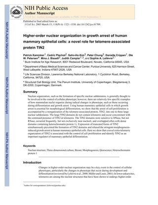
13. Fig. 1.
Large TIN2 domains are present in HMECs organized into acini. (A) Immunostaining for TIN2
(green) and PNA FISH for telomeres (red) in S1 acini in 3D culture. (B) Immunostaining for
TIN2 (green) in a biopsy from normal breast tissue. (C) Dual immunostaining for TIN2 (red)
and TRF2 (green) in the nuclei of proliferating 184 HMECs. Colocalization of TIN2 and TRF2
appears yellow. (D) Dual immunostaining for TIN2 (red) and V5 (green) in the nuclei of acini
formed by 184 HMECs expressing V5-tagged TIN2 in 3D culture. Nuclei are counterstained
with DAPI (blue) in A, C and D, and propidium iodide (red) in B. Images in B and D are
confocal sections of acini containing several nuclei. Arrowheads indicate large TIN2 domains.
Bar, 5 μm.
Kaminker et al. Page 13
J Cell Sci. Author manuscript; available in PMC 2010 September 3.
NIH-PAAuthorManuscriptNIH-PAAuthorManuscriptNIH-PAAuthorManuscript
14. Fig. 2.
Large TIN2 domains are often perinucleolar and colocalized with dense HP 1γ foci. (A) Dual
immunostaining for TIN2 (green) and nucleolin (red) in S1 acini in 3D culture. The image is
a confocal section of an acinus containing several nuclei, illustrated by the drawing on the left.
(B) Dual staining for TIN2 (green) and PML (red) in S1 acini. 3D cultures were untreated
(control) or treated for 30 minutes with DNase I (DNase) or RNase A (RNase) prior to
immunostaining. (C) Dual immunostaining for HP 1γ (red) and TIN2 (green) in S1 acini.
Arrows indicate HP 1γ domains that overlap with large TIN2 domains, arrowheads indicate
large TIN2 domains and dashed lines delineate the nuclear periphery. Bar, 5 μm.
Kaminker et al. Page 14
J Cell Sci. Author manuscript; available in PMC 2010 September 3.
NIH-PAAuthorManuscriptNIH-PAAuthorManuscriptNIH-PAAuthorManuscript
15. Fig. 3.
Formation of large TIN2 domains in growth-arrested cells is independent of the differentiation
status. (A) Large TIN2 domains (arrowheads) revealed by immunostaining (red) in correctly
polarized S1 cells cultured in 3D laminin-rich ECM (S1-Matrigel), S1 cells cultured in 3D
collagen I (S1-Collagen I) that display altered polarity, and growth-arrested (EGF-deprived)
S1 cells cultured as a monolayer on plastic. Nuclei are counterstained with DAPI (blue). (B)
Immunostaining for TIN2 (yellow) in growth-arrested 184 cells cultured as a monolayer on
plastic. The superimposed phase-contrast image shows the nucleoli as dark gray subnuclear
structures. Arrows indicate large TIN2 domains located next to nucleoli. (C) Dual
immunostaining for HP 1γ (green) and TIN2 (red) in growth-arrested 184 cells cultured as a
Kaminker et al. Page 15
J Cell Sci. Author manuscript; available in PMC 2010 September 3.
NIH-PAAuthorManuscriptNIH-PAAuthorManuscriptNIH-PAAuthorManuscript
16. monolayer on plastic. Arrowheads indicate overlapping (yellow) HP 1 staining and large TIN2
domains. Nuclei were counterstained with DAPI (blue). Bar, 5 μm.
Kaminker et al. Page 16
J Cell Sci. Author manuscript; available in PMC 2010 September 3.
NIH-PAAuthorManuscriptNIH-PAAuthorManuscriptNIH-PAAuthorManuscript
17. Fig. 4.
Formation of large TIN2 domains is independent of telomeres and TRF proteins. (A) HMECs
(strain 184) were cultured as a monolayer on plastic and growth-arrested before fixation and
dual staining for TIN2 (red) and telomeres (PNA FISH; green) (upper panels), TIN2 (red) and
TRF1 (green) (middle panels), and TIN2 (red) and TRF2 (green) (lower panels). (B) Higher
magnification images of dual staining for TIN2 (red) and TRF2 (green) showing colocalization
at small foci (yellow), indicating telomeric localization. Nuclei were counterstained with DAPI
(blue). Arrowheads indicate large TIN2 domains. Bar, 5 μm.
Kaminker et al. Page 17
J Cell Sci. Author manuscript; available in PMC 2010 September 3.
NIH-PAAuthorManuscriptNIH-PAAuthorManuscriptNIH-PAAuthorManuscript
18. Fig. 5.
Formation of large TIN2 domains correlates with exit from the cell cycle. (A) Percentage of
S1 cells with large TIN2 domain(s) and Ki-67 staining. S1 cells were cultured in 3D for 3 days
to obtain a mixed population of cycling Ki-67 positive (Ki-67+) and growth-arrested Ki-67
negative (Ki-67−) cells, and then fixed and dual immunostained for TIN2 and Ki-67. Shown
is the percentage of cells containing large TIN2 domains (TIN2 clusters) and Ki-67 positive
(filled bars) or negative (open bars) staining. Error bars show the s.e.m. for three different
experiments. *P<0.001 when compared with Ki-67+ cells. (B) Dual immunostaining for TIN2
(green) and Ki-67 (red) in S1 cells after 3D culture for 3 days. The different phases of the cell
cycle were identified by the pattern of Ki-67 staining. The percentage of cells showing large
TIN2 domains (TIN2 clusters) in each phase of the cell cycle is given below each panel, and
is the mean±s.e.m. of three different experiments. Arrowheads indicate large TIN2 domains
and dashed lines delineate the nuclear periphery. (C) Histogram of the percentage of
synchronized 184 HMECs with large TIN2 domains (TIN2 clusters) as a function of the cell
cycle. Nuclei showing large TIN2 domains were counted as a percentage of total nuclei
(revealed by DAPI counterstaining) during exponential (EXP), G0, S and G2-M phases of the
cell cycle. Percentages of Ki-67-positive nuclei in each phase are shown. Bar, 2.5 μm.
Kaminker et al. Page 18
J Cell Sci. Author manuscript; available in PMC 2010 September 3.
NIH-PAAuthorManuscriptNIH-PAAuthorManuscriptNIH-PAAuthorManuscript
19. Fig. 6.
TIN2 controls growth arrest in mammary epithelial cells. (A) Schematic of wild-type TIN2
(WT TIN2), and N-terminally (TIN2-13) and C-terminally (TIN2-15) truncated forms of TIN2.
(B) Expression of TIN2 and TIN2-13 in control and TIN2-13 expressing cells shown on
western blots. Lanes contained cells used for infection (control), cells expressing TIN2-13
(TIN2-13), cells infected with empty vector (control vector), cells overexpressing wild-type
TIN2 (TIN2), control HT1080 fibroblasts expressing exogenous TIN2 and TIN2-13 (TIN2
+TIN2− 13 mixture in HT1080). Arrows indicate the location of the respective bands for TIN2
and TIN2-13. β-catenin was used as a loading control. (C) TIN2-15 expression in monolayer
and 3D culture shown by anti-myc immunostaining (green). Nuclei were counterstained with
DAPI. Magnification ×1200. (D) Vector control, TIN2-13 and TIN2-15 infected S1 cells were
cultured in 3D for 10 days. Shown are phase-contrast images of acini formed by vector control
S1 cells and abnormal looking colonies formed by TIN2-13 and TIN2-15 S1 cells. The arrows
indicate enlarged and/or irregular multicellular structures. (E) Non-infected S1 cells (control)
and vector control, TIN2-13, or TIN2-15 infected S1 cells were cultured in 3D for 10 days.
Acini were classified according to six diameter ranges (6–15 μm, 16–25 μm, 26–35 μm, 36–
45 μm, 46–55 μm, 56–65 μm). Shown is the percentage of acini in each diameter range out of
a total of 400 acini observed in each independent experiment. Three experiments were
performed. (F) Immunostaining for the endogenous basement membrane component collagen
IV (red) and Ki-67 (green) in vector control or TIN2-15 S1 cells cultured in 3D for 10 days.
When proper morphogenesis occurs, acini are surrounded by a continuous basement membrane
and >90% of the cells arrest proliferation. One nucleus positive for Ki-67 is seen out of ten
nuclei in vector control; five nuclei positive for Ki-67 are seen out of 14 nuclei in TIN2-15.
Arrows indicate the absence of collagen IV around part of the TIN2-15 colony. (G) GFP-S1
cells organized in an acinus (left panel). Immunostaining for the endogenous basement
membrane component collagen IV (red) (central panel) and α6-integrin (green) and β-catenin
(red) (right panel) in hTERT-expressing S1 cells cultured in 3D for 10 days. When proper
Kaminker et al. Page 19
J Cell Sci. Author manuscript; available in PMC 2010 September 3.
NIH-PAAuthorManuscriptNIH-PAAuthorManuscriptNIH-PAAuthorManuscript
20. morphogenesis occurs, in addition to the continuous basement membrane, acini display the
localization of α6-integrin at the basal cell membrane (against the basement membrane) and
β-catenin at cell-cell junctions. (H) Immunostaining for TIN2 (red) in control or TIN2-15 S1
cells. In the control acinus, eight of nine nuclei, identified by DAPI staining, have a large TIN2
domain (arrowheads). In the acinar structure formed by TIN2-15-expressing cells, four of
thirteen nuclei show one or two large TIN2 domains (arrowheads) and one nucleus shows
completely fragmented TIN2 domains (arrow). Bar, 25 μm.
Kaminker et al. Page 20
J Cell Sci. Author manuscript; available in PMC 2010 September 3.
NIH-PAAuthorManuscriptNIH-PAAuthorManuscriptNIH-PAAuthorManuscript