More Related Content
Similar to poster for Bio dept UMB 36x40 rev 5
Similar to poster for Bio dept UMB 36x40 rev 5 (20)
poster for Bio dept UMB 36x40 rev 5
- 1. samples
were then
sonicated
and were
frozen on -
80°C
temperatur
e for 24
hours.
Total protein
concentration
s in samples
were
determined
by BCA
(bicinchoninic
- acid) Protein
Assays
Samples
then
underwent
electrophor
esis on
BioRad-
Criterion™
TGX stain-
Free™
Precast gels
Membrane
was cut into 3
pieces and
each piece
was incubated
with a
different
concentration
of DYRK1A
antibody.
Protein from
criterion gel
were
transferred
into
nitrocellulose
membrane
Gel was
imaged using
chemilumine
scent device
Membrane
was
incubated
for another
5 minutes in
Bio Rad ECL
luminol and
peroxide
mixture
Membrane
was imaged
using
chemilumin
escent
device
Determining DYRK1A and dopamine cell count on untreated male
and female rats, feminized male rats, and masculinized female rat
brain to obtain further information about its role in sexual
differentiation of the brain
3 different brain
from 19 days old
rats were cut 0.35
µm thick and
stored in sucrose
for cryo
protection.
Sliced brain
tissues were
washed with
0.05M phosphate
buffered saline
(PBS) and
incubated for 48
hours using
Rabbit polyclonal
DYRK1A (Abcam
ab65220)
6 different
concentration of
DYRK1A antibody
were prepared
and each brain
section was
washed and
labelled with its
designated
DYRK1A
concentration.
DYRK1A
incubated brain
tissues were
incubated with
secondary
antibody and will
ends with Ni (II)-
DAB
Labelled
brain
sections
were
mounted on
a glass slide
and
coverslipped.
Brain sections
were analyzed
under the
microscope to
identify DYRK1A
expressing cells in
the AVPV of P19
rat brain
Rationale Method and Results
Acknowledgement
References
DYRK1A protein is present in male and female juvenile rat brain: possible role in cell death of dopaminergic
cells in the developing hypothalamus
Rudy Matheson1 and Susan Zup, PhD2
1. Department of Biology, University of Massachusetts Boston , 2. Department of Psychology, University of Massachusetts Boston
This project is funded by McCone and Alumni Research Fund
(MARF) and University of Massachusetts Boston fund for
undergraduate researchAY 2015-2016.
I would like to express my undying gratitude for Dr Susan Zup
for the opportunity to conduct this experiment and guidance
from my fellow labmates, Amanda Madden and Kimbra Wagner,
also Andrew Bartlett (Hunter’s lab) and Brooke Plotkin (Park’s
lab).
• A part of the hypothalamus, the anteroventral periventricular
nucleus (AVPV) is larger in females than males, due mainly to a
higher number of dopaminergic neurons (DA) present in
females.4,6
• Testosterone, or its estrogenic metabolite, will promote cell
death, called apoptosis, of the DA neurons 2. .
• enzyme tyrosine-(Y)-phosphorylation regulated kinase 1A
(DYRK1A) is a protein kinase that has reported to facilitate
androgen receptor (AR) activation mechanism via endogenic
RNA interference3.
• Previous work has shown that DYRK1A increases survival of DA
neurons in other parts of the developing brain by inhibiting the
activity of the intrinsic apoptotic pathway, specifically by
phosphorylating caspase 9, which inhibits caspase 33.
• We hypothesize that DYRK1A in the female AVPV protects DA
neurons from cell death. Therefore, dopaminergic neurons in
the female AVPV should express more DYRK1A than those
neurons in the AVPV of males or testosterone treated females
Future direction
DYRK1A is observed to be present in COS-7 cells as speckles using
GFP labelling under fluorescence microscopy (Pictures from
Alvarez, Estivill and de la Luna, 2003)
DYRK1A expressing
cell
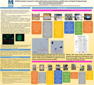
Immunohistochemistry on P19 rat brain
Western blot on P5 male and female rat brain
1:30,000
20 X
DYRK1A expressing
cell
40 X
DYRK1A speckles
Immunohistochemistry labeling in the AVPV of P19 rats
indicated that DYRK1A-expressing cells are present but
sparse. Within each cell, DYRK1A is localized as several
clusters, presumed to be nuclear speckles.
Western blot result shows that DYRK1A is
present in pre-optic area and striatum of the
brain and possibly in different concentration
in P5 male and female rat brain
1. Álvarez, M, Estivill, X and de la Luna, S, “DYRK1A accumulates in splicing speckles through a novel
targeting signal and induces speckle disassembly” Journal of CellScience © 2003.
2. Barallobre MJ, Perier C, Bové J, Laguna A, Delabar JM, Vila M and Arbonés ML, “DYRK1A promotes
dopaminergic neuron survival in the developing brain and in a mouse model of Parkinson’s disease” Cell
Death and Disease (2014)
3. Sitz, JH, Tigges M, Baumgärtel K, Khaspekov LG., and Lutz B. “Dyrk1A Potentiates Steroid Hormone-
Induced Transcription via the Chromatin Remodeling Factor Arip4” Molecular and Cellular Biology, July
2004,
4. Tsukahara S. “Sex Differences and the Roles of Sex Steroids in Apoptosis of Sexually Dimorphic Nuclei of
the Preoptic Area in Postnatal Rats” Journal of Neuroendocrinology 2009..
5. Waters EM, and Simerly RB. ” Estrogen Induces Caspase-Dependent Cell Death during Hypothalamic
Development”. The Journal of Neuroscience, 2009
6. Zup SL, Carrier H, Waters EM, Tabor A, Bengston L, Rosen GJ, Simerly RB, and Forger NG.
“Overexpression of Bcl-2 Reduces Sex Differences in Neuron Number in the Brain and Spinal Cord” The
Journal of Neuroscience, March 15, 2003