
Recommended
More Related Content
What's hot
What's hot (20)
Similar to Cardiac Pacemakers - Basics
Similar to Cardiac Pacemakers - Basics (20)
Recently uploaded
Recently uploaded (20)
Cardiac Pacemakers - Basics
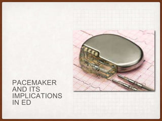
- 2. THE PACEMAKER • A pacemaker is a small, battery-operated device. • Senses when your heart is beating irregularly or too slowly. • It sends a signal to your heart that makes your heart beat at the correct pace.
- 3. PARTS OF PACEMAKER • The pulse generator. This small metal container houses a battery and the electrical circuitry that regulates the rate of electrical pulses sent to your heart. • Leads (electrodes). One to three flexible, insulated wires are each placed in a chamber, or chambers, of your heart and deliver the electrical pulses to adjust your heart rate.
- 4. TYPES OF PACEMAKERS • Single chamber pacemaker • This type of pacemaker usually carries electrical impulses from the pulse generator to the right ventricle of your heart. • Dual chamber pacemaker • A dual chamber pacemaker carries electrical impulses from the pulse generator to both the right ventricle and the right atrium of your heart. The impulses help control the timing of contractions between the two chambers. • Biventricular pacemaker • A biventricular pacemaker is a treatment option for people with heart failure whose hearts' electrical systems have been damaged. Unlike a regular pacemaker, a biventricular pacemaker stimulates both of the lower chambers of the heart (the right and left ventricles) to make the heart beat more efficiently. • A biventricular pacemaker paces both ventricles so that all or most of the ventricular muscle pumps together. This allows your heart to pump blood more effectively. Because this treatment resets the ventricles' pumping mechanism, it's also referred to as cardiac resynchronization therapy (CRT).
- 5. COMMON INDICATIONS • 3rd degree or advanced 2nd degree (Mobitz II) AV block with any of the following • Symptomatic bradycardia • Ventricular dysrhythmia resulting from block • Symptomatic bradycardia resulting from 2nd degree AV block • Chronic bifascicular or trifascicular block with intermittent 3rd degree or 2nd degree AV block • Exertional 2nd or 3rd degree AV block
- 7. SENSING RESPONSE • Triggered (T): Sensed intrinsic depolarisation will result in the pacemaker discharging (this setting not used in current generation pacemakers) • Inhibited (I): Sensed intrinsic depolarisation will result in inhibition of the pacemaker • Dual (D): Dual inhibition of both atrial and ventricular pacing in response to intrinsic ventricular depolarisation • None (O): Does not trigger or inhibit regardless of the native activity
- 8. • Example #1: DDD • Chamber paced: Atrium and ventricle can be paced • Chamber sensed: Intrinsic depolarization of the atrium and ventricle is sensed • Sensory response: Can inhibit pacemaker in response to an intrinsic ventricular depolarization or trigger in response to an atrial intrinsic depolarization without a ventricular response • Example #2: VVI • Device has a single lead in the ventricle that senses ventricular activity and can pace the ventricle • A ventricular event outside the refractory period will be inhibited
- 14. PROBLEMS WITH PACING • Failure to Sense • Definition: No paced stimulus is generated from the device resulting in decreased or absent pacemaker function • Causes: Oversensing, Lead displacement, Fracture of pacing wires • Failure to Capture • Definition: Electrical stimulus does not result in depolarization of the myocardium (no QRS complex generated) • Causes: Exit block, myocardial infarction, lead displacement, wire fracture, electrolyte abnormalities
- 15. UNDER SENSING • Under sensing occurs when the pacemaker fails to sense native cardiac activity. • Results in asynchronous pacing. • Causes include increased stimulation threshold at electrode site (exit block), poor lead contact, new bundle branch block or programming problems. • ECG findings may be minimal, although presence of pacing spikes within QRS complexes is suggestive of undersensing. • Lead fracture, Lead Displacement and Break in insulation.
- 16. UNDER SENSING
- 17. OVERSENSING • Definition: Pacemaker mistakes electrical signals as native cardiac activity and thus, pacemaker function is inhibited • Sources: Large P or T wave, pectoralis muscle contraction, cell phone signal (typically when on ipsilateral ear) • Abnormal signals may not be evident on ECG. • Reduced pacemaker output / output failure may be seen on ECG monitoring if the patient stimulates their rectus or pectoral muscles (due to oversensing of muscle activity).
- 20. PACEMAKER INDUCED DYSRHYTHMIAS • Pacemaker mediated tachycardia (PMT) (aka endless loop tachycardia) • Re-entry Tachycardia: Ventricular depolarization conducts retrograde into the atria leading atrial lead to detect activity as incoming P wave resulting in ventricular depolarization (viscous cycle develops) • Treatment • Administer AV blocker (adenosine, beta blocker, calcium channel blocker) • Apply magnet over pacemaker
- 22. RUNAWAY PACEMAKER • Potentially life-threatening malfunction of older-generation pacemakers - related to low battery voltage • Delivers paroxysms of pacing spikes at 2000 bpm - may provoke ventricular fibrillation. • Paradoxically, there may be failure to capture — causing bradycardia • Application of a magnet can be life saving but definitive treatment requires replacement of the pacemaker.
- 24. FAILURE TO CAPTURE • Failure to capture occurs when a pacing artifact is not followed by an atrial or a ventricular complex. This may be due to the following: • Lead fracture • Lead dislodgement • Fractured lead insulation • Elevated pacing threshold • MI at the lead tip • Drugs (eg, flecainide) • Metabolic abnormalities (eg, hyperkalemia, acidosis, alkalosis) • Cardiac perforation • Poor lead connection at the takeoff from the generator • Improper amplitude or pulse-width settings
- 25. FAILURE TO CAPTURE • Consideration given to treating metabolic abnormalities • Potential myocardial infarction (MI). • Temporary pacing is used to stabilise the patient until an electrophysiology technician or cardiologist can further evaluate the pacemaker.
- 26. PACEMAKER SYNDROME • Caused by improper timing of atrial and ventricular contractions resulting in AV dyssynchrony and loss of atrial “kick”. • Variety of clinical symptoms including fatigue, dizziness, palpations, pre-syncope. • Associated decrease in systolic blood pressure > 20 mmHg during change from native rhythm to paced rhythm.
- 28. MAGNET INHIBITION • Features of magnet inhibition are as follows: • In most devices, placing a magnet over a permanent pacemaker temporarily "reprograms" the pacer into an asynchronous pacing mode; it does not turn the pacemaker off • If the device company parameters are known, application of a magnet can determine whether the pacer's battery needs to be replaced • Although many different branded pacemaker/ ICD magnets are available, in general, any pacemaker/ICD magnet can be used to inhibit the device • Magnet use inhibits pacing • In some devices, "magnet function" can be disabled
- 29. THANK YOU!