Gout - types,causes,treatment
•Download as PPT, PDF•
3 likes•1,523 views

gout topic include their basic introduction, causes, types, symptoms, types of gout and their treatments

Recommended
More Related Content
What's hot
Similar to Gout - types,causes,treatment
Similar to Gout - types,causes,treatment (20)
More from Ravish Yadav
More from Ravish Yadav (20)
Recently uploaded
Recently uploaded (20)
Gout - types,causes,treatment
- 2. INTRODUCTION: • GOUT is known as the “disease of kings “ and “rich man’s disease”. • Gout (also known as podagra when it involves the big toe) • it is a medical condition usually characterized by recurrent attacks of acute inflammatory arthritis-red, tender, hot, swollen joints • Gout is a kind of arthritis that occurs when uric acid builds up in blood and causes joint inflammation. • Gout effects more men then woman in them occurs after menopause 2
- 3. CAUSES • Hyperuricemia is the underlying cause of gout. • diet, genetic predisposition, or underexcretion of urate • Low uric acid level in blood • the exact cause is unknown. • Partly genetic cause in the genes contributing to about 60% of variability in uric acid level • Three genesSLC2A9, SLC22A12 and ABCG2 have been found to commonly be associated with gout, and variations in them can approximately double the risk • Loss of function mutations in SLC2A9 and SLC22A12 cause hereditary hypouricaemia by reducing urate absorption and unopposed urate secretion
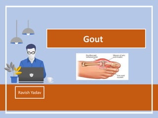
- 4. SYMPTOMS • gouty arthritis • Acute gouty arthritis in big toe(podagra) • Kidney stones • Acute pain in joints • Uric acid crystal depositon in the form of tophi • Tophi in ear lobe, achilles ankle and elbow • Fatigue • Mailase • High uric acid levels
- 5. 5
- 6. TYPES OF GOUT • Depending upon the symptoms and severity of disease gout is classified into • Acute gout • Chronic gout 6
- 7. ACUTE GOUT • Acute gout is a painful condition that typically affects one joint • Symptoms usually involve only one or a few joints. The big toe, knee, or ankle joints are most often affected. • throbbing, crushing, or excruciating pain • joint appears warm and red • fever. • The attack may go away in a few days, but may return from time to time. Additional attacks often last longer. • After a first gouty attack, people will have no symptoms. Half of patients have another attack.
- 8. Crystal-induced inflammation PMN (polymorphonuclear leukocytes)is critical component of crystal- induced inflammation crystal deposition hyperuricemia protein binding receptor binding cytokine release influx of PMN’s crystals engulfed inflammation
- 9. CHRONIC GOUT • Those with chronic arthritis symptoms include: • joint damage and • loss of motion in the joints. • joint pain and other symptoms most of the time. • Tophi below the skin around joints or in other places. Tophi usually develop only after a patient has had the disease for many years.
- 10. DIAGNOSTIC TESTS • Synovial fluid analysis (shows uric acid crystals) • Uric acid – blood • BUN (blood urea nitrogen • Joint x-rays (may be normal) • Synovial biopsy • Uric acid – urine • Creatnine level 10
- 11. BIOCHEMICAL TESTS FOR GOUT: 11
- 12. EXAMINATION OF SYNOVIAL FLUID •ASPIRATION: •The health care provider uses a needle attached to a syringe to draw out fluid from the affected joint. •LAB ANALYSIS: •The fluid sample is sent to a laboratory for analysis. Testing can reveal the presence of monosodium urate (MSU) crystals, which will nearly always confirm a diagnosis of gout. The laboratory can also test the sample for infection. •The procedure itself can cause infection, though this occurs in less than 0.1% of patients. Aspiration sometimes eases the patient's symptoms by reducing swelling and pressure on the tissue surrounding the joint. •
- 13. 13
- 14. Monosodium urate crystals polarized light red compensator needle shape negative birefringence
- 15. Drugs used to treat gout allopurinol probenecid febuxostat? steroids NSAID’s colchicine Acute Arthritis Drugs Urate Lowering Drugs rest + analgesia + time
- 16. TREATMENT • NSAID,s • Colchicine • Uricosuric agents • Allopurinol 16 COLCHICINE Produces its anti-inflammatory effects by binding to the intracellular protein tubulin, preventing its polymerization leading to the inhibition of leukocyte migration into affected area. Inhibits the synthesis & release of leukotrienes.
- 17. CONTINUED URICOSURIC AGENTS: • Probenecid & Sulfinpyrazone • They are weak organic acids .Sulfinpyrazone is a metabolite of • phenylbutazone. NSAIDS: Inhibits pain & inflammation. Inhibits urate crystal phagocytosis by decreasing the migration of granulocytes into the inflammatory area. Indomethacin ALLOPURINOL Inhibits synthesis of uric acid by inhibiting xanthine oxidase enzyme 17
Editor's Notes
- Podagra is a genus of moths of the Noctuidae family. The propensity of gout for the foot was recognised by the ancient Greeks who referred to it as podagra, literally “foot-grabber” [3]. The name “gout” derives from humoral theory and the Latin word gutta or “drop”, Gout is the disease of rich and famous as people use to drink a lot of alcohol and food intake is the major reason for gout It effects more men then woman as men are more execcisve user for podagra being thought to arise as a result of the bodily humours falling to the affected body part. Although our current understanding of the pathogenesis of gout is
- Hyperuricemia: high calicum monosodium urate concentration which is due to low uric acid levels in the blood and body fails to remove the uric acid solute carrier family 2, facilitated glucose transporter member 9 is a protein that in humans is encoded by the SLC2A9 gene.[1 SLC2A9 has also recently been found to transport uric acid, and genetic variants of the transporter have been linked to increased risk of development of both hyperuricemia and gout SLC22A The protein encoded by this gene is a urate transporter and urate-anion exchanger which regulates the level of urate in the blood. This protein is an integral membrane protein primarily found in kidney. Two transcript variants encoding different isoforms have been found for this gene
- gouty arthritis is typically the sudden onset of a hot, red, swollen joint. ric acid crystals can form outside joints. Collections of these crystals, known as tophi, can be found in the earlobe, elbow, andAchilles tendon (back of the ankle), or in other tissues. Typically, these tophi are not painful but can be a valuable clue for the diagnosis as the crystals that form them can be removed with a small needle for microscopic examination. Microscopic evaluation of a tophus reveals a nest-like accumulation of uric acid crystals embedded with white blood cells of inflammation.
- BUN concentrations may be elevated when there is excessive protein breakdown (catabolism), significantly increased protein in the diet, or gastrointestinal bleeding (because of the proteins present in the blood). Low BUN levels are not common and are not usually a cause for concern. They may be seen in severe liver disease,malnutrition, and sometimes when a patient is overhydrated (too much fluid volume), but the BUN test is not usually used to diagnose or monitor these conditions. Examination of Synovial Fluid Synovial fluid examination is the most accurate method for diagnosing gout. The synovial fluid is the lubricating liquid that fills the synovium (the membrane that surrounds a joint and creates a protective sac). The fluid cushions joints and supplies nutrients and oxygen to the cartilage surface that coats the bones. This exam also helps detect gout during intercritical periods. The health care provider uses a needle attached to a syringe to draw out fluid from the affected joint. This is called aspiration. Local anesthesia is not used because it can reduce the effectiveness of the procedure. However, the procedure is usually only mildly uncomfortable. Afterwards, there can be some minor discomfort in the area where the needle was inserted, but it usually goes away quickly. The fluid sample is sent to a laboratory for analysis. Testing can reveal the presence of monosodium urate (MSU) crystals, which will nearly always confirm a diagnosis of gout. The laboratory can also test the sample for infection. The procedure itself can cause infection, though this occurs in less than 0.1% of patients. Aspiration sometimes eases the patient's symptoms by reducing swelling and pressure on the tissue surrounding the joint. http://www.umm.edu/patiented/articles/what_risk_factors_gout_000093_5.htm#ixzz2BBsc1QLS Examination of Synovial Fluid Synovial fluid examination is the most accurate method for diagnosing gout. The synovial fluid is the lubricating liquid that fills the synovium (the membrane that surrounds a joint and creates a protective sac). The fluid cushions joints and supplies nutrients and oxygen to the cartilage surface that coats the bones. This exam also helps detect gout during intercritical periods. The health care provider uses a needle attached to a syringe to draw out fluid from the affected joint. This is called aspiration. Local anesthesia is not used because it can reduce the effectiveness of the procedure. However, the procedure is usually only mildly uncomfortable. Afterwards, there can be some minor discomfort in the area where the needle was inserted, but it usually goes away quickly. The fluid sample is sent to a laboratory for analysis. Testing can reveal the presence of monosodium urate (MSU) crystals, which will nearly always confirm a diagnosis of gout. The laboratory can also test the sample for infection. The procedure itself can cause infection, though this occurs in less than 0.1% of patients. Aspiration sometimes eases the patient's symptoms by reducing swelling and pressure on the tissue surrounding the joint. http://www.umm.edu/patiented/articles/what_risk_factors_gout_000093_5.htm#ixzz2BBsc1QLS Examination of Synovial Fluid Synovial fluid examination is the most accurate method for diagnosing gout. The synovial fluid is the lubricating liquid that fills the synovium (the membrane that surrounds a joint and creates a protective sac). The fluid cushions joints and supplies nutrients and oxygen to the cartilage surface that coats the bones. This exam also helps detect gout during intercritical periods. The health care provider uses a needle attached to a syringe to draw out fluid from the affected joint. This is called aspiration. Local anesthesia is not used because it can reduce the effectiveness of the procedure. However, the procedure is usually only mildly uncomfortable. Afterwards, there can be some minor discomfort in the area where the needle was inserted, but it usually goes away quickly. The fluid sample is sent to a laboratory for analysis. Testing can reveal the presence of monosodium urate (MSU) crystals, which will nearly always confirm a diagnosis of gout. The laboratory can also test the sample for infection. The procedure itself can cause infection, though this occurs in less than 0.1% of patients. Aspiration sometimes eases the patient's symptoms by reducing swelling and pressure on the tissue surrounding the joint. http://www.umm.edu/patiented/articles/what_risk_factors_gout_000093_5.htm#ixzz2BBsc1QLS
- Uric acid in urine Is low due to hyperurecmia as kidneys are unable to excrete the uric acid
- The synovial fluid is the lubricating liquid that fills the synovium (the membrane that surrounds a joint and creates a protective sac). The fluid cushions joints and supplies nutrients and oxygen to the cartilage surface that coats the bones. This exam also helps detect gout during intercritical periods.