

Recommended
More Related Content
What's hot
What's hot (20)
Similar to Gout and Pseudogout
Similar to Gout and Pseudogout (20)
More from Dr Usha (Physio)
More from Dr Usha (Physio) (20)
Recently uploaded
Recently uploaded (20)
Gout and Pseudogout
- 1. Gout and Pseudo Gout
- 2. Gout • Gout is a metabolic disorder manifesting in the primary or secondary forms characterized by hyperuricaemia and joint lesions. • Primary gout: It occurs in conditions where there are no obvious causes for hyperuricaemia such as drugs and myeloproliferative disorders. • Secondary gout: It occurs in conditions causing secondary hyperuricaemia.
- 3. Aetiopathology • Gout is a disease due to an inborn error of uric acid (purine) metabolism. • In this condition, sodium urate crystals are deposited on the articular cartilage, synovial membrane and in the periarticular tissues such as tendons and bursae. • This provokes an inflammatory reaction in these tissues. • These deposits often increase in size and burst through the skin to form sinuses discharging a chalky white material.
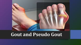
- 4. Clinical Features • It occurs mostly in men, in the third or fourth decade of life. The condition may present clinically as an acute attack or as chronic gout. • The most commonly affected joint in acute gout is the metatarsophalangeal joint of the big toe. • The attack is characterized by a sudden onset of excruciating pain at night with marked swelling and redness of the joint of the big toe. • The attack may subside in about a week. The arthritis shows remissions and exacerbations and gradually becomes chronic.
- 5. • In the chronic stage of the disease, there is a deposition of urate salts (SUM) in the periarticular tissues of the involved joint or in the subcutaneous tissues of the ear or in the olecranon bursa. • These lumps are called gouty tophi. It also occurs in the knee. • After a few attacks, the patient may have persistent pain in some joints and the joints develop features of secondary osteoarthrosis. Other features are the formation of urate calculi in the kidneys with colic and hematuria ending in renal failure.
- 6. Laboratory Findings • The main finding is the raised level of serum uric acid from the normal of 3-6 mg%. • The synovial fluid will show the typical slender needle- shaped urate crystals negatively birefringent on examination under the polarised microscope.
- 7. Radiological Features • Radiographs in the chronic stage may show the characteristic punched-out appearance at the periphery of the articular cartilage in the metatarsophalangeal joints. • Tophi may be seen as radio-opaque shadows in the periarticular tissues. • In later stages, degenerative changes will be seen in the joints with narrowing of joint spaces and sclerosis.
- 8. Treatment • Rest is given to the joint. • Anti-inflammatory drugs such as indomethacin in high doses are given. Colchicine is a specific drug for acute gout. • Drugs are given either to diminish the formation of uric acid (Allopurinol) or to act as uricosuric agents to increase the excretion of uric acid in the urine (Probenecid). • At the acute stage, the joint is rested in a splint. • Large gouty tophi may need surgical curetting or excision.
- 9. Treatment of gout can be summarized as follows: • Acute gout: NSAIDs and Colchicine • Chronic gout: Allopurinol and Probenecid and NSAIDs • Tophaceous gout: Excision of tophi, if necessary
- 10. Secondary Gout • Serum uric acid may be high whenever there is excessive destruction of cells and their nuclei as in multiple myeloma, leukaemia and other malignant diseases. • This is found during the chemotherapeutic treatment of malignant tumours with cytotoxic drugs. • The joint condition in such cases is called secondary gout.
- 11. Pseudo-Gout • Pseudo-gout is a form of arthritis due to deposition of crystals of calcium pyrophosphate dehydrate in the synovium or the articular cartilage or menisci. • Clinically, the acute attacks resemble gout but the serum uric acid level is normal. • The knee is the most common joint involved.
- 12. • Radiograph shows calcified spots in the menisci and articular cartilage. • The synovial effusion shows rhomboid-shaped crystals, positively birefringent under the polarized microscope. The treatment consists of the following: • Rest to the joint • High-dose NSAIDs • Aspiration of the joint and corticosteroid injection
- 13. Thank You