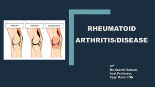
RHEUMATOID ARTHRITIS SYMPTOMS, STAGES AND TREATMENT
•Download as PPTX, PDF•
17 likes•1,921 views

Rheumatoid arthritis is a chronic autoimmune disease characterized by inflammation of the joints that can lead to long-term joint damage and disability. It is caused by the immune system attacking the synovial membrane and joint lining, causing swelling and stiffness. Common symptoms include pain, swelling, and stiffness in the small joints of the hands and feet. While the exact cause is unknown, genetic and environmental factors are believed to play a role. Treatment focuses on reducing inflammation and preventing further joint damage through medications, physical therapy, exercise, and sometimes surgery.

Recommended
More Related Content
What's hot
What's hot (20)
Similar to RHEUMATOID ARTHRITIS SYMPTOMS, STAGES AND TREATMENT
Similar to RHEUMATOID ARTHRITIS SYMPTOMS, STAGES AND TREATMENT (20)
More from keerthi samuel
More from keerthi samuel (20)
Recently uploaded
Recently uploaded (20)
RHEUMATOID ARTHRITIS SYMPTOMS, STAGES AND TREATMENT
- 3. INTRODUCTION • Rheumatoid arthritis is a chronic disease characterized by periods of disease flares and remissions with an unknown cause. • In RA multiple joints are involved but not always symmetrical. • It affects all the ages. • Damage to the joints occurs early and doesnot correlate with the severity of symptoms. • The rheumatoid factor is an antibody that can be found in the blood of 80% people with rheumatoid arthritis.
- 4. DEFINITION ■ Rheumatoid arthritis is an autoimmune disease caused by chronic inflammation of unknown etiology marked by symmetric , peripheral polyarthritis which results in joint damage &physical disability. ■ It is a progressive disease of synovial lining of peripheral joints characterized by symmetrical inflammation leading to potentially deforming polyarthritis. It is the most common systemic inflammatory disease characterized by symmetrical joint involvement
- 5. INCIDENCE ■ New cases of RA are typically two to three ties higher in women than men. ■ People with a genetic component ,inherited traits are at higher risk. ■ Acc to CDC from 2013-2015 an estimated of 54.4million US adults annually were affected. ■ 49.6% of people above 65 years, 41.3 million Non- Hispanic whites are affected. ■ Adults aged 18 years or older who are overweight or obese report doctor-diagnosed arthritis more often than adults with a lower body mass index (BMI)
- 6. ETIOLOGY ■ Unknown cause,Believed that it is hereditary. 1. ENVIRONMENTAL INFLUENCES : Such as infection, trauma, 2. GENETIC MARKERS: HLA-DR4 triggers RA. Such factors are not considered as diagnosis because half of the people who posses this antigen donot develop RA 3. ANTIGEN DEPENDENT ACTIVTION OF T LYMPHOCYTES: leads to proliferation of synovial memebrane. Activation of pro inflammatory cells from the bone marrow , cytokinins and auto antibody production. ENVIRONMENTAL FACTORS GENETIC MARKERS ANTIGEN DEPENDENT ACTIVATION OF T LYMPHOCYTES ANTI-CITRULLINATED PROTEINS TUMOR NECROSIS FACTOR (TNF) SYNOVITTIS
- 7. ETIOLOGY 4. ANTI-CITRULLINATED PROTEINS: these are peptides highly specific for RA 5. TUMOR NECROSIS FACTOR: IL-1,IL-6 and growth factors propogate the inflammatory process, and agents found to alter these cytokines reduces pain and deformity. 6. SYNOVITIS- hallmark in pathogenesis of RA. Synovium proliferates abnormally ,groeing into the joint space and into the bone forming a PANNUS. The pannus migrates to the articular cartilage and subchondral bone leading to destruction of cartilage, bone tendons and blood vessels. ENVIRONMENTAL FACTORS GENETIC MARKERS ANTIGEN DEPENDENT ACTIVATION OF T LYMPHOCYTES ANTI-CITRULLINATED PROTEINS TUMOR NECROSIS FACTOR (TNF) SYNOVITTIS
- 8. ETIOLOGY 4. ANTI-CITRULLINATED PROTEINS: these are peptides highly specific for RA 5. TUMOR NECROSIS FACTOR: IL-1,IL-6 and growth factors propogate the inflammatory process, and agents found to alter these cytokines reduces pain and deformity. 6. SYNOVITIS- hallmark in pathogenesis of RA. Synovium proliferates abnormally ,groeing into the joint space and into the bone forming a PANNUS. The pannus migrates to the articular cartilage and subchondral bone leading to destruction of cartilage, bone tendons and blood vessels. ENVIRONMENTAL FACTORS GENETIC MARKERS ANTIGEN DEPENDENT ACTIVATION OF T LYMPHOCYTES ANTI-CITRULLINATED PROTEINS TUMOR NECROSIS FACTOR (TNF) SYNOVITTIS
- 9. PREDISPOSING FACTORS 1. GENDER: women before the menopause are affected three times more often than men. After the menopause the frequency of onset is similar between the sexes, suggesting an etiological role of male sex hormone. The use of oral contraceptives delay the onset of disease but has no effect on RA 2. FAMILIAL: Increased incidence in first degree relatives and high risk in monozygotic twins (15%)than dizygotic twins (3.5%). It affects families for many generations.
- 11. PATHOPHYSIOLOGY I. TRIGGER ■ The combination of etiological factors sends a trigger to the body to create antibodies – known as autoantibodies that seek out joint linings. ■ These autoantibodies include rheumatoid factor (RF) and anti-cyclic citrullinated peptide antibody (anti-CCP). II.INFLAMMATION ■ This results in the production of chemicals being released including tumour necrosis factor alpha (TNF-α), Interleukin (IL)-1, IL- 6, IL-8, transforming growth factor beta (TGF-β), fibroblast growth factor (FGF) and platelet-derived growth factor (PDGF). ■ Increased levels of cytokines are present. Cytokines play a central role in the perpetuation of synovial inflammation.
- 12. PATHOPHYSIOLOGY III.JOINT &TISSUE DESTRUCTION ■ These chemicals inflame and damage the body’s cartilage, bone, tendons, and ligaments which causes extravasation of leucocytes. ■ HYPERPLASIA of the synovial membrane with extensive angiogenesis. ■ There is an increased number of both type synoviocytes and is infiltrated with immune and inflammatory cells: particularly macrophages, B- and T-lymphocytes, plasma cells and dendritic cells. ■ The persistence of the chronic inflammatory response in conjunction with ongoing joint destruction (is finding in many patients with RA despite the use of effective anti-inflammatory agents and disease- modifying drugs).
- 13. STAGES OF RA I. SYNOVITIS ■ Stage 1 is early stage RA. ■ Many people feel joint pain, stiffness, or swelling. During Stage 1, ■ there is inflammation inside the joint. ■ The tissue in the joint swells up. With no damage to the bones, but ,synovium, is inflamed. ■ Can progress to bone erosion
- 14. STAGES OF RA II. PANNUS FORMATION ■ Moderate stage RA. ■ Synovitis causes damage to the joint cartilage. When cartilage is damaged, there will be pain and loss of mobility. ■ Range of motion in the joints may become limited. ■ Inflammation and exuberant proliferation of the synovium leads to formation of pannus and destruction of cartilage, bone, tendons, ligaments, and blood vessels. Basically, the hypertrophied synovium is called PANNUS
- 15. STAGES OF RA III. FIBROUS ANKYLOSIS ■ Stage 3, it is considered severe. ■ damage extends not only to the cartilage but to the bones due to increased friction between the bones. Pain and swelling increases causing FIBROUSANKYLOSIS with bone erosion. ■ Fibrous ankylosis is a fibrous connective tissue process which results in decreased range of motion. Symptoms present as bony ankylosis, in which osseous tissue fuses two bones together reducing mobility, which is why fibrous ankylosis is also known as false ankylosis. BONEANKYLOSIS FIBROUSANKYLOSIS
- 16. STAGES OF RA IV. BONY ANKYLOSIS ■ At Stage 4, there’s no longer inflammation in the joint. ■ This is end-stage RA, when joints no longer work. ■ In end-stage RA, people may still experience pain, swelling, stiffness, and mobility loss. There may be reduced muscle strength. The joints may become destroyed and the bones fused together (ankylosis). ■ Bony ankylosis is the union of the bones of a joint by loss of articular cartilage, resulting in complete immobility.
- 17. STAGES OF RHEUMATOID ARTHRITIS
- 18. CLINICAL FEATURES ■ Early rheumatoid arthritis tends to affect smaller joints first — particularly the joints that attach your fingers to your hands and your toes to your feet later spreads to the wrists, knees, ankles, elbows, hips and shoulders leading to POLYARTHRITIS. ■ Swollen, warm, tender and stiff joints limits movements particularly early in the morning on waking or prolonged inactivity. ■ The deformities seen are: – Buttonhole deformity – Subluxation of metacarpophalangeal joint/ULNAR DRIFT – Z thumb deformity – Swan neck deformity – Hammer toe deformity – Arthritis mutilans 1. JOINT
- 19. 'Z deformity' may occur in rheumatoid arthritis. It is seen at the thumb and consists of hyperextension of the interphalangeal joint, and fixed flexion and subluxation of the metacarpophalangeal joint Swan-neck deformity/BOUTONNIERE DEFORMITY/BUTTON HOLE is a bending in (flexion) of the base of the finger, a straightening out (extension) of the middle joint, and a bending in (flexion) of the outermost joint. Swollen,red, tender joints
- 20. A hammer toe or contracted toe is a deformity of the muscles and ligaments of the proximal interphalangeal joint of the second, third, or fourth toe causing it to be bent, resembling a hammer Ulnar deviation, also known as ulnar drift, is a hand deformity in which the swelling of the metacarpophalangeal joints causes the fingers to become displaced, tending towards the little finger. Arthritis mutilans is a rare medical condition involving severe inflammation damaging the joints of the hands and feet, and resulting in deformation and problems with moving the affected areas; it can also affect the spine
- 21. CLINICAL FEATURES ■ The rheumatoid nodule or NECROTIZING GANULOMA, which is sometimes in the skin, is the most common non- joint feature .The typical rheumatoid nodule may be a few millimetres to a few centimetres in diameter and is usually found over bony prominences, such as the elbow, the heel, the knuckles, or other areas that sustain repeated mechanical stress. ■ Nodules are associated with a positive RF (rheumatoid factor) titer, and severe erosive arthritis. ■ Rheumatoid vasculitis can thus commonly present with skin ulceration and vasculitic nerve infarction known as mononeuritis multiplex. The most common presentation is due to involvement of small- and medium-sized vessels 2.SKIN
- 22. CLINICAL FEATURES ■ Sweet syndrome is a rare disorder characterized by fever and the sudden onset of a rash, which consists of multiple tender, red or bluish-red bumps or lesions. These lesions usually occur on the arms, legs, trunk, face or neck. In some cases, additional systems of the body can become involved including the musculoskeletal system such as inflammation of the joints (arthritis). ■ Diffuse alopecia areatais seen in RA. 2.SKIN
- 24. DIAGNOSTIC TESTS 1.RHEUMATOID FACTOR: ■ Found in60% of patients with RA. however 5% of healthy individuals will have elevated levels of RF. ■ If initially negative the test can be repeated in 6-12 months. ■ RF is not an accurate measure of disease progression. 2.ERYTHROCYTE SEDIMENTATION RATE: ■ They are markers of inflammation ad are usually elevated in RA. ■ It helps to indicate the activity of the disease but they don’t indicate the severity of the disease.
- 25. DIAGNOSTIC TESTS 3. ANTICYCLIC CITRULLINATED PEPTIDE ANTIBODIES(ACPA): ■ These are found in patients with RA and useful in predictive erosive disease. 4.RADIOGRAPHIC EXAMINATION: ■ This can reveal the extent of bone erosion and cartilage loss. ■ An MRI can detect proliferative pannus
- 26. OTHER BLOOD TESTS ■ C- Reactive protein ■ Complete blood count ■ Renal function test – uric acid ■ Liver enzymes and other immunological tests like anti nuclear antibodies. ■ Elevated ferritin – can be a sign of RA and seronegative RA.
- 27. EULAR -(European League against Rheumatism) If the score is greater than or equal to 6 it indicates definite rheumatism.
- 28. MANAGEMENT ■ GOALS: – to control inflammation – relieve pain, and – reduce disability associated with RA.
- 29. MANAGEMENT Medical management Physical and occupatonal therapy Surgical management Cognitive therapy Natural therapy
- 30. MEDICAL MANAGEMENT 1. NSAIDS – NON STEROIDAL ANTI INFLAMAMTORY DRUGS: Commonly used to treat RA. They help to manage chronic pain, inflammation and swelling. They do not slow down the disease. Most people with RA also take other types of medications, such as methotrexate or biologics, to help prevent further joint damage. EX: Aspirin, celecoxib, diclofenac, ibuprofen, ketoprofen, ketorolac MECHANISM OF ACTION: They block cyclic oxygenase enzymes which cuts pain and stiffness. SIDE EFFECT: Increased BP, gastric irritation , headaches, anemia, rash, tinnitus, heart attack and stroke.
- 31. MEDICAL MANAGEMENT 2. DMARD’S: DISEASE MODIFYING ANTI RHEUMATIC DRUGS: • Commonly used are methotrexate, hydroxychloroquine, sulfasalazine. • Anti-cytokine agents: anti TNF drugs include infliximab • IL-1 receptor antagonist : which depletes peripheral B cells. Ex: rituximab. 3.IMMUNOSUPPRESIVE AGENTS: • Less frequently used. • Ex: Azathioprine, D- penicillamine, Gold (auranofin), cyclophosphamide, cyclosporin 4.COMBIANTION THERAPY: • Cyclosporine + methotrexate • Methotrexate + Sulfasalazine and hydroxychloroquine. 5.GLUCOCORTICOIDS: low dose prednisolone
- 32. PHYSICAL AND OCCUPATIONAL THERAPY ■ For people with RA, physiotherapy may be used together with medical management. This may include cold and heat application, electronic stimulation, and hydrotherapy. ■ Physiotherapy promotes physical activity. In RA, physical activity like exercise in the appropriate dosage (frequency, intensity, time, type, volume, progression) and physical activity promotion is effective in improving cardiovascular fitness, muscle strength, and maintaining a long term active lifestyle. Physical activity promotion according to the public health recommendations should be an integral part of standard care for people with RA and other arthritic diseases.
- 34. SURGICALMANAGEMENT- ARTHROSCOPY ■ It is a type of joint surgery in which a thin tube with a light source (called an arthroscope) is inserted into the joint through a small incision (cut) in the skin, allowing to see the inside of the joint. ■ Instruments are inserted through other small cuts to work on the joint. Surgery will not cure rheumatoid arthritis or stop the disease's progress. But it may improve function and provide some pain relief.
- 35. SURGICALMANAGEMENT- SYNOVECTOMY ■ Synovectomy surgery is done to remove inflamed joint tissue (synovium) that is causing unacceptable pain or is limiting your ability to function or your range of motion. Ligaments and other structures may be moved aside to access and remove the inflamed joint lining. ■ Following knee synovectomy, knee will be immobilized in a removable cast. And physical therapy is started after 1 to 2 days. Synovectomy does not cure the disease. But it may relieve symptoms temporarily. ■ There may also be a loss in the range of motion of the joint, or the inflammation in the joint may return.
- 36. SURGICALMANAGEMENT-ARTHRODESIS ■ Also known as arthrodesis, joint fusion is a surgical procedure for the treatment of severe arthritis pain. It involves fusing together the bones in your aching joint to create one solid bone. The fused bone is often more stable and results in decreased pain. ■ Ankle fusion may also be carried out to treat a severe foot deformity, like club foot, high-arched or flat foot which has damaged the ankle joint or made it unstable.
- 37. SURGICALMANAGEMENT-ARTHRODESIS ■ It can take up to three months before you’re walking again after surgery. This can be a frustrating and difficult time, especially if you’re used to being active. Most people resign themselves to weeks of hobbling around on crutches. Fortunately, there are now some alternatives which can make the process a lot easier for you. Here are the main options(RECENTADVANCEMENT) ■ Conventional Crutches ■ Knee Scooters or “KneeWalkers” ■ Hands-FreeCrutch:The iWALK 2.0
- 38. SURGICALMANAGEMENT- JOINT REPLACEMENT ■ The definitive treatment for advanced joint destruction in the late stages of rheumatoid arthritis can be successfully treated with total joint arthroplasty. Total knee arthroplasty has been shown to be a well-proven modality that can provide pain relief and restoration of mobility for those with debilitating knee arthritis. ■ The knee is one of the most commonly affected joints in patients suffering from chronic rheumatoid arthritis (RA) ■ In advanced disease, when synovectomy is of no benefit, total knee arthroplasty (TKA) has proven to be the most successful intervention that reduces knee pain and improves physical function in RA ■ Total hip replacement is another option
- 39. COGNITIVE THERAPY ■ This review concludes that cognitive behavioral therapy (CBT) is the most efficacious treatment for pain management in RA; however, there are indications that mindfulness may have particular benefits for patients with a history of depression. CBT is most effective when administered early in the course of the disease especially to manage chronic pain. ■ One of the major challenges is ensuring access to effective interventions for patients, particularly early on in the course of the disease, with a view to preventing physical and psychological morbidity.
- 40. NATURAL THERAPY
- 41. RECENT ADVANCEMENTS ■ IMMUNO ADSORPTION APHEREIS: ■ Immunoadsorption is a selective apheresis method for the removal of specific antibodies and immune complexes, leaving other plasma components and obviating the need for plasma replacement. • Extracorporeal immune adsorption of plasma over columns containing inert silica matrix and covalently attached staphylococcal protein is done. • Used in patients who failed other therapies. • Used in join pains, swellings