SlideShare a Scribd company logo
1 of 39
Download to read offline
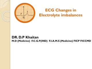
ECG Changes in
Electrolyte imbalances
DR. D.P Khaitan
M.D (Medicine) F.C.G.P(IND) F.I.A.M.S (Medicine) FICP FICCMD
Discussion on ECG changes in electrolyte imbalances under
the following subheads:
 Arrhenius theory of ‘Electrolyte dissociation’
 Electrolytes move across the cardiac membrane
 A bird’s eye view on ECG changes in electrolyte imbalances
 Electrophysiology with ECG changes in
o Hyperkalemia
o Hypokalemia
o Hypercalcaemia
o Hypocalcaemia
 Illustration by ECGs
 Arrhythmogenicity
 Home take message
My talk on ‘ECG changes in Electrolyte imbalances’ is
dedicated to the genius of Arrhenius –
Father of electrolytes
Arrhenius's main contribution is his theory of
‘Electrolytic dissociation’ (1887) –
the Nobel prize in chemistry 1903 ………..
To conduct electricity one must have free-moving
ions.
He noticed that the solution of acid conducts
electricity by dissolving the substance in the solution,
which dissociates into ions. This theory is known as
“Electrolytic dissociation.”
Svante August Arrhenius
(1859-1927)
Swedish Physicist and
Physical chemist
A concept of electrolytes
As per Arrhenius's theory of ‘electrolyte dissociation’ :
The word 'electro' denotes the state of electrolytes being in charged ionic state
and the word 'lyte' (lytos) indicates the capability of electroytes to undergo lysis
when dissolved in a solvent – electrolyte dissociation.
An Electrolyte is a chemical substance when
dissolved in aqueous solvent, it gets dissociated into
cations and anions which are capable of producing
electricity
Electrolytes move across the cardiac membrane
Phase 0 Phase 1 Phase 2
Extracellular
Phase 3 Phase 4
Na+
K+ K+
Ca+
K+ Na+
K+
Intracellular
QRS J-Point ST segment T-wave
 Sodium and calcium ions predominantly exist in the
extracellular space and potassium ion exists mainly
in the intracellular space
 Normal range (conventional units)
Normal serum potassium = 3.5-5.5 mEq/L
Normal serum calcium = 8.5-10.5 mg/dL
(Ionized serum calcium = 4.5-5.6 mg/dL)
The Cardiac Action Potential is a series of brief changes in voltage across
the cardiac cell membrane, brought about by fluxes of ions through ion
channels (free-moving ions)
Cardiac membrane
Phase 0 Phase 1 Phase 2
Extracellular
Phase 3 Phase 4
Na+
K+ (transient)) K+ (Ikr)
Ca+
K+ (Iks & Iki) Na+
K+
R
QT Interval
P
T
Intracellular (rapid)
A bird’s eye view on ECG changes in electrolyte imbalances
S
Q
 In suspected cases of electrolyte disturbances one should carefully see the ‘QTc’ interval
• Shortening of QTc interval , mainly due to shortening of Phase 2 (Ca+ - K+ exchange)
Hyperkalemia : QTc shortening with peaked tall T-wave.
Hypercalcaemia : QTc shortening with complete absence of ST segment with
wide based T-wave.
• Prolongation of QTc interval
Hypokalemia : The merging of U-wave with T forming ‘TU’ complex
Hypocalcaemia : Due to the lengthening of ST segment
(unlike , hypokalemia the ST segment is not displaced from the baseline , i.e. it is
straight)
 Other ECG changes associated with hyperkalemia should also be looked into :
peaked tall T , ongoing atrial paralysis , SA nodal and AV nodal dysfunctioning with its
appendages involvement, diffuse intraventricular conduction delay, + sine wave .
QRS J-Point ST segment T-wave
Hyperkalemia changes
Electrophysiology in hyperkalemia
SA Node
Atria
AV node
Purkinje fiber
Ventricle
4
0
3
0
4
1 2
3
Ongoing atrial paralysis •P wave broadening /flattening
•PR prolongation
•Eventually complete disappearance of P wave
SA node is less sensitive to hyperkalemia and so is the
interconnective link in between SA node and AV node
Sinus bradycardia / Sinus arrest (intact sinoventricular conduction)
Impaired AV conduction different degree AV block
Secondary pacemaker capability of Purkinje fibers is supressed
Infra nodal escape pacemaker is unreliable in the
presence of heart block frank asystole
Sine wave
Ventricular fibrillation
(diffuse intraventricular conduction delay)
Red signal sign
Nodal system is less sensitive to hyperkalemia
 Sinus node itself is less sensitive to hyperkalemia because of its low negative
membrane potential with more steeping rise during Phase 4
(equipped with more stable automaticity)
 Sinoventricular condution – hyperkalemic resistence to the internodal tracts
connecting the SA node to the AV node.
 AV node is also relatively less sensitive to hyperkalemia
(an important secondary pacemaker station , with lesser steep rise during Phase 4)
Phase 0 Phase 1 Phase 2
Extracellular
Phase 3 Phase 4
Na+
K+ (transient)) K+ (Ikr)
Ca+
K+ (Iks & Iki) Na+
K+
Intracellular (rapid)
ECG changes in Hyperkalemia
T
T
ST
 Extracellular surplus K+ Decrease phase 2 shortening
of QT (QTc) interval T-wave near to QRS complex
  Electrochemical gradient (surplus K+) Peaked Tall T
Surplus potassium
Normal Tall peaked T ST depression Broad low P
with prolonged
PR interval
Atrial arrest
(No P wave)
Diffuse
intraventricular
conduction delay
sine wave
ECG Changes as per potassium level (mEq/L)
Potassium
level (mEq/L)
Mechanism ECG changes
5.5 -6.5  K+ electrochemical gradient
during phase 3
Peaked and tall T waves
6.5-7.0 Ongoing atrial paralysis P wave broadening /flattening
PR prolongation
Eventually complete disappearance of P wave
7.0-9.0 Conducting pathways
abnormalities
Conduction pathway abnormalities with
Bradyarrhytmias in association with + other evidences
of hyperkalemia , serum potassium level should be
estimated for the purpose
 Suppression of SA Node – Sinus Bradycardia
 Different degree of AV block of high grade
nature with slow junctional /ventricular
escape rhythm.
 Bundle branch block , hemiblocks
>9.0 Fewer Na+ channels in
operation due to its marked
inactivation + very early onset
of repolarization
Sine-wave (diffuse intraventricular conduction delay)
Pulseless electrical activity with wide Bizarre shaped
QRS complex.
Hypokalemia changes
Electrophysiology : Hypokalemia
T U
Phase 0 Phase 1 Phase 2
Extracellular
Phase 3 Phase 4
Na+
K+ (transient)) K+ (Ikr)
Ca+
K+ (Iks & Iki) Na+
K+
Intracellular (rapid)
Normal Low U wave Prominent
U wave
• ST depression
• Fusion of T with U
• Prolongation of QT
(prominent P and
prolonged PR)
Normally Synchronized repolarization of
cardiac myocytes and Purkinje fibres
In hypokalemia dichotomized repolarization :
delayed and prolonged repolarization through
purkinje fibres T-U complex
fusion of T with U QT prolongation
Hypercalcaemia changes
Electrophysiology : Hypercalcaemia
Phase 0 Phase 1 Phase 2
Extracellular
Phase 3 Phase 4
Na+
K+ (transient)) K+ (Ikr)
Ca+
K+ (Iks & Iki) Na+
K+
Intracellular (rapid)
 Shortening of Phase 2 Shortening of ST segment Shortening of QT interval
(ST segment may be completely absent and is replaced by T wave having
widened base to counteract the shorten QT interval – a compensatory effect of
hypercalcaemia).
Since the QT interval is shortened in hypercalcaemia , initial upstroke of the T
wave may immediately start after the QRS complex mimicking the hyper acute
phase of myocardial infarction , especially when the T waves are rapid and
taller than usual
When the upstroke of T-wave starts immediately after the QRS complex , it
indicates more rapid entry of calcium ions inside the myocardial cells. To
equalize the electrochemical gradient there is somewhat exaggerated K+ exit in
phase 1 resulting in the augmentation of the J-wave – termed as
Osborn’s wave.
ECG changes in hypercalcaemia
Elevation of the J point with shortened QT interval , followed by T wave
having widened base (so counteracting the shortened QT interval - a
compensatory effect).
Hypocalcaemia changes
Electrophysiology : Hypocalcaemia
Phase 0 Phase 1 Phase 2
Extracellular
Phase 3 Phase 4
Na+
K+ (transient)) K+ (Ikr)
Ca+
K+ (Iks & Iki) Na+
K+
Intracellular (rapid)
 Prolongation of QTc interval due to prolongation of ST segment that tends
to be in straight line.
 In hypokalemia the ST segment is depressed from the baseline with QT
interval prolongation due to QTU assembling.
II
Hypocalcaemia : QTc prolongation with straight ST segment
ST
Pertinent points to be considered
 Hypomagnesemia
The ECG changes are similar to hypokalemia. This should be stated here
that if potassium supplementation tends not to normalize the QTc interval
, hypomagnesemia must be suspected.
 Hypermagnesemia
The ECG findings are similar to hyperkalemia but no definite criteria has
been laid down.
 Uremia
Uremia on ECG is recognized by hyperkalemia + hypocalcaemia with
prolongation of QTc interval
Illustration by ECGs
ECG changes in
Hyperkalemia
 The earliest ECG manifestation of hyperkalemia is seen with Serum
potassium near about 6 mEq/L by the presence of high peaked tall tented
T-waves , seen in all the leads especially over the precordial leads. With the
further rise in serum K+ level , its amplitude and peaking character decreases
but width increases. At much higher levels , the T-waves becomes broad with
bizarre QRS running together to inscribed sine wave.
 Hyperkalemia cannot be diagnosed with certainty on the basis of T-waves
changes along. Braun et al. found that the typical T-wave change on
hyperkalemia were present in 22% of patients.
(Chou’s Electrocardiography in Clinican Practice – Sixth Edition – P 532)
Some important consideration related to T-wave in hyperkalemia :
 Junctional rhythm with retro negative P in inferior leads with heart rate
44 bpm
 RBBB pattern (see V1)
 Peaked tented P seen over V3 to V6 and over leads I , II , III and aVF
A diabetic male aged 45years with Serum creatinine 2.5 mg and K 6.5 mEq/L .
ECG findings :
ECG 1
 ECG showing ACS with hyperkalemia ( tall peaked T wave and absence of
P wave with wide QRS and bradycardia mimicking nodal rhythm). There is
one capture P where seen following 1st QRS complex in leads II and III
Diabetic middle aged male with chest pain since 3 hours
Trop I positive , Serum K 6.5 mEq/L , Serum Creatinine 3.5 mg
ECG 2
Male aged 41 years with h/o giddiness and fainting attack
P
R T
Flattened P with Mobitz type 2 AV block having
ventricular rate 25 bpm , Tall T
Paradoxical shortening of QT interval
Peaked and tall T-wave
Serum potassium >7 mEq/L
P P
ECG 3
P
S
r T P P P
RBBB
AV block ( Mobitz type 2)
Possibly LAFB
Tall peaked T
ECG suggestive of hyperkalemia
ECG 4
A male aged 45 years with h/o fainting attack
Serum K 7.4 mEq/L
 One should suspect hyperkalemia in the presence of conduction pathway
abnormality e.g. CHB associated with tall and peaked T-wave
 Inferior MI (Non-STEMI)
Middle aged Diabetic male with severe chest pain since 3 hours
(Blood sugar 200mg, Serum K 5.7, Serum Creatinine 1.5 Trop I positive)
ECG 5
Patient aged 50 years in unconscious state
BP = 180/110 mmHg with deep rapid breathing
Sine wave (diffuse intraventricular conduction delay) with RBBB pattern in V1 with
extreme width 220 ms.
A fewer Na+ channels availability to be activated during depolarization contributes
to widened QRS plus early initiation of repolarization phase having more widened T
fusion of T-wave with the preceding QRS complex.
Serum potassium = 8.0 mEq/L
ECG 6 (A)
Serum K corrected to normal = 4.5 mEq/L
ECG 6 (B)
The same patient’s ECG after recovery
A 24 years lady presented with breathlessness (ESRD with Serum K 7.88 mEq/L)
This ECG is also indicative of sine wave , as discussed with the preceding ECG
ECG 7
ECG changes in
Hypercalcaemia and
Hypocalcaemia
74 years old man , a chronic smoker with h/o Polyuria , Polydysia ,
Fatigue and Pain abdomen. Physical examination was unremarkable ,
Chest X-ray normal , Blood sugar , urea , serum catenin , Na+ , Cl- and
HCO3 , Parathyroid hormone , Vitamin D – all were normal (no
evidence of nephrocalcinosis or renal stone)
 QTc (X ST seg. ,
widened T-wave)
T
Osborn’s wave with
 ST seg.
ECG features are suggestive of Hypercalcaemia
Serum calcium 12 mg
Familial Hypercalcaemia
ECG 8
ECG 9
45 years Female presented to the emergency department with recurrent seizures
since 1 day. She was diagnosed with epilepsy 6 years back and was put on sodium
valproate 300 mg bd. No proper workup for seizure was done. Her ECG shows
prolonged QT interval (QTc interval exceeding half of the RR interval).
Serum calcium-7.5mg/dl
Serum albumin-4.5gm/ml
Prolonged QTc interval with straight ST segment
Hypocalcaemia
Arrhythmogenicity
 Severe hyperkalemia can lead to heart block , asystole pulseless
electrical activity and ventricular arrhythmias including ventricular
fibrillation.
 With hypokalemia
• Frequent supraventricular ectopic may be caused by myocardial
hyperexcitability.
• Supraventricular tachyarrhythmias : AF , atrial flutter, atrial tachycardia
• Even moderate hypokalemia may inhibit the sodium potassium pump in
myocardial cells promoting spontaneous early afterdepolarizations that
lead to ventricular tachycardia/fibrillation. At times a few isolated
ventricular extrasystoles.
Arrhythmogenicity (contd.)
 Cardiac arrhythmias are uncommon in patients with
hypercalcemia. Hypercalcaemia decreases ventricular conduction
velocity and shortens the effective refractory period. Ventricular
arrhythmias ranging from VPCs to Frank Ventricular fibrillation can be
encountered in severe hypercalcaemia
 In hypocalcaemia arrhythmias are uncommon , although atrial fibrillation
has been reported
Torsades de pointes may occur , but is much less common than hypokalemia
and hypomagnesaemia.
HOME TAKE MESSAGE
 The word 'electro' denotes the state of electrolytes being in charged ionic
state and the word 'lyte' (lytos) indicates the capability of electroytes to
undergo lysis when dissolved in a solvent – electrolyte dissociation.
 The Cardiac Action Potential is a series of brief changes in voltage across
the cardiac cell membrane, brought about by fluxes of ions through ion
channels.
Phase 0 Phase 1 Phase 2
Extracellular
Phase 3 Phase 4
Na+
K+ K+
Ca+
K+ Na+
K+
Intracellular
QRS J-Point ST segment T-wave
 Normal range (conventional units)
Normal serum potassium = 3.5-5.5 mEq/L
Normal serum calcium = 8.5-10.5 mg/dL
(Ionized serum calcium = 4.5-5.6 mg/dL)
 Nodal system is less sensitive to hyperkalemia
• Sinus node itself is less sensitive to hyperkalemia because of its low
negative membrane potential and more steeping rise during Phase 4
(equipped with more stable automaticity)
• Sinoventricular condution – hyperkalemic resistence to the internodal tracts
connecting the SA node to the AV node.
• AV node is also relatively less sensitive to hyperkalemia
(an important secondary pacemaker station , with lesser steep rise with
Phase 4)
 Electrophysiology in hyperkalemia
 Extrcellular surplus K+ Decrease phase 2 shortening
of QT (QTc) interval T-wave near to QRS complex
  Electrochemical gradient (surplus K+) Peaked Tall T
Normal Tall peaked T ST depression Broad low P
with prolonged
PR interval
Atrial arrest
(No P wave)
Diffuse
intraventricular
conduction delay
sine wave
ECG Changes as per potassium level (mEq/L)
Potassium
level (mEq/L)
Mechanism ECG changes
5.5 -6.5  K+ electrochemical gradient
during phase 3
Peaked and tall T waves
6.5-7.0 Ongoing atrial paralysis P wave broadening /flattening
PR prolongation
Eventually complete disappearance of P wave
7.0-9.0 Conducting pathways
abnormalities
Conduction pathway abnormalities with
Bradyarrhytmias in association with + other evidences
of hyperkalemia , serum potassium level should be
estimated for the purpose
 Suppression of SA Node – Sinus Bradycardia
 Different degree of AV block of high grade
nature with slow junctional /ventricular
escape rhythm.
 Bundle branch block , hemiblocks
>9.0 Fewer Na+ channels in
operation due to its marked
inactivation + very early onset
of repolarization
Sine-wave (diffuse intraventricular conduction delay)
Pulseless electrical activity with wide Bizarre shaped
QRS complex.
 Hypokalemia
In hypokalemia dichotomized repolarization : delayed and prolonged repolarization
through purkinje fibres T-U complex fusion of T with U QT prolongation
 Hypercalcaemia : Shortening of ST segment Shortening of QT interval
with widened T base + initial upstrokeT-wave may start just after the QRS
complex + Osborn’s wave.
 Hypocalcaemia : QTc prolongation with straight ST segment
 Arrhythmogenicity
o Severe hyperkalemia : different grades of AV block , asystole , PEA ,
ventricular arrhythmias including ventricular fibrillation.
o With hypokalemia : Supraventricular ectopi / tachyarrhythmias ,
Ventricular arrhythmias
o Cardiac arrhythmias are uncommon in patient with hypercalcaemia and
hypocalcemia
ECG Changes in Electrolyte Imbalances

More Related Content

Similar to ECG Changes in Electrolyte Imbalances

Ecg findings in life threatening conditions
Ecg findings in life threatening conditionsEcg findings in life threatening conditions
Ecg findings in life threatening conditionsMEEQAT HOSPITAL
 
Cardiology 2.1. ECG or EKG - by Dr. Farjad Ikram
Cardiology 2.1. ECG or EKG - by Dr. Farjad IkramCardiology 2.1. ECG or EKG - by Dr. Farjad Ikram
Cardiology 2.1. ECG or EKG - by Dr. Farjad IkramFarjad Ikram
 
Antiarrhythmic drugs class Bidya.pdf
Antiarrhythmic drugs class Bidya.pdfAntiarrhythmic drugs class Bidya.pdf
Antiarrhythmic drugs class Bidya.pdfSaishDalvi
 
Electrolytes imbalance and ECG changes presentation 2005
Electrolytes imbalance and ECG changes presentation 2005Electrolytes imbalance and ECG changes presentation 2005
Electrolytes imbalance and ECG changes presentation 2005Idrees Muhammad
 
ECG approach to arrhythmias 2017
ECG approach to arrhythmias 2017ECG approach to arrhythmias 2017
ECG approach to arrhythmias 2017Ashutosh Pakale
 
Mimicks Masks of Myocardial Infarction.pptx
Mimicks Masks of Myocardial Infarction.pptxMimicks Masks of Myocardial Infarction.pptx
Mimicks Masks of Myocardial Infarction.pptxRajeshPonnada3
 
Ecg update(basic cardiology)
Ecg update(basic cardiology)Ecg update(basic cardiology)
Ecg update(basic cardiology)Lee Oi Wah
 
Introduction to Electrocardiography
Introduction to ElectrocardiographyIntroduction to Electrocardiography
Introduction to ElectrocardiographyRanjeet Dalvi
 
Use of ECG in myocardial infarction
Use of ECG in myocardial infarctionUse of ECG in myocardial infarction
Use of ECG in myocardial infarctionsantiagobutcher
 
E.C.G. UNDERSTANDING AND INTERPRETATION
E.C.G. UNDERSTANDING AND INTERPRETATION E.C.G. UNDERSTANDING AND INTERPRETATION
E.C.G. UNDERSTANDING AND INTERPRETATION DrMalathiVenketesham
 
2020.06.05 ECG basics
2020.06.05 ECG basics2020.06.05 ECG basics
2020.06.05 ECG basicsdrsrb
 
base-110816084037-phpapp02.pdf
base-110816084037-phpapp02.pdfbase-110816084037-phpapp02.pdf
base-110816084037-phpapp02.pdfssuser61d4e0
 

Similar to ECG Changes in Electrolyte Imbalances (20)

Ecg findings in life threatening conditions
Ecg findings in life threatening conditionsEcg findings in life threatening conditions
Ecg findings in life threatening conditions
 
ECG: Hyperkalemia
ECG: HyperkalemiaECG: Hyperkalemia
ECG: Hyperkalemia
 
ECG Basics
ECG BasicsECG Basics
ECG Basics
 
Ecg made easy
Ecg made easyEcg made easy
Ecg made easy
 
Cardiology 2.1. ECG or EKG - by Dr. Farjad Ikram
Cardiology 2.1. ECG or EKG - by Dr. Farjad IkramCardiology 2.1. ECG or EKG - by Dr. Farjad Ikram
Cardiology 2.1. ECG or EKG - by Dr. Farjad Ikram
 
Antiarrhythmic drugs class Bidya.pdf
Antiarrhythmic drugs class Bidya.pdfAntiarrhythmic drugs class Bidya.pdf
Antiarrhythmic drugs class Bidya.pdf
 
Electrolytes imbalance and ECG changes presentation 2005
Electrolytes imbalance and ECG changes presentation 2005Electrolytes imbalance and ECG changes presentation 2005
Electrolytes imbalance and ECG changes presentation 2005
 
Genesis of cardiac
Genesis of cardiacGenesis of cardiac
Genesis of cardiac
 
ECG approach to arrhythmias 2017
ECG approach to arrhythmias 2017ECG approach to arrhythmias 2017
ECG approach to arrhythmias 2017
 
ecg.ppt
ecg.pptecg.ppt
ecg.ppt
 
Mimicks Masks of Myocardial Infarction.pptx
Mimicks Masks of Myocardial Infarction.pptxMimicks Masks of Myocardial Infarction.pptx
Mimicks Masks of Myocardial Infarction.pptx
 
Ecg update(basic cardiology)
Ecg update(basic cardiology)Ecg update(basic cardiology)
Ecg update(basic cardiology)
 
Introduction to Electrocardiography
Introduction to ElectrocardiographyIntroduction to Electrocardiography
Introduction to Electrocardiography
 
Ecg interpretation
Ecg interpretationEcg interpretation
Ecg interpretation
 
Use of ECG in myocardial infarction
Use of ECG in myocardial infarctionUse of ECG in myocardial infarction
Use of ECG in myocardial infarction
 
Ecg basics
Ecg basicsEcg basics
Ecg basics
 
E.C.G. UNDERSTANDING AND INTERPRETATION
E.C.G. UNDERSTANDING AND INTERPRETATION E.C.G. UNDERSTANDING AND INTERPRETATION
E.C.G. UNDERSTANDING AND INTERPRETATION
 
2020.06.05 ECG basics
2020.06.05 ECG basics2020.06.05 ECG basics
2020.06.05 ECG basics
 
Basic ecg
Basic ecgBasic ecg
Basic ecg
 
base-110816084037-phpapp02.pdf
base-110816084037-phpapp02.pdfbase-110816084037-phpapp02.pdf
base-110816084037-phpapp02.pdf
 

Recently uploaded

The basics of sentences session 2pptx copy.pptx
The basics of sentences session 2pptx copy.pptxThe basics of sentences session 2pptx copy.pptx
The basics of sentences session 2pptx copy.pptxheathfieldcps1
 
A Critique of the Proposed National Education Policy Reform
A Critique of the Proposed National Education Policy ReformA Critique of the Proposed National Education Policy Reform
A Critique of the Proposed National Education Policy ReformChameera Dedduwage
 
How to Make a Pirate ship Primary Education.pptx
How to Make a Pirate ship Primary Education.pptxHow to Make a Pirate ship Primary Education.pptx
How to Make a Pirate ship Primary Education.pptxmanuelaromero2013
 
Hybridoma Technology ( Production , Purification , and Application )
Hybridoma Technology  ( Production , Purification , and Application  ) Hybridoma Technology  ( Production , Purification , and Application  )
Hybridoma Technology ( Production , Purification , and Application ) Sakshi Ghasle
 
MENTAL STATUS EXAMINATION format.docx
MENTAL     STATUS EXAMINATION format.docxMENTAL     STATUS EXAMINATION format.docx
MENTAL STATUS EXAMINATION format.docxPoojaSen20
 
Organic Name Reactions for the students and aspirants of Chemistry12th.pptx
Organic Name Reactions  for the students and aspirants of Chemistry12th.pptxOrganic Name Reactions  for the students and aspirants of Chemistry12th.pptx
Organic Name Reactions for the students and aspirants of Chemistry12th.pptxVS Mahajan Coaching Centre
 
URLs and Routing in the Odoo 17 Website App
URLs and Routing in the Odoo 17 Website AppURLs and Routing in the Odoo 17 Website App
URLs and Routing in the Odoo 17 Website AppCeline George
 
Presentation by Andreas Schleicher Tackling the School Absenteeism Crisis 30 ...
Presentation by Andreas Schleicher Tackling the School Absenteeism Crisis 30 ...Presentation by Andreas Schleicher Tackling the School Absenteeism Crisis 30 ...
Presentation by Andreas Schleicher Tackling the School Absenteeism Crisis 30 ...EduSkills OECD
 
Contemporary philippine arts from the regions_PPT_Module_12 [Autosaved] (1).pptx
Contemporary philippine arts from the regions_PPT_Module_12 [Autosaved] (1).pptxContemporary philippine arts from the regions_PPT_Module_12 [Autosaved] (1).pptx
Contemporary philippine arts from the regions_PPT_Module_12 [Autosaved] (1).pptxRoyAbrique
 
Introduction to ArtificiaI Intelligence in Higher Education
Introduction to ArtificiaI Intelligence in Higher EducationIntroduction to ArtificiaI Intelligence in Higher Education
Introduction to ArtificiaI Intelligence in Higher Educationpboyjonauth
 
Industrial Policy - 1948, 1956, 1973, 1977, 1980, 1991
Industrial Policy - 1948, 1956, 1973, 1977, 1980, 1991Industrial Policy - 1948, 1956, 1973, 1977, 1980, 1991
Industrial Policy - 1948, 1956, 1973, 1977, 1980, 1991RKavithamani
 
Kisan Call Centre - To harness potential of ICT in Agriculture by answer farm...
Kisan Call Centre - To harness potential of ICT in Agriculture by answer farm...Kisan Call Centre - To harness potential of ICT in Agriculture by answer farm...
Kisan Call Centre - To harness potential of ICT in Agriculture by answer farm...Krashi Coaching
 
Introduction to AI in Higher Education_draft.pptx
Introduction to AI in Higher Education_draft.pptxIntroduction to AI in Higher Education_draft.pptx
Introduction to AI in Higher Education_draft.pptxpboyjonauth
 
Crayon Activity Handout For the Crayon A
Crayon Activity Handout For the Crayon ACrayon Activity Handout For the Crayon A
Crayon Activity Handout For the Crayon AUnboundStockton
 
Solving Puzzles Benefits Everyone (English).pptx
Solving Puzzles Benefits Everyone (English).pptxSolving Puzzles Benefits Everyone (English).pptx
Solving Puzzles Benefits Everyone (English).pptxOH TEIK BIN
 
Paris 2024 Olympic Geographies - an activity
Paris 2024 Olympic Geographies - an activityParis 2024 Olympic Geographies - an activity
Paris 2024 Olympic Geographies - an activityGeoBlogs
 
Employee wellbeing at the workplace.pptx
Employee wellbeing at the workplace.pptxEmployee wellbeing at the workplace.pptx
Employee wellbeing at the workplace.pptxNirmalaLoungPoorunde1
 
Separation of Lanthanides/ Lanthanides and Actinides
Separation of Lanthanides/ Lanthanides and ActinidesSeparation of Lanthanides/ Lanthanides and Actinides
Separation of Lanthanides/ Lanthanides and ActinidesFatimaKhan178732
 

Recently uploaded (20)

The basics of sentences session 2pptx copy.pptx
The basics of sentences session 2pptx copy.pptxThe basics of sentences session 2pptx copy.pptx
The basics of sentences session 2pptx copy.pptx
 
A Critique of the Proposed National Education Policy Reform
A Critique of the Proposed National Education Policy ReformA Critique of the Proposed National Education Policy Reform
A Critique of the Proposed National Education Policy Reform
 
How to Make a Pirate ship Primary Education.pptx
How to Make a Pirate ship Primary Education.pptxHow to Make a Pirate ship Primary Education.pptx
How to Make a Pirate ship Primary Education.pptx
 
Hybridoma Technology ( Production , Purification , and Application )
Hybridoma Technology  ( Production , Purification , and Application  ) Hybridoma Technology  ( Production , Purification , and Application  )
Hybridoma Technology ( Production , Purification , and Application )
 
TataKelola dan KamSiber Kecerdasan Buatan v022.pdf
TataKelola dan KamSiber Kecerdasan Buatan v022.pdfTataKelola dan KamSiber Kecerdasan Buatan v022.pdf
TataKelola dan KamSiber Kecerdasan Buatan v022.pdf
 
Model Call Girl in Bikash Puri Delhi reach out to us at 🔝9953056974🔝
Model Call Girl in Bikash Puri  Delhi reach out to us at 🔝9953056974🔝Model Call Girl in Bikash Puri  Delhi reach out to us at 🔝9953056974🔝
Model Call Girl in Bikash Puri Delhi reach out to us at 🔝9953056974🔝
 
MENTAL STATUS EXAMINATION format.docx
MENTAL     STATUS EXAMINATION format.docxMENTAL     STATUS EXAMINATION format.docx
MENTAL STATUS EXAMINATION format.docx
 
Organic Name Reactions for the students and aspirants of Chemistry12th.pptx
Organic Name Reactions  for the students and aspirants of Chemistry12th.pptxOrganic Name Reactions  for the students and aspirants of Chemistry12th.pptx
Organic Name Reactions for the students and aspirants of Chemistry12th.pptx
 
URLs and Routing in the Odoo 17 Website App
URLs and Routing in the Odoo 17 Website AppURLs and Routing in the Odoo 17 Website App
URLs and Routing in the Odoo 17 Website App
 
Presentation by Andreas Schleicher Tackling the School Absenteeism Crisis 30 ...
Presentation by Andreas Schleicher Tackling the School Absenteeism Crisis 30 ...Presentation by Andreas Schleicher Tackling the School Absenteeism Crisis 30 ...
Presentation by Andreas Schleicher Tackling the School Absenteeism Crisis 30 ...
 
Contemporary philippine arts from the regions_PPT_Module_12 [Autosaved] (1).pptx
Contemporary philippine arts from the regions_PPT_Module_12 [Autosaved] (1).pptxContemporary philippine arts from the regions_PPT_Module_12 [Autosaved] (1).pptx
Contemporary philippine arts from the regions_PPT_Module_12 [Autosaved] (1).pptx
 
Introduction to ArtificiaI Intelligence in Higher Education
Introduction to ArtificiaI Intelligence in Higher EducationIntroduction to ArtificiaI Intelligence in Higher Education
Introduction to ArtificiaI Intelligence in Higher Education
 
Industrial Policy - 1948, 1956, 1973, 1977, 1980, 1991
Industrial Policy - 1948, 1956, 1973, 1977, 1980, 1991Industrial Policy - 1948, 1956, 1973, 1977, 1980, 1991
Industrial Policy - 1948, 1956, 1973, 1977, 1980, 1991
 
Kisan Call Centre - To harness potential of ICT in Agriculture by answer farm...
Kisan Call Centre - To harness potential of ICT in Agriculture by answer farm...Kisan Call Centre - To harness potential of ICT in Agriculture by answer farm...
Kisan Call Centre - To harness potential of ICT in Agriculture by answer farm...
 
Introduction to AI in Higher Education_draft.pptx
Introduction to AI in Higher Education_draft.pptxIntroduction to AI in Higher Education_draft.pptx
Introduction to AI in Higher Education_draft.pptx
 
Crayon Activity Handout For the Crayon A
Crayon Activity Handout For the Crayon ACrayon Activity Handout For the Crayon A
Crayon Activity Handout For the Crayon A
 
Solving Puzzles Benefits Everyone (English).pptx
Solving Puzzles Benefits Everyone (English).pptxSolving Puzzles Benefits Everyone (English).pptx
Solving Puzzles Benefits Everyone (English).pptx
 
Paris 2024 Olympic Geographies - an activity
Paris 2024 Olympic Geographies - an activityParis 2024 Olympic Geographies - an activity
Paris 2024 Olympic Geographies - an activity
 
Employee wellbeing at the workplace.pptx
Employee wellbeing at the workplace.pptxEmployee wellbeing at the workplace.pptx
Employee wellbeing at the workplace.pptx
 
Separation of Lanthanides/ Lanthanides and Actinides
Separation of Lanthanides/ Lanthanides and ActinidesSeparation of Lanthanides/ Lanthanides and Actinides
Separation of Lanthanides/ Lanthanides and Actinides
 

ECG Changes in Electrolyte Imbalances

  • 1. ECG Changes in Electrolyte imbalances DR. D.P Khaitan M.D (Medicine) F.C.G.P(IND) F.I.A.M.S (Medicine) FICP FICCMD
  • 2. Discussion on ECG changes in electrolyte imbalances under the following subheads:  Arrhenius theory of ‘Electrolyte dissociation’  Electrolytes move across the cardiac membrane  A bird’s eye view on ECG changes in electrolyte imbalances  Electrophysiology with ECG changes in o Hyperkalemia o Hypokalemia o Hypercalcaemia o Hypocalcaemia  Illustration by ECGs  Arrhythmogenicity  Home take message
  • 3. My talk on ‘ECG changes in Electrolyte imbalances’ is dedicated to the genius of Arrhenius – Father of electrolytes Arrhenius's main contribution is his theory of ‘Electrolytic dissociation’ (1887) – the Nobel prize in chemistry 1903 ……….. To conduct electricity one must have free-moving ions. He noticed that the solution of acid conducts electricity by dissolving the substance in the solution, which dissociates into ions. This theory is known as “Electrolytic dissociation.” Svante August Arrhenius (1859-1927) Swedish Physicist and Physical chemist
  • 4. A concept of electrolytes As per Arrhenius's theory of ‘electrolyte dissociation’ : The word 'electro' denotes the state of electrolytes being in charged ionic state and the word 'lyte' (lytos) indicates the capability of electroytes to undergo lysis when dissolved in a solvent – electrolyte dissociation. An Electrolyte is a chemical substance when dissolved in aqueous solvent, it gets dissociated into cations and anions which are capable of producing electricity
  • 5. Electrolytes move across the cardiac membrane Phase 0 Phase 1 Phase 2 Extracellular Phase 3 Phase 4 Na+ K+ K+ Ca+ K+ Na+ K+ Intracellular QRS J-Point ST segment T-wave  Sodium and calcium ions predominantly exist in the extracellular space and potassium ion exists mainly in the intracellular space  Normal range (conventional units) Normal serum potassium = 3.5-5.5 mEq/L Normal serum calcium = 8.5-10.5 mg/dL (Ionized serum calcium = 4.5-5.6 mg/dL) The Cardiac Action Potential is a series of brief changes in voltage across the cardiac cell membrane, brought about by fluxes of ions through ion channels (free-moving ions) Cardiac membrane
  • 6. Phase 0 Phase 1 Phase 2 Extracellular Phase 3 Phase 4 Na+ K+ (transient)) K+ (Ikr) Ca+ K+ (Iks & Iki) Na+ K+ R QT Interval P T Intracellular (rapid) A bird’s eye view on ECG changes in electrolyte imbalances S Q  In suspected cases of electrolyte disturbances one should carefully see the ‘QTc’ interval • Shortening of QTc interval , mainly due to shortening of Phase 2 (Ca+ - K+ exchange) Hyperkalemia : QTc shortening with peaked tall T-wave. Hypercalcaemia : QTc shortening with complete absence of ST segment with wide based T-wave. • Prolongation of QTc interval Hypokalemia : The merging of U-wave with T forming ‘TU’ complex Hypocalcaemia : Due to the lengthening of ST segment (unlike , hypokalemia the ST segment is not displaced from the baseline , i.e. it is straight)  Other ECG changes associated with hyperkalemia should also be looked into : peaked tall T , ongoing atrial paralysis , SA nodal and AV nodal dysfunctioning with its appendages involvement, diffuse intraventricular conduction delay, + sine wave . QRS J-Point ST segment T-wave
  • 8. Electrophysiology in hyperkalemia SA Node Atria AV node Purkinje fiber Ventricle 4 0 3 0 4 1 2 3 Ongoing atrial paralysis •P wave broadening /flattening •PR prolongation •Eventually complete disappearance of P wave SA node is less sensitive to hyperkalemia and so is the interconnective link in between SA node and AV node Sinus bradycardia / Sinus arrest (intact sinoventricular conduction) Impaired AV conduction different degree AV block Secondary pacemaker capability of Purkinje fibers is supressed Infra nodal escape pacemaker is unreliable in the presence of heart block frank asystole Sine wave Ventricular fibrillation (diffuse intraventricular conduction delay) Red signal sign
  • 9. Nodal system is less sensitive to hyperkalemia  Sinus node itself is less sensitive to hyperkalemia because of its low negative membrane potential with more steeping rise during Phase 4 (equipped with more stable automaticity)  Sinoventricular condution – hyperkalemic resistence to the internodal tracts connecting the SA node to the AV node.  AV node is also relatively less sensitive to hyperkalemia (an important secondary pacemaker station , with lesser steep rise during Phase 4)
  • 10. Phase 0 Phase 1 Phase 2 Extracellular Phase 3 Phase 4 Na+ K+ (transient)) K+ (Ikr) Ca+ K+ (Iks & Iki) Na+ K+ Intracellular (rapid) ECG changes in Hyperkalemia T T ST  Extracellular surplus K+ Decrease phase 2 shortening of QT (QTc) interval T-wave near to QRS complex   Electrochemical gradient (surplus K+) Peaked Tall T Surplus potassium Normal Tall peaked T ST depression Broad low P with prolonged PR interval Atrial arrest (No P wave) Diffuse intraventricular conduction delay sine wave
  • 11. ECG Changes as per potassium level (mEq/L) Potassium level (mEq/L) Mechanism ECG changes 5.5 -6.5  K+ electrochemical gradient during phase 3 Peaked and tall T waves 6.5-7.0 Ongoing atrial paralysis P wave broadening /flattening PR prolongation Eventually complete disappearance of P wave 7.0-9.0 Conducting pathways abnormalities Conduction pathway abnormalities with Bradyarrhytmias in association with + other evidences of hyperkalemia , serum potassium level should be estimated for the purpose  Suppression of SA Node – Sinus Bradycardia  Different degree of AV block of high grade nature with slow junctional /ventricular escape rhythm.  Bundle branch block , hemiblocks >9.0 Fewer Na+ channels in operation due to its marked inactivation + very early onset of repolarization Sine-wave (diffuse intraventricular conduction delay) Pulseless electrical activity with wide Bizarre shaped QRS complex.
  • 13. Electrophysiology : Hypokalemia T U Phase 0 Phase 1 Phase 2 Extracellular Phase 3 Phase 4 Na+ K+ (transient)) K+ (Ikr) Ca+ K+ (Iks & Iki) Na+ K+ Intracellular (rapid) Normal Low U wave Prominent U wave • ST depression • Fusion of T with U • Prolongation of QT (prominent P and prolonged PR) Normally Synchronized repolarization of cardiac myocytes and Purkinje fibres In hypokalemia dichotomized repolarization : delayed and prolonged repolarization through purkinje fibres T-U complex fusion of T with U QT prolongation
  • 15. Electrophysiology : Hypercalcaemia Phase 0 Phase 1 Phase 2 Extracellular Phase 3 Phase 4 Na+ K+ (transient)) K+ (Ikr) Ca+ K+ (Iks & Iki) Na+ K+ Intracellular (rapid)  Shortening of Phase 2 Shortening of ST segment Shortening of QT interval (ST segment may be completely absent and is replaced by T wave having widened base to counteract the shorten QT interval – a compensatory effect of hypercalcaemia). Since the QT interval is shortened in hypercalcaemia , initial upstroke of the T wave may immediately start after the QRS complex mimicking the hyper acute phase of myocardial infarction , especially when the T waves are rapid and taller than usual When the upstroke of T-wave starts immediately after the QRS complex , it indicates more rapid entry of calcium ions inside the myocardial cells. To equalize the electrochemical gradient there is somewhat exaggerated K+ exit in phase 1 resulting in the augmentation of the J-wave – termed as Osborn’s wave.
  • 16. ECG changes in hypercalcaemia Elevation of the J point with shortened QT interval , followed by T wave having widened base (so counteracting the shortened QT interval - a compensatory effect).
  • 18. Electrophysiology : Hypocalcaemia Phase 0 Phase 1 Phase 2 Extracellular Phase 3 Phase 4 Na+ K+ (transient)) K+ (Ikr) Ca+ K+ (Iks & Iki) Na+ K+ Intracellular (rapid)  Prolongation of QTc interval due to prolongation of ST segment that tends to be in straight line.  In hypokalemia the ST segment is depressed from the baseline with QT interval prolongation due to QTU assembling. II Hypocalcaemia : QTc prolongation with straight ST segment ST
  • 19. Pertinent points to be considered  Hypomagnesemia The ECG changes are similar to hypokalemia. This should be stated here that if potassium supplementation tends not to normalize the QTc interval , hypomagnesemia must be suspected.  Hypermagnesemia The ECG findings are similar to hyperkalemia but no definite criteria has been laid down.  Uremia Uremia on ECG is recognized by hyperkalemia + hypocalcaemia with prolongation of QTc interval
  • 21. ECG changes in Hyperkalemia  The earliest ECG manifestation of hyperkalemia is seen with Serum potassium near about 6 mEq/L by the presence of high peaked tall tented T-waves , seen in all the leads especially over the precordial leads. With the further rise in serum K+ level , its amplitude and peaking character decreases but width increases. At much higher levels , the T-waves becomes broad with bizarre QRS running together to inscribed sine wave.  Hyperkalemia cannot be diagnosed with certainty on the basis of T-waves changes along. Braun et al. found that the typical T-wave change on hyperkalemia were present in 22% of patients. (Chou’s Electrocardiography in Clinican Practice – Sixth Edition – P 532) Some important consideration related to T-wave in hyperkalemia :
  • 22.  Junctional rhythm with retro negative P in inferior leads with heart rate 44 bpm  RBBB pattern (see V1)  Peaked tented P seen over V3 to V6 and over leads I , II , III and aVF A diabetic male aged 45years with Serum creatinine 2.5 mg and K 6.5 mEq/L . ECG findings : ECG 1
  • 23.  ECG showing ACS with hyperkalemia ( tall peaked T wave and absence of P wave with wide QRS and bradycardia mimicking nodal rhythm). There is one capture P where seen following 1st QRS complex in leads II and III Diabetic middle aged male with chest pain since 3 hours Trop I positive , Serum K 6.5 mEq/L , Serum Creatinine 3.5 mg ECG 2
  • 24. Male aged 41 years with h/o giddiness and fainting attack P R T Flattened P with Mobitz type 2 AV block having ventricular rate 25 bpm , Tall T Paradoxical shortening of QT interval Peaked and tall T-wave Serum potassium >7 mEq/L P P ECG 3
  • 25. P S r T P P P RBBB AV block ( Mobitz type 2) Possibly LAFB Tall peaked T ECG suggestive of hyperkalemia ECG 4 A male aged 45 years with h/o fainting attack Serum K 7.4 mEq/L
  • 26.  One should suspect hyperkalemia in the presence of conduction pathway abnormality e.g. CHB associated with tall and peaked T-wave  Inferior MI (Non-STEMI) Middle aged Diabetic male with severe chest pain since 3 hours (Blood sugar 200mg, Serum K 5.7, Serum Creatinine 1.5 Trop I positive) ECG 5
  • 27. Patient aged 50 years in unconscious state BP = 180/110 mmHg with deep rapid breathing Sine wave (diffuse intraventricular conduction delay) with RBBB pattern in V1 with extreme width 220 ms. A fewer Na+ channels availability to be activated during depolarization contributes to widened QRS plus early initiation of repolarization phase having more widened T fusion of T-wave with the preceding QRS complex. Serum potassium = 8.0 mEq/L ECG 6 (A)
  • 28. Serum K corrected to normal = 4.5 mEq/L ECG 6 (B) The same patient’s ECG after recovery
  • 29. A 24 years lady presented with breathlessness (ESRD with Serum K 7.88 mEq/L) This ECG is also indicative of sine wave , as discussed with the preceding ECG ECG 7
  • 30. ECG changes in Hypercalcaemia and Hypocalcaemia
  • 31. 74 years old man , a chronic smoker with h/o Polyuria , Polydysia , Fatigue and Pain abdomen. Physical examination was unremarkable , Chest X-ray normal , Blood sugar , urea , serum catenin , Na+ , Cl- and HCO3 , Parathyroid hormone , Vitamin D – all were normal (no evidence of nephrocalcinosis or renal stone)  QTc (X ST seg. , widened T-wave) T Osborn’s wave with  ST seg. ECG features are suggestive of Hypercalcaemia Serum calcium 12 mg Familial Hypercalcaemia ECG 8
  • 32. ECG 9 45 years Female presented to the emergency department with recurrent seizures since 1 day. She was diagnosed with epilepsy 6 years back and was put on sodium valproate 300 mg bd. No proper workup for seizure was done. Her ECG shows prolonged QT interval (QTc interval exceeding half of the RR interval). Serum calcium-7.5mg/dl Serum albumin-4.5gm/ml Prolonged QTc interval with straight ST segment Hypocalcaemia
  • 33. Arrhythmogenicity  Severe hyperkalemia can lead to heart block , asystole pulseless electrical activity and ventricular arrhythmias including ventricular fibrillation.  With hypokalemia • Frequent supraventricular ectopic may be caused by myocardial hyperexcitability. • Supraventricular tachyarrhythmias : AF , atrial flutter, atrial tachycardia • Even moderate hypokalemia may inhibit the sodium potassium pump in myocardial cells promoting spontaneous early afterdepolarizations that lead to ventricular tachycardia/fibrillation. At times a few isolated ventricular extrasystoles.
  • 34. Arrhythmogenicity (contd.)  Cardiac arrhythmias are uncommon in patients with hypercalcemia. Hypercalcaemia decreases ventricular conduction velocity and shortens the effective refractory period. Ventricular arrhythmias ranging from VPCs to Frank Ventricular fibrillation can be encountered in severe hypercalcaemia  In hypocalcaemia arrhythmias are uncommon , although atrial fibrillation has been reported Torsades de pointes may occur , but is much less common than hypokalemia and hypomagnesaemia.
  • 35. HOME TAKE MESSAGE  The word 'electro' denotes the state of electrolytes being in charged ionic state and the word 'lyte' (lytos) indicates the capability of electroytes to undergo lysis when dissolved in a solvent – electrolyte dissociation.  The Cardiac Action Potential is a series of brief changes in voltage across the cardiac cell membrane, brought about by fluxes of ions through ion channels. Phase 0 Phase 1 Phase 2 Extracellular Phase 3 Phase 4 Na+ K+ K+ Ca+ K+ Na+ K+ Intracellular QRS J-Point ST segment T-wave  Normal range (conventional units) Normal serum potassium = 3.5-5.5 mEq/L Normal serum calcium = 8.5-10.5 mg/dL (Ionized serum calcium = 4.5-5.6 mg/dL)
  • 36.  Nodal system is less sensitive to hyperkalemia • Sinus node itself is less sensitive to hyperkalemia because of its low negative membrane potential and more steeping rise during Phase 4 (equipped with more stable automaticity) • Sinoventricular condution – hyperkalemic resistence to the internodal tracts connecting the SA node to the AV node. • AV node is also relatively less sensitive to hyperkalemia (an important secondary pacemaker station , with lesser steep rise with Phase 4)  Electrophysiology in hyperkalemia  Extrcellular surplus K+ Decrease phase 2 shortening of QT (QTc) interval T-wave near to QRS complex   Electrochemical gradient (surplus K+) Peaked Tall T Normal Tall peaked T ST depression Broad low P with prolonged PR interval Atrial arrest (No P wave) Diffuse intraventricular conduction delay sine wave
  • 37. ECG Changes as per potassium level (mEq/L) Potassium level (mEq/L) Mechanism ECG changes 5.5 -6.5  K+ electrochemical gradient during phase 3 Peaked and tall T waves 6.5-7.0 Ongoing atrial paralysis P wave broadening /flattening PR prolongation Eventually complete disappearance of P wave 7.0-9.0 Conducting pathways abnormalities Conduction pathway abnormalities with Bradyarrhytmias in association with + other evidences of hyperkalemia , serum potassium level should be estimated for the purpose  Suppression of SA Node – Sinus Bradycardia  Different degree of AV block of high grade nature with slow junctional /ventricular escape rhythm.  Bundle branch block , hemiblocks >9.0 Fewer Na+ channels in operation due to its marked inactivation + very early onset of repolarization Sine-wave (diffuse intraventricular conduction delay) Pulseless electrical activity with wide Bizarre shaped QRS complex.
  • 38.  Hypokalemia In hypokalemia dichotomized repolarization : delayed and prolonged repolarization through purkinje fibres T-U complex fusion of T with U QT prolongation  Hypercalcaemia : Shortening of ST segment Shortening of QT interval with widened T base + initial upstrokeT-wave may start just after the QRS complex + Osborn’s wave.  Hypocalcaemia : QTc prolongation with straight ST segment  Arrhythmogenicity o Severe hyperkalemia : different grades of AV block , asystole , PEA , ventricular arrhythmias including ventricular fibrillation. o With hypokalemia : Supraventricular ectopi / tachyarrhythmias , Ventricular arrhythmias o Cardiac arrhythmias are uncommon in patient with hypercalcaemia and hypocalcemia