This presentation contains the detailed description about the courses, branches and supply of the Trigeminal Nerve, contains variations of maxillary nerve & Mandibular Nerve, and the detail about trigeminal Neurolgia and its managements
Call Girls in Lucknow Just Call 👉👉7877925207 Top Class Call Girl Service Avai...
Trigeminal nerve ppt
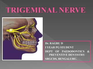
1. Dr. RAGHU D
I YEAR PG STUDENT
DEPT OF PAEDODONTICS &
PREVENTIVE DENTISTRY
SRGCDS, BENGALURU.
2. Introduction
Elementary structure of Neuron
12 Cranial Nerves
Embryology of the Nerve
Trigeminal Ganglion
Associated roots, branches and relations
Division of Trigeminal nerve
1. Ophthalmic nerve
2. Maxillary nerve
3. Mandibular nerve
3. Description of each nerve
Variations of maxillary nerve
Variations in mandibular canal & foramina
Ganglion associated with Trigeminal nerve
Applied aspects
› Trigeminal neurolgia
› Herpes zoster ophthalmicus
References
4. Nerve : A bundle of fibers that uses chemical
and electrical signals to transmit Sensory and
Motor information from one part of the body
to the another.
Neurons : Neurons are the specialized cells
that constitute functional units of the nervous
system and have a special property of being
able to conduct impulses rapidly.
5. Nervous system is the
most complicated
system in the body, it
is responsible for:
• Behaviour
• Thought
• Action
• Emotion reflects its
activity.
6. Neuron consists of a cell
body also called as soma
or perikaryon.
It gives off a variable
number of processes
called as neurites.
They are 2 types
• Dendrites
• Axon
7. Unipolar/ Pseudo-Unipolar : single pole both
axon and dendrites arise from single pole
Bipolar : 2 poles -1 for axon and 1 for
Dendrite
Multipolar : Many poles- 1 for axon and rest
all for dendrites . 2types
• Golgi type 1 Neurons
• Golgi type 2 Neurons
11. Mixed nerve: both fibers
1. Trigeminal nerve
2. Facial nerve
3. Glossopharyngeal nerve
4. Vagus nerve
Cranial nerve XIII is also known as the
“zero nerve” or “nerve N”.
(First discovered in 1870 in sharks and other
types of fish, it was initially referred to as the nerve
of pinkus)
Bordoni and Zanier Cranial nerves XIII and XIV: nerves in the
shadows
Journal of Multidisciplinary Healthcare 2013:6 87–91
12. Cranial nerve XIV was first identified in 1563,
but it was not until 1777 that it was mentioned
in a textbook as the nerve of Wrisberg.
[In modern textbooks, it is referred to as the
nervus intermedius or “intermediary
nerve”. Its name is consistent with its
intermediary location between the facial
nerve (cranial nerve VII) and the superior
section of the vestibulocochlear nerve
(cranial nerve VIII)]
13. The trigeminal nerve is
so called because of its
three main divisions i.e.
the Ophthalmic,
Maxillary &
Mandibular nerves.
It is the largest of the
cranial nerves.
It is the fifth cranial
nerve.
It is a mixed nerve.
14. It is sensory to the greater part of the scalp, the
teeth, and the oral and nasal cavities.
Motor supply is to the MOM (muscles of
mastication). Proprioceptive nerve fibers arise
from the masticatory and extra-ocular muscles.
15. During the
development of the
embryo, the
Pharyngeal arches
appear in the fourth
and fifth week.
It gives rise to six
pharyngeal arches, of
which the 5th arch
disappears.
16. Each arch characterized by its own:
Muscular component
Nerve component
Arterial component
Skeletal component
Trigeminal nerve is derived from 1st
Pharyngeal arch.
17. Muscles of mastication :
Temporalis
Masseter
Pterygoids
Anterior belly of digastric
Mylohyoid
Tensor tympani
Tensor palatini
Nerve supply to these muscles is provided by
mandibular division of Trigminal nerve.
18. It has got 4 nuclei:
1. Main Sensory nuclei
2. Spinal nuclei Purely Sensory
3. Mesencephalic nuclei
4. Motor nuclei - Motor
19. Occupies a cavity (Meckel’s cave) in
Duramater that contains the trigeminal
impression near the apex of the petrous part
of the Temporal bone.
20. It is somewhat cresentic or semilunar in
shape, with its convexity directed anterio
medially
The 3 divisions of the Trigeminal nerve
emerges from this convexity.
Neurons are of Pseudounipolar type.
21. The central processes of the ganglion cells
forms the large sensory root of the trigeminal
nerve, which is attached to pons at its
junction with the middle cerebellar peduncle.
The peripheral processes form the three
divisions of the Trigeminal nerve.
22. Small motor root of the trigeminal nerve is
attached to the pons superomedially to the
sensory root.
It passes the ganglion from its medial to the
lateral side and joins the mandibular nerve at
the foramen ovale.
23. Medially: Internal carotid artery and
posterior part of cavernous sinus
Laterally: Middle meningeal artery
Superiorly: Parahippocampal gyrus
Inferiorly: Motor root of trigeminal nerve,
greater petrosal nerve, apex of the petrous
temporal bone and foramen lacerum.
25. Motor nerve/root
It consists of fibers that have their origin in the
motor nucleus located in the upper pons.
These filaments pass from the pons, along the medial
side of the semilunar ganglion.
Then passes below to the foramen ovale, through
which it passes to join the mandibular division
immediately below the base of skull.
26.
27. Motor fibers of the trigeminal nerve supply the
following muscles:
Masticatory – masseter
- temporalis
- medial pterygoid
- lateral pterygoid
Mylohyoid
Anterior belly of digastric
Tensor tympani
Tensor veli palatini
28. The fibers of the sensory root of the trigeminal nerve
arise from the semilunar [gasserian] ganglion.
They enter the brain stem through the side of the pons.
Semilunar ganglion is located in Meckel’s cavity.
The ganglion is crescent shaped.
The ganglion with its unipolar neurons forms central &
peripheral processes.
29. The central branches are the sensory roots of the
trigeminal nerve.
These central branches leave the semilunar
ganglion & pass back & enter the pons, where
they divide into ascending & descending fibers.
The ascending fibers terminate in the upper
sensory nucleus in the pons lateral to the motor
nucleus.
30. It consists of afferent fibers that accompany the
fibers of the motor root.
Entering the pons from the peripheral distribution
of the mandibular division of the trigeminal
nerve, these fibers ascend to the mesencephalic
nucleus of the trigeminal nerve.
31. This nucleus serve as an afferent station that
receives proprioceptive impulses from the
temporomandibular joint, the periodontal
membrane, the maxillary & mandibular teeth &
the hard palate.
32. Three large nerves proceed from the convex
border of the semilunar ganglion
- ophthalmic nerve[V1]
- maxillary nerve [V2]
- mandibular nerve [V3]
33. It is superior and smallest
division
Wholly sensory
Arises from the
anteromedial end of the
trigeminal ganglion
It passes forward in the
lateral wall of the
cavernous sinus, below
the occulomotor and
trochlear nerves
34. The nerve joined by the filaments from the
internal carotid sympathetic plexus.
It communicates with the Oculomotor,
trochlear and abducent nerve
35. Before or just entering
the orbit through the
superior orbital fissure it
divides into:
1. Lacrimal ( smallest)
2. Nasocilliary (Intermediate)
Internal Nasal
External Nasal
Long ciliary
Infra trochlear
Posterior ethamoidal
37. Smallest of main Ophthalmic branches
Enters the orbit through the lateral part of the
superior orbital fissure
Runs along the upper border of the rectus
lateralis with the lacrimal artery
Receives twig from the Zygomaticotemporal
branch of maxillary nerve, which contains
Lacrimal secretomotor fibres
38. Supplies the lacrimal gland and adjoining
conjunctiva.
Pierces the orbital septum
Ends in upper eyelid, where it joins filaments
of the facial nerve
39. Largest branch of
Ophthalmic division
Enters the orbit through
the lateral part of the
superior orbital fissure.
Runs above the levator
palpebrae superioris and
divides into:
• Supra trochlear
• Supra orbital
40. It supplies :
• Conjunctiva
• Skin of the upper
eyelid
• Skin over the lower
fore head near the
midline
41. Transverses the
supraorbital foramen
It supplies :
• Frontal air sinus
• Upper eyelid
• Forehead
• Scalp till vertex
42. Intermediate in size between frontal and
lacrimal. Deeply placed in the orbit
Enters the orbit through the lateral part of the
superior orbital fissure and lie between the
two rami of the occulomotor nerve
Runs on the medial wall of the orbit between
superior oblique & medial rectus muscle
43. Anterior Ethmoidal
a. Middle or anterior ethmoidal sinus
b. Medial internal nasal
c. Lateral internal nasal
Posterior ethmoidal:
a. Posterior ethmoidal air sinus
b. Sphenoidal air sinus
Long cilliary ganglionic branches
a. Iris of cornea
44. External nasal
a. Skin of the ala
b. Tip of the nose
Infratrochlear
a. Both eyelids side of the nose
b. Lacrimal sac
45. It is intermediate
division of Trigeminal
nerve
Wholly sensory
Origin:
It leaves the ganglion
between the
Ophthalmic and
mandibular division as
a flat plexiform hand
46. Passes slightly medial to lateral wall of the
cavernous sinus
Gives a sensory branch to the duramater within
the cranium
Then it leaves the cranium through foramen
rotandum, which is located in the greater wing
of spenoid bone.
Once outside the cranium, it crosses the
uppermost part of the pterygopalatine fossa
47. As it crosses the pterygopalatine fossa it
gives of branches :
• Sphenopalatine ganglionic branches
• Posterior superior alveolar nerve
• Infra orbital nerve
• Zygomatic branches
48. 1. Within Cranial Cavity
a. Meningeal nerve (Dura
mater)
2. Ganglionic branches
a. Orbital
b. Palatine
c. Nasal
d. Pharyngeal
e. Lacrimal
3. Zygomatic
a. Zygomatico Temporal
b. Zygomatico Facial
49. 4. Infraorbital
a. Middle Superior Alveolar
b. Anterior Superior Alveolar
c. Face
• i. Palpebral
• ii. Nasal
• iii. Superior Labial
5. Posterior Superior Alveolar
50. Also known as nervus meningeus medius.
It lies within the cranium.
It receives a ramus from the internal carotid
sympathetic plexus and accompanies the
middle meningeal artery to supply the
duramater.
51. Starts in pterygopalatine fossa
Enters the orbit through the inferior orbital
fissure.
Runs along the lateral wall to reach
zygomatic bone.
Just before/after entering zygomatic bone it
gives two branches:
› Zygomaticotemporal
› Zygomaticofacial
52. Zygomaticotemporal:
A communicating
secretomotor fibers
given to the lacrimal
gland through the
lacrimal nerve
Zygomaticofacial: it
supplly to Skin over
the zygomatic
prominence and to the
anterior part of the
temple.
53. It descends from main trunk of maxillary
division in the pterygopalatine fossa.
Through pterygopalatine fossa it reaches
posterior surface of the maxilla
From here it enters the maxilla through PSA
canal.
54.
55. Travels down to the posteriolateral wall of
maxillary sinus
Provides Sensory innervation to the
maxillary sinus
Continuing downwards it provides sensory
innervation to the alveoli, periodontal
ligament and pulpal tissues of the maxillary
3rd, 2nd and 1st molar teeth
56. Applied anatomy: During nerve block there
is a greater risk of hematoma formation
57. This ganglion is also known sphenopalatine
ganglion or ganglion of Hay fever
The ganglionic branches of maxillary nerve
suspend the ganglion in the pterygopalatine
fossa
It is the largest peripheral parasymphathetic
ganglion
It serves as a relay station for secretomotor
fibres to the lacrimal gland
58. Topographically related
to maxillary nerve, but
functionally it is related
to facial nerve(through
greater petrosal
branch)
59. 1. Orbit
2. Nasal
a. Superior posterior
nasal
i. Medial
ii. Lateral
b. Nasopalatine
60. 3. Palate
a. Greater (anterior
palatine)
b. Lesser (middle &
Posterior)
4. Pharynx
5. Lacrimal
63. Emerges on the hard
palate through the
greater palatine
foramen( usually
located about 1cm
towards the palatal
midline, just distal to
the second molar)
64. The nerve courses anteriorly supplying
sensory innervation to the palatal soft tissues
and bone as far as the first premolar, where
it communicates with the terminal fibres of
the nasopalatine nerve.
It provides sensory innervation to some part
of the soft palate
65. Emerges from the lesser palatine foramen
along with the posterior palatine nerve.
Provides sensory innervation to the mucous
membrane of soft palate
The posterior palatine nerve: Innervates the
tonsillar region
66. It is a small nerve
Passes through the pharyngeal canal and
distributed to the mucous membrane of the
nasal part of the pharynx posterior to the
auditory tube.
67. Enters the orbit through IOF
Runs forward on the floor of the orbit
First in the infraorbital groove, then in the canal
. Here it gives 2 branches
› Anterior superior alveolar
› Middle superior alveolar
The nerve terminates by emerging on the face
through infraorbital foramen giving out its
terminal branches
› Lower palpebral
› Lateral nasal
› Superior labial
68.
69. Arises from the infraorbital nerve
Provides sensory innervation to two
maxillary premolars and periodntal tissues,
buccal soft tissues and bone in the premolar
region
Traditionally it has been stated that the MSA
is absent in 30% to 50% of individuals
In its absence the usual innervations are
provided by either the PSA or the ASA nerve.
70.
71. It is a relatively large branch
Given off from the infraorbital nerve at
approximately 6 to 10mm before it exit from
the infra orbital foramen
It provides pulpal innervation to the :
› Central and lateral incisors
› Canine
› Periodontal tissues
› Buccal bone
› Mucous membrane of these teeth
72. It emerges from the
infraorbital foramen onto
the face by dividing into
its terminal branches
1. Inferior palpebral :
supplying the skin of the
lower eyelid
2. The External nasal
branch : providing
sensory innervation to
skin of lateral part of the
nose
73. 3. Superior labial branch : supplying the skin
and mucous membrane of the upper lip.
74. Knowledge of the anatomical variations of the
maxillary nerve is necessary for a surgeon while
performing maxillofacial surgery and regional block
anesthesia.
Infraorbital nerve:
Infraorbital foramen is usually a single foramen
but several studies have proven to have two or
three foramen. A low percentage (4.7%) was
observed during a study on 1064 skulls, with a
higher frequency on the left side, both in male
and in female skulls. The distance from the
infraorbital foramen to the inferior border of the
orbital rim is from 4.6 to 10.4 mm
75. Posterior superior alveolar nerve:
Mc Daniel found that posterior superior alveolar
nerve had one branch in 21%, two branch in
30% and three branches in 25% of specimens.
Branching pattern of this nerve should be
considered during anesthetic procedure in this
nerve, the different origins of the posterior
superior alveolar nerve compared to the middle
and the anterior branches offers the possibility to
anesthetize only the posterior branch.
76. Anterior superior alveolar nerve:
The anterior superior alveolar nerve was present
as a single trunk in 75%, of cases as reported by
Mc Daniel; in 35% there was a diffuse fine plexus
of the anterior superior alveolar nerve branches
overlying the canine fossa.
The presence of a superior dental plexus
appears to be favoured by multiple posterior
branches and by the presence of a middle
branch or an anterior branch with multiple main
branches.
77. Middle superior alveolar nerve:
Middle superior alveolar nerve arises from infra
orbital nerve when it is the infraorbital canal.
McDaniel reported that the middle superior
alveolar nerve followed the classical description
in only 30% of examined cases whilst the
majority of middle branch entered the formation
of a nerve plexus that supplied the teeth.
When the middle branch was absent, the
innervation of the premolar teeth may be
provided by secondary branches of the anterior
superior alveolar nerve, by the posterior superior
alveolar nerve or by a nervous plexus between
these two nerves.
78. Nasopalatine:
Gray (1980) reports that the nerve innervates the
mucous membrane in the anterior part of the
hard palate and that it communicates with the
anterior palatine nerves.
Cunningham (1981) suggests that the anterior
palatine nerves supply the gingivae and
supporting structures of the upper teeth only as
far forward as the canines and that the
nasopalatine nerve innervates the mucosa in the
incisor region.
Last (1984) states that it supplies the incisive
gum of the hard palate.
79. Dixon (1986) is more specific, stating that the
nasopalatine nerve may supply an area of mucous
membrane in the region of the incisive papilla and
may also help to supply the supporting structures of
the central and often lateral incisor teeth
Sai Pavithra .R et al. Maxillary Nerve Variations and Its Clinical
Significance,, J. Pharm. Sci. & Res. Vol. 6(4), 2014, 203-205
80. Largest division of
Trigeminal nerve
It is mixed in nature
Has a large sensory
root & small Motor root
81. Sensory root originates from trigeminal
ganglion whereas the motor root originates
in the pons & medulla oblongata
The two root emerges from the cranium
separately through the foramen ovale
The motor root lying medial to sensory root
They unite just outside the skull & from the
main trunk of 3rd division
85. Meningeal branch
Enters the skull through foramen spinosum (
along with the middle meningeal artery)
Supply the duramater of the middle cranial
fossa
The nerve is also called Nervous spinosus
86. It is a motor nerve to
medial pterygoid
muscle
87. Motor branch to: The
muscles of mastication
Buccal nerve: Sensory
innervation to the
mucous membrane of
the cheek and buccal
mucous membrane of
the mandibular molars
The anterior division is
smaller than the
posterior division
88. Under the lateral pterygoid nerve it gives off
some branches i,e.
1. Deep temporal nerve: to the temporal
muscle
2. Masseteric nerve: providing motor
innervation to masseter muscle
3. Lateral pterygoid nerve: Providing motor
innervation to the lateral pterygoid muscle
89. It is also called as long
buccal nerve
Usually passes
between 2 heads of
the lateral pterygoid
Reaches the external
surface of the muscle
Follows the inferior
part of the temporal
muscle
90. Then it emerges under the anterior border of
the masseter muscle
At the level of occlusal plane of the
mandibular 3rd and 2nd molar
Crosses in front of the ramus
Enters the cheek through buccinator muscle
91. It provides sensory innervation to:
1. Skin over the anterior part of buccinator
2. Buccal gingiva of mandibular molars
3. Mucobucca fold in that region
Buccal nerve does not innervate the
buccinator muscle, but the facial nerve
does.
92. Larger division
Mainly sensory
Divides into 3
branches
1. Auriculotemporal
2. Lingual nerve
3. Inferior alveolar nerve
(only motor)
I. Mylohyoid
II. Anterior digastric
93. It has 2 roots:
Encircles the middle meningeal artery
Runs back under lateral pterygoid on the
surface of tensor veli palatini to pass between
the sphenomandibular joint in relation with the
upper part of the parotid gland
Emerging from behind the joint it ascends
posterior to the superficial temporal vessels
over posterior root of the zygoma
Divides into superficial temporal branches
94. 1. Two anterior auricular branch : supply the skin
of the tragus and sometimes small part of
adjoining helix and the temporomandibular
joint
2. Two branches to external acoustic meatus:
Supply the skin of the meatus and the
tympanic membrane
3. Superficial temporal branch: supply skin in the
temporal region and connects with the facial
and zygomaticotemporal nerves
95. It communicates with facial nerve providing
sensory fibres to the skin over the areas of
innervation of motor brances of facial nerve
It communicates with the otic ganglion
providing sensory, secretory and vasomotor
fibres to parotid gland
96. 2nd branch of the posterior division of the
mandibular nerve
Runs between the tensor veli palatini and
lateral pterygoid, where it is joined by chorda
tympani branch of facial nerve from here
It descends to rest between the ramus and
medial pterygoid muscle in the
pterygomandibular space
97. Then it runs anteriorly and medially to the
inferior alveolar nerve whose path is parallel to
it
It then continues to reach side of the base of
the tongue slightly below and behind the
mandibuar 3rd molar
Here it lies just below the mucous membrane in
the lateral lingual sulcus
Then it proceeds anteriorly across the muscles
of the tongue
Looping medial to submandibular duct
(wharton’s duct) to deep surface of
submandibular and sublingual glands where it
breaks up into terminal branches
98.
99. Mucosa of the floor of the mouth, lingual
gingivae
Mucosa of anterior 2/3rd of the tongue
Also carries postganglionic fibers from
submandibular ganglion to sublingual and
anterior lingual glands
Applied anatomy:
Lingual nerve is at the greatest risk during surgical
removal of the impacted 3rd molar
During removal of the submandibular salivary gland,
the duct must be dissected from Lingual nerve
100. Largest branch of the mandibular division
Descends medial to the lateral pterygoid muscle
and posterior to lingual nerve
Passes between the sphenomandibular
ligament and mandibular ramus to enter the
mandibular canal via mandibular foramen
Through out its path it is accompanied by
inferior alveolar artery and inferior alveolar vein
Nerve travels anteriorly in the canal till it
reaches the mental foramen
101. 2 branches :
1. Mental nerve
2. Incisive nerve
Applied aspects:
Lower lip and tongue is also anaesthetized
during Inferior alveolar nerve block. Hence
young child or physically or mentally
handicapped patients should be informed
prior to administration to avoid soft tissue
injury
102. Incisive nerve :
Continues forward in the body of canal giving
off branches to :
Premolar
Canine
Incisors
Associated labial gingiva
103. Mental Nerve:
Exit the canal through the mental foramen
between and just below the apices of the
premolars and divides into three branches
that innervates:
Skin of the chin
Skin of lower lip
Buccal mucous membrane from 2nd premolar to
the midline i,e. Central incisor region
104.
105. Just before the mandibular canal, the inferior
alveolar nerve gives off a small Mylohyoid
branch
It pierces the sphenomandibular ligament
and enters a shallow groove on medial
surface of mandible
It is a mixed nerve
106. It provides motor
innervation to:
Mylohyoid & anterior
belly of digastric
Sensory fibers to inferior
and anterior surface of
mental protuberence
Mandibular incisors
(sometimes)
108. Connected to maxillary
nerve in infratemporal
fossa
Sensory to orbital
septum, orbicularis
and nasal cavity,
maxillary sinus , palate
and nasopharynx
109. It lies between the
trunk of mandibular
nerve and tensor
palatini
Nerve to medial
pterygoid passes
through but does not
relapse in the ganglion
110. It related to lingual
nerve, rest on
hypoglossus
Supplies posterior
ganglionic
parasympathetic
secretomotor fibres to
submandibular and
sublingual gland
111. Chavez et al. suggested that during embryonic
development three canals fuse to form a single
nerve canal. Failure of these canals to fuse can
explain presence of multiple canals in some
individuals.
The location and configuration of the
mandibular canal are important in surgical
procedures involving the mandible.
Bifid mandibular canals (BMC) and trifid
mandibular canals (TMC) are variations on the
normal anatomy with incidences ranging from
0.08% to 65.0%
112. The clinical relevance of bifid and trifid mandibular canals
Oral Maxillofac Surg. 2012 Mar; 16(1): 147–151
113. The incidence of bifid mandibular foramina
has been estimated by many studies to vary
from 0.05% to 65% in the general
population.
Bifid mandibular foramina can occur
unilaterally or bilaterally on the mandible
The presence of multiple mandibular
foramens might contribute to the failure of
inferior alveolar blocks.
114. In most cases of bifid mandibular canals
more anesthetic injections are needed
leaving a higher chance of increased
anesthetic neurotoxicity and or injuring the
inferior alveolar neurovascular bundles.
Thus, the presence of bifid mandibular
canals should be considered as a risk factor
for inferior alveolar paresthesia and should
be taken into consideration in third molar
extraction, mandibular surgery, and implant
placement.
117. Trigeminal neuralgia (TN) is also called tic
douloureux
Trigeminal neuralgia is defined as sudden,
usually unilateral, severe, brief, stabbing
recurrent episodes of pain within the
distribution of one or more branches of the
trigeminal nerve, which has a profound effect
on quality of life.
Majeed M H, Arooj S, Khokhar M, et al. (December 18, 2018)
Trigeminal Neuralgia: A Clinical Review for the General Physician .
118.
119. TN is characterized by an abrupt onset and
short-lived unilateral shock-like pain, limited to
the distribution of the trigeminal nerve.
Triggers for classical TN (CTN) usually include
mastication (73%),
touch (69%),
tooth brushing (66%),
eating (59%),
talking (58%), and
cold wind on the face (50%).
120. Trigger zones are present in more than 90%
of the patients, with touch and vibrations
being the most common stimuli in provoking
pain.
Pain is usually distributed along the V2 and
V3 branches
Pain occurs slightly more often (59% to 66%)
on the right side of the face and rarely (3%
to 5%) is bilateral.
121. Classical
Secondary
Idiopathic
Majeed M H, Arooj S, Khokhar M, et al. (December 18, 2018) Trigeminal
Neuralgia: A Clinical Review for the General Physician . C
122. Herpes Zoster infestation of the trigeminal
ganglion of the ophthalmic nerve
Chronic paroxysmal hemicrania
Tolosa-Hunt syndrome
Migraine
Cluster headache
Glossopharyngeal neuralgia are among the
differential diagnoses of TN
123. Advanced age.
› The risk of TN is higher among older people,
especially between 50 to 60 years of age.
› Age related changes, such as hardening and
elongation of blood vessels and sagging of the brain
(just like aged skin) can cause blood vessel-nerve
contact where there was none before—resulting in
irritable and sensitive nerves.
› Advancement of age also causes degenerative
changes in nerves resulting in loss of myelin sheath,
making the nerves susceptible to irritation.
124. Female sex. Women are at a higher risk
than men to be affected by TN.
Multiple sclerosis. TN is known to be
associated with multiple sclerosis, a
condition that causes degeneration of the
myelin sheath of nerves.
Trigeminal Neuralgia Causes and Risk Factors, By Rob
D. Dickerman, DO, PhD, FACOS
125. First line treatment is typically :
Sodium channel blockers, either carbamazepine
or oxcarbazepine.
(The European Federation of Neurological Societies
and the Quality Standards Subcommittee of the
American Academy of Neurology consider
carbamazepine (CBZ) as the drug of choice for the
treatment of TN)
(The typical starting dose is 100 to 200 mg twice
daily and then is gradually increased to 200 mg. The
usual maintenance dose is 600 to 1200 mg in divided
doses with a desired therapeutic blood level of 4 to
12 ug/ml)
Majeed M H, Arooj S, Khokhar M, et al. (December 18, 2018) Trigeminal
Neuralgia: A Clinical Review for the General Physician . C
126. However, as a result of intolerable side
effects, such treatment may fail.
Surgical treatment microvascular
decompression (MVD) then becomes the next
choice if neurovascular contact has been
demonstrated.
MVD : It involves open surgery and separation
of the trigeminal nerve root entry zone from the
offending vessel by virtue of Teflon sponge. It
maintains the integrity of the trigeminal nerve
following surgery. So the postoperative facial
numbness and dysaesthesia are rarely seen.
Trigeminal Neuralgia – Diagnosis and Treatment Option
Aug 02, 2016 | By neurologicalsurgery
127. Botulinum toxin-A injection at the site of pain the
trigger point using a 1 mL syringe with a 0.45×16
mm needle, while for multiple site injections,15m
mintervals were measured between injection sites,
with 5 U at each site
Radiofrequency thermo coagulation is a safe and
proven means of treating trigeminal neuralgia.
It uses radiofrequency to heat up a small
part of the nerve tissue so that the pain signals are
interrupted.
Shouyi Wu1 Yajun Lian1 .Botulinum Toxin Type A for refractory
trigeminal neuralgia in older patients: a better therapeutic effect,
Journal of pain research 2019
Trigeminal Neuralgia – Diagnosis and Treatment Option
Aug 02, 2016 | By neurologicalsurgery
129. Varicella represents the primary infection in the
nonimmune or incompletely immune person.
During the primary infection, the virus gains
entry into the sensory dorsal root ganglia.
How the virus enters the sensory dorsal root
ganglia and whether it resides in neurons or
supporting cells are not completely understood.
The varicella-zoster virus genome has been
identified in the trigeminal ganglia of nearly all
seropositive patients
130. Hyperesthesia , paresthesias, burning
dysaesthesias or pruritis along the affected
dermatome(s)
Pain is the most common complaint for which
patients with herpes zoster seek medical care.
The pain may be described as “burning” or
“stinging” and is generally unrelenting.
Indeed, patients may have insomnia because of
the pain.
Although any vertebral dermatome may be
involved.
131.
132. The vesicles eventually become hemorrhagic or
turbid and crust over within seven to 10 days.
As the crusts fall off, patients are generally left
with scarring and pigmentary changes.
Ocular complications occur in approximately
one half of patients with involvement of the
ophthalmic division of the trigeminal nerve.
These complications include mucopurulent
conjunctivitis, episcleritis, keratitis and anterior
uveitis.
134. Trigeminal nerve, courses and its branches are very
important from Dentist point of view as inadvertent
procedures may lead to trigeminal nerve injury.
Disorders of trigeminal nerve is not rare, knowing
about it will help in diagnosis and treatment thus
achieving the best possible recovery of trigeminal
nerve function
Nerve blocks given for carrying various dental
procedures involves the various branches of
trigeminal nerve, hence to avoid any one need to
have a knowledge about course and branches of
Trigeminal nerve
135. B D Chourasia’s. Human Anatomy for Dental students, 2nd
ed. 2012.
Inderbir singh. G P Pal, Human Embryology, 9th ed. 2012
Sperber. Craniofacial Development, 2001
Wheeler’s. Dental Anatomy, Physiology and Occlusion, 9th
ed. 2013
Guyton and Hall. Textbook of Medical Physiology. 9th ed.
1996
Dr. A P Krishna. Textbook Of Physiology, 7th ed. 2010
Shafer’s. Textbook of oral pathology, 6th ed. 2009