
Recommended
More Related Content
What's hot
What's hot (20)
Similar to Juvenile nasopharyngeal angiofibroma
Similar to Juvenile nasopharyngeal angiofibroma (20)
More from Raafi Ul Zargar
Recently uploaded
Recently uploaded (20)
Juvenile nasopharyngeal angiofibroma
- 1. JUVENILE NASOPHARYNGEAL ANGIOFIBROMA R A A F I U L B A S H E E R Z A R G A R
- 2. INTRODUCTION It is also known as nasopharyngeal fibroma . It is a rare tumor but commonest of all benign tumor of nasopharynx.
- 3. ETIOLOGY Idiopathic Mostly seen in adolescent males in 2nd decade of life. Hormonal Theory • Testosterone dependent • Patients have a hamartomatous nidus of vascular tissue and this is activated to form angiofibroma when male sex hormone appears.
- 4. SITE Posterior part of nasal cavity close to the superior margin of sphenopalatine foramen
- 5. GROWTH Locally invasive benign tumors. From site of origin to nasal cavity, nasopharynx and into the pterygopalatine fossa, running behind the posterior wall of maxillary sinus which is pushed forward as the tumor grows. Laterally it extends into pterygomaxillary fossa then to infratemporal fossa and cheek.
- 6. PATHOLOGY Severe bleeding as the vessels lose ability to contract Made up of vascular and fibrous tissue mostly the vessels are just endothelium lined spaces with no elastic or muscle coat
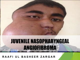
- 8. EXTENSION Nasal cavity- Causing nasal obstruction, epistaxis and nasal discharge Paranasal sinuses- Maxillary, sphenoid and ethmoid sinuses can all be invaded Pterygomaxillary fossa, infratemporal fossa and cheek Orbits-proptosis, frog face deformity
- 9. EXTENSION CRANIAL CAVITY • ANTERIOR CRANIAL FOSSA through roof of ethmoids or cribriform plate • MIDDLE CRANIAL FOSSA through erosion of floor of middle cranial fossa or indirectly by invading the sphenoid sinus and sella tunica.
- 10. SYMPTOMS SEX-MOSTLY IN MALES AGE-10-20YRS PROFUSE, RECURRENT AND PAINLESS EPISTAXIS LEAD TO ANAEMIC DUE TO REPEATED BLOOD LOSS.
- 11. SYMPTOMS Progressive nasal obstruction and denasal speech Conductive hearing loss and otitis media with effusion Mass in the nasopharynx Broadening of nasal bridge Proptosis Swelling of cheek Conductive hearing loss and serous otitis media due to obstruction of Eustachian tube
- 12. SIGNS Anterior Rhinoscopy – 1. Pink or purplish nasopharyngeal mass 2. sessile, lobulated or smooth 3. obstructs one or both choanae 4. Consistency is firm but digital palpation is never done because it can result in profuse bleeding.
- 13. INVESTIGATIONS CT Scan • Investigation of choice • The extent of tumour, bony destruction or displacements can be seen. • Hollman-Miller Sign Anterior bowing of maxilla and Posterior bowing of ptyerigoid. Contrast CT scan juvenile nasopharyngeal angiofibroma. Note the pterygopalatine fossa and infratemporal fossa extension
- 14. INVESTIGATIONS MRI Carotid angiography shows the extent of tumors
- 15. STAGING Sessions’s classification, modified by Radkowski reflects IA Tumor limited to nose and nasopharyngeal vault IB Extension to paranasal sinuses IIA Minimal extension to pterygomaxillary fissure (PMF) IIB Full extension to PMF and/or erosion of orbital bones IIC Extension to infratemporal fossa and/or cheek or posterior to pterygoid plates IIIA Erosion of skull base: minimal intracranial IIIB Extensive intracranial and/or cavernous sinus extension
- 16. MEASURES TO REDUCE THE VASCULARITY OF THE TUMORREDUCE THE VASCULARITY OF TUMOR LARITY OF TUMOR • . • Embolization of the feeding vessels. • Estrogen therapy: Stilboestrol 2.5 mg three times a day for 3 weeks. Not preferred currently. • Preoperative radiation: Generally not favored. • Cryotherapy.
- 17. TREATMENT(OPEN SURGICAL EXCISION) Surgical approaches Transpalatine: Tumours confined to nasopharynx, nasal cavity and sphenoid sinus. Le Fort 1 osteotomy approach: For extension to paranasal sinuses, pterygopalatine fossa and infratemporal fossa. Medial maxillectomy: It provides access to orbit, ethmoid and sphenoid sinuses and anterior skull base. Sardana’s approach: Transpalatine + Sublabial. Extended lateral rhinotomy Extended Denker’s approach Intracranial-extracranial Infratemporal fossa
- 18. TREATMENT
- 19. TREATMENT RADIATION THERAPY • For advanced tumors with intracranial extension
- 20. COMPLICATIONS Secondary malignancy Abnormal craniofacial development Cataracts Optic atrophy Osteoradionecrosis.
- 21. MANAGEMENT OF RECURRENCE Radiotherapy • 3000-3500 cGy of radiation given in 15 to 20 sittings. Chemotherapy • Drugs like Vincristine, Doxorubicin , Dacarbazine The above methods can arrest the growth and cause tumor regression but not total tumor eradication
- 22. PROGNOSIS Excellent prognosis on complete surgical removal.
- 23. THANK YOU