Determination of primary structure of proteins
•Download as PPTX, PDF•
35 likes•37,617 views

Biochemistry

Recommended
Recommended
More Related Content
What's hot
What's hot (20)
Similar to Determination of primary structure of proteins
Similar to Determination of primary structure of proteins (20)
More from Pradeep Singh Narwat
More from Pradeep Singh Narwat (20)
Recently uploaded
Recently uploaded (20)
Determination of primary structure of proteins
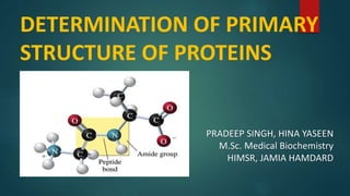
- 1. DETERMINATION OF PRIMARY STRUCTURE OF PROTEINS PRADEEP SINGH, HINA YASEEN M.Sc. Medical Biochemistry HIMSR, JAMIA HAMDARD
- 2. INTRODUCTION Proteins are polymers composed of hundreds or even thousands of amino acids linked by peptide bonds. Proteins are most abundant biomolecules which makes about 75% of total body weight. Carbon, hydrogen, oxygen and nitrogen are major components of proteins. Average molecular weight of proteins range between 5kDa- 220kDa.
- 3. ORGANISATION OF PROTEIN Proteins are usually large molecules with a complex three dimensional structure. Four levels of proteins are commonly defined as: 1. Primary structure 2. Secondary structure 3. Tertiary structure 4. Quaternary structure
- 4. Primary Structure of Proteins It is a linear polymer formed by linking the α-carboxyl group of one amino acid to the α- amino group of another amino acid . This type of linkage is called a peptide bond or amide bond forming polypeptide chain.
- 5. Dipeptide is formed by two amino acids. Similarly, tripeptide is formed by three amino acids. Eg: Glutathione is a tripeptide. Each amino acid unit in a polypeptide is called residue. The sequence of amino acids in a polypeptide chain is written starting with the amino terminal residue called as N-terminal residue.
- 6. A polypeptide chain consist of a regularly repeating part, called main chain or backbone, and a variable part, comprising the distinct side chain (R1 , R2 , R3 etc). The linear polypeptide chain is cross linked and the most common cross-links are disulphide bonds.
- 7. PEPTIDE BOND All amino acids are linked by peptide bond. N terminal end- free NH2 group of amino acid. C terminal end- free COOH group of amino acid. Amino acid are sequenced from N- terminal end to the C- terminal end.
- 8. CHARACTERISTICS OF PEPTIDE BOND 1. Partial double bond character. 2. Rigid and planer. 3. C-N bond is trans in nature. 4. Uncharged but polar. 5. This prevents free rotation around the bond between the carbonyl carbon and the nitrogen of the peptide bond.
- 9. Secondary Structure Secondary structure are formed by a regular pattern of H-bonds and other non covalent interactions between the side chain residues of amino acids that are near to one another in the linear sequence. Example: Myoglobin Types of secondary structure: 1. Alpha Helix 2. Beta Pleated Sheet 3. Loops 4. Bends
- 10. α- helix β - pleated sheet • Right handed spiral structure. • H- bonding interaction. • Proline and hydroxyproline will not allow the formation of alpha helix • Almost fully extended • Stabilized by h- bonding. • Zig- zag or pleated pattern • Adjacent strands in a sheet can run in the same direction or opposite direction.
- 11. Tertiary Structure The entire three dimension conformation of a polypeptide is referred to as tertiary structure. Tertiary structure is maintained by weak bonds (H-bonds, electrostatic forces and other non covalent interactions). Tertiary structure represent the lowest energy and greatest stability state of a polypeptide chain . Example: Enzymes are globular proteins with a specific tertiary structure. Folding of primary structure to form tertiary structure in enzymes
- 12. Quaternary Structure Two or more peptides combine together to form the quaternary structure of protein. These peptides chains are linked to one another by non covalent interactions. Example- Haemoglobin
- 13. Types of protein on the basis of structure Proteins fall into three basic classes according to shape and solubility: 1. Fibrous Protein – Collagen 2. Globular Protein – Myoglobin, Hemoglobin 3. Membrane Proteins
- 14. • Regular linear structures • Play structural roles in the cell (matrix formation) • Often water Insoluble 1. Fibrous Protein 2. Globular Protein 3. Membrane Protein • Roughly spherical in shape • Often water soluble • Hydrophobic amino acid side chains present in the interior of the molecule while hydrophilic side chains are outside exposed to the solution • Hydrophobic amino acid side chains present outside • Play role in the cellular transport • Often water insoluble but soluble in detergents Collagen Myoglobin GPCR
- 15. DETERMINATION OF PRIMARY STRUCTURE OF PROTEIN
- 16. Why we Need to Determine the Primary Structure of Protein? Protein play important role as “the building block of life” Enzyme Hormones and hormone receptors Sensing device: such as rhodopsin Role in immune system Expression of genetic information (transcription) Constituent of important body part: collagen Transporters: Albumin, Myoglobin & Hb. To locate the gene of interest in the host cell. To artificial synthesis the above products by using the applications of biotechnology, we need the determine the primary structure of protein.
- 17. PURIFICATION OF PROTEINS Step 1: Solubilization of protein A. Homogenization B. Centrifugation C. Filtration Step 2: Stabilization of proteins Step 3: Purification of proteins by various techniques including chromatography, Gel Electrophoresis etc. Step 4: Digestion of proteins into smaller peptides Step 5: Sequencing of individual peptide.
- 18. DETERMINATION OF THE AMINO ACID COMPOSITION OF A POLYPEPTIDE The first step in determining the primary structure of a polypeptide is to identify and quantitate its constituent amino acids. Cleavage of disulphide bonds by the 2-Ercaptoethanol. A purified sample of the polypeptide to be analyzed is first hydrolyzed by strong acid (6M HCl) at 110°C for 24 hours. This treatment cleaves the peptide bonds and releases the individual amino acids, which can be separated by cation-exchange chromatography.
- 19. The separated amino acids contained in the eluate from the column are quantitated by heating them with Ninhydrin—a reagent that forms a purple compound with most amino acids, ammonia, and amines. The amount of each amino acid is determined by spectrophotometrically by measuring the amount of light absorbed by the Ninhydrin derivative.
- 20. Determination of amino acid composition Several Methods are used in combination to get the final composition- ACID HYDROLYSIS: Purified Sample to be analysed is first hydrolysed by 6N HCL and heated at 110-120 °C for 24hrs. in sealed vessel . The peptide bonds are broken and the hydrolysate is analysed by HLPC to determine the composition. It degrades serine, threonine, tyrosine, tryptophan.
- 21. ALKALINE HYDROLYSIS: A Purified sample to be analysed is hydrolysed with a strong base e.g: NaOH and hydrolysate is examined. It does not destroy tyrosine , tryptophan and glutamine but it destroys Serine, threonine, arginine. ENZYMATIC HYDROLYSIS: To cleave the proteins into a small number of peptide fragments. Cyanogen Bromide (CNBr) splits polypeptide chain only on the carboxylic side of methionine residues.
- 23. End group analysis Identification of N- terminal and C- terminal amino acids in a polypeptide chain is called End group analysis.
- 24. Identification of N- terminal 1) Sanger’s Method 2) Edman’s Degradation Technique 3) Dansyl Chloride Method
- 25. Sanger’s Method This was the first technique to determine the sequence of proteins. Sanger’s Reagent – 2,4- Dinitroflouro Benzene. Sanger’ s Reagent derivatizes the amino terminal residues. The first proteins to be sequenced by the method is Insulin by Fredrick Sanger. He got Noble Prize in 1958. Only dipeptides or tripeptides can be sequenced.
- 26. Steps: 2,4- dinitro fluorobenzene is reacted with amino group of a peptide or a proteins to form 2,4- dinitrophenyl derivative of N- terminal amino acid which is yellow in colour. The treated peptide is then subjected to acid hydrolysis which cleaves all the peptide bonds except the bond between 2,4- DNF and NH2 group which is resistant to acid hydrolysis . Separated by Chromatography.
- 27. Steps: +
- 28. Edman’s Degradation Method This process was developed by Pehr Edmen. Edman ‘s Reagent – Phenyl isothiocyanate It is a technique for identifying specific amino acids at each position in the peptide chain beginning at the amino terminal end. Edman’s technique can sequence many residues (5-40)of a single polypeptide sample.
- 29. METHOD Phenyl iso thiocyanate (PITC) react with amino group of a polypeptide under mild alkaline conditions to form a corresponding phenylthiol- carbamoyl-peptide. It involves a controlled stepwise cleavage of the polypeptide. ADVANTAGE: This method over sanger’s Method is that the remaining peptide after the removal of the N-terminal amino acid is not hydrolysed and can be used again to detect the next amino acid.
- 31. DANSYL CHLORIDE METHOD Dansyl chloride- 5’-dimethyl-1-naphthalene-sulphonyl-chloride NH2 –terminal is reacted with dansyl chloride which is a fluorescent compound to form a fluorescent dansyl amino acid derivative. This derivative is removed from polypeptide by hydrolysis. Amino terminal residue is separated by Chromatography. ADVANTAGE: It is a sensitive method and can detect picomole quantity
- 33. Identification of C- Terminal amino acid Akabori Method By Carboxy Peptidase
- 34. AKABORI METHOD Akabori method - Treatment with hydrazine In Akabori method, identification of c-terminus amino acids, involves the heating of a linear peptide in the presence of anhydrous hydrazine in a sealed tube for several hours The amino group of each peptide bond react with hydrazine to form the corresponding amino acid hydrazide. Identified by chromatography.
- 35. The application of microwave irradiation to the Akabori reaction was investigated . It was observed that microwave irradiation reduced the time required for the completion of the Akabori reaction from hours to minutes. This approach provided information not only about the C-terminus but also the N-terminus.
- 37. BY CARBOXYPEPTIDASES Carboxy peptidases cleave a polypeptide form C-terminal, removing one amino acid at a time by cleaving the peptide bond. Removal of one amino acid leaves behind a new polypeptide which is the target of carboxy peptidase . Carboxypeptidases that have a stronger preference for those amino acids containing aromatic or branched hydrocarbon chains are called carboxypeptidase A
- 38. Carboxypeptidases that cleave positively charged amino acids (arginine, lysine) are called carboxypeptidase B Plants contain carboxypeptidase C that liberates the amino acid proline as well as being able to release many of the other protein amino acids.
- 39. CLEAVAGE OF THE POLYPEPTIDE INTO SMALLER FRAGMENTS
- 40. ENZYMATIC METHOD To cleave the proteins into a small no. of pure fragments. Example- Cyanogen Bromide (CNBr) splits polypeptide chain only on the carboxylic side of methionine residues. Highly specific cleavage is also obtained by trypsin, a proteolytic enzyme secreted by pancreas. Trypsin cleaves polypeptide chain on the carboxylic side of arginine residues. Peptides obtained specific chemical or enzymatic cleavage are separated by some type of chromatography.
- 41. Reagent Chemical cleavage Cleavage site Cyanogen bromide COOH side of methionine residue hydroxylamine Asparagine - glycine bond O-iodosobenzoate COOH side of tryptophan residue 2- Nitro-5- thiocyanobenzoate NH2 side of cysteine residue Enzymatic cleavage Trypsin Lysin and arginine residue Chymotrypsin Tyrosine, tryptophan, phenylalanine, leucine and methionine Thrombin Arginine Carbopeptidase A C-terminal amino acid clostripain Arginine residue
- 43. • When exposed to trypsin • When expose to chymotrypsin
- 46. PRINCIPLE- A mass spectrometer generates multiple ions from the sample under investigation, it then separates them according to their specific mass-to-charge ratio (m/z), and then records the relative abundance of each ion type. Detect ions-
- 47. For small organic molecules the MW can be determined to within 5 ppm or 0.0005% which is sufficiently accurate to confirm the molecular formula from mass alone For large biomolecules the MW can be determined within an accuracy of 0.01% (i.e. within 5 Da for a 50 kD protein) Recall 1 dalton = 1 atomic mass unit (1 amu)
- 48. Mass spectrometry is a technique for analyzing ionized forms of molecules in the gas phase. It is most readily applied to gases or to volatile liquids that easily release gas-phase ions. Mass measurement obtained by determining how readily ion is accelerated in an applied electric field.
- 50. METHODS OF MS Methods are widely used:- ESI-QTOF Electrospray ionization source + quadrupole mass filter + time-of-flight mass analyzer MALDI-QTOF Matrix-assisted laser desorption ionization + quadrupole + time-of-flight mass analyzer Tandem LC and Tandem MS Separates by HPLC, ID’s by mass and AA sequence.
- 51. Different Ionization Methods Electron Impact small molecules, 1-1000 Daltons Fast Atom Bombardment peptides, sugars, up to 6000 Daltons Electrospray Ionization peptides, proteins, up to 200,000 Daltons Matrix Assisted Laser Desorption peptides, proteins, DNA, up to 500 kD
- 52. MALDI Sample is ionized by bombarding sample with laser light Sample is mixed with a UV absorbent matrix (sinapinic acid for proteins, 4-hydroxycinnaminic acid for peptides) Light wavelength matches that of absorbance maximum of matrix so that the matrix transfers some of its energy to the analyte (leads to ion sputtering)
- 53. Tandem Mass Spectrometry Purpose is to fragment ions from parent ion to provide structural information about a molecule Also allows mass separation and AA identification of compounds in complex mixtures Uses two or more mass analyzers/filters separated by a collision cell filled with Argon or Xenon Collision cell is where selected ions are sent for further fragmentation
- 54. Advantages of Tandem Mass Spectrometry FAST No Gels Determines MW and AA sequence Can be used on complex mixtures-including low copy # Can detect post-translational modif.-ICAT High-thoughput capability
- 56. Enzyme linked immunosorbent assays Western blot of SDS- polyacrylamide gel Immunofluorescence microscopy Other Techniques To Investigate The Protein
- 57. Sickle Cell Disease It is an genetic inherited blood disorder. It occur due to a single amino acid substitution, in which glutamate at position 6th has been replaced with Valine.
- 58. Replacement of the charged glutamate with the nonpolar valine forms a protrusion on the β-globin that fits into a complementary site on the β chain of another hemoglobin molecule in the cell. Sickled cells frequently block the flow of blood in the narrow capillaries. Sickled cells have a decreased ability to deform and an increased tendency to adhere to vessel walls, and so have difficulty moving through small vessels.
- 59. Thank You
Editor's Notes
- Nitrogen is main characteristic of pr. 16% of N.
- Disulphide bonds are formed by oxidation of cysteine residues. Extracellular proteinss often have several disulphide bonds. ICP lacks
- Glutathione is formed by Glutamine, Cysteine & Glycine.
- Proline imino grp. Good h-bond doner. S-s bond is a covalent linkage formed the sulfhydryl e.G insulin and imminogloblin. Cystein to cystine formatin,oxydation
- Dig.
- Regular arrangement of amino acid that are located near to each other.
- Secondary amino group is not geometrically compatible with the right handed spiral.
- The analysis described above is performed using an amino acid analyzer—an automated machine whose components
- Also it converts Asparagine and glutamine to aspartate and glutamate
- 100 times more sensitive than sanger’s React with wide verity of bases.
- Two type carboxypeptidase A B
- Analytical method to measure the molecular or atomic weight of samples
- Production of ion in gas phase Acceleration of ine to specific velocity Separation through the mass analyser. Detection each species by m/z ratio.