Lecture chronic pancreatitis
•Download as PPT, PDF•
21 likes•4,127 views

chronic pancreatitis for 4th year medical students.. created by Dr ONYI

Recommended
More Related Content
What's hot
What's hot (20)
Similar to Lecture chronic pancreatitis
Similar to Lecture chronic pancreatitis (20)
Recently uploaded
Recently uploaded (20)
Lecture chronic pancreatitis
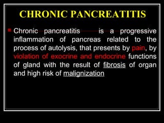
- 1. CHRONIC PANCREATITIS Chronic pancreatitis is a progressive inflammation of pancreas related to the process of autolysis, that presents by pain, by violation of exocrine and endocrine functions of gland with the result of fibrosis of organ and high risk of malignization
- 2. ETIOLOGY AND PATHOGENESIS Gallstone disease is the most frequent cause of chronic pancreatitis (70%). Pathogenesis of cholangiogenic pancreatitis is hypertension in pancreatic duct and reflux of infected bile or secretion of duodenum. Spasms and stenosis of the Vater's papilla are instrumental in causing reflux. As result occur activates the enzymes of pancreas and progress inflammation. Development of pancreatitis potentiates infection.
- 3. CAUSES The CAUSES of such violations are - due to the attack of acute pancreatitis in past, alcoholism, traumas of gland, pathology of its vessels, gastroduodenal ulcers, gastritis or duodenostasis, hyperparathyroidism, hyperlipidemia, virus infections, idiopathic pancreatitis.
- 4. CLASSIFICATION AND ETIOLOGY CHRONIC CALCIFIC PANCREATITIS CHRONIC OBSTRUCTIVE PANCREATITIS CHRONIC INFLAMMATORY PANCREATITIS CHRONIC AUTOIMMUNE PANCREATITIS ASYMPTOMATIC PANCREATIC FIBROSIS ALCOHOL PANCREATIC TUMORS UNKNOWN Autoimmune disorders (primary sclerosing cholangitis) CHRONIC ALCOHOLIC HEREDITARY DUCTAL STRICTURE SJOGREN'S SYNDROME Endemic in asymptomatic residents in tropical climates TROPICAL GALLSTONE OR TRAUMA- INDUCED Primary biliary cirrhosis HYPERLIPIDE MIA PANCREAS DIVISUM HYPERCALC EMIA DRUG-INDUCED IDIOPATHIC
- 5. CLASSIFICATION (by O.O. Shalitnov) Chronic fibrous pancreatitis without violation of patency of main pancreatic duct. Chronic fibrous pancreatitis with violation of patency of main pancreatic duct and hypertension of pancreatic juice. Chronic fibrous-degenerative pancreatitis. TAKING INTO ACCOUNT CLINICAL FEATURES Chronic recurrent pancreatitis. Chronic pain pancreatitis Chronic painless (latent) pancreatitis. Chronic pseudo tumor-like pancreatitis. Chronic cholecystocholangiopancreatitis (cholangiogenic pancreatitis). Chronic indurative pancreatitis.
- 6. PATHOMORPHOLOGY The morphological changes in pancreas in chronic pancreatitis are mainly due to the development of degenerative process and atrophy of parenchyma
- 7. CLINICAL MANAGEMENT As the progress of the disease has cyclic character with the periodic changes of remission and acute exacerbations. Violation of exocrine and endocrine functions of pancreas, determine polymorphism of symptoms that are characteristic of the period of exacerbations pancreatitis
- 8. PAIN Patients with chronic pancreatitis complaining on dull pain that is in the epigastric and radiates to the back
- 9. PAIN The pathophysiology of the pain associated with increase intraductal pressures, neural inflammation, formation of pseudocysts, bile duct strictures, and duodenal obstruction.
- 10. MALABSORPTION With sufficient loss of functional exocrine pancreas, diarrhea, steatorrhea, and azotorrhea can develop. Because of the 10-fold reserve of exocrine pancreaticenzymes, malabsorption occurs only after 90% of the functioning exocrine cell mass is lost. Pancreatic insufficiency resulting from alcohol-induced chronic pancreatitis usually takes 10 to 20 years to develop. The secretion of lipase is usually diminished earlier than the secretion of the proteolytic enzymes, and as a result, steatorrhea precedes protein-aqueous diarrhea.
- 11. CHRONIC UPPER ABDOMINAL PAIN AND WEIGHT LOSS should suggest a diagnosis of chronic pancreatitis. Weight loss occurs with malabsorption, and of the fat-soluble vitamins develop. Postprandial pancreatic bicarbonate secretion is diminished. The duodenal pH may decrease (pH<4) and an acidic milieu with precipitation of bile salts and inactivation of pancreatic enzymes results in a decrease intestinal digestion.
- 12. ENDOCRINE INSUFFICIENCY Glucose intolerance frequently develops early Endocrine insufficiency develops in up to 60% of patients, but in general not until after the diagnosis of chronic pancreatitis has been made.
- 13. STOOL EXAMINATION Steatorrhea and creatorrhea are characteristic for Chronic Pancreatitis (plenty of muscle fibres). Examination of endocrine function includes: 1) determination of sugar in blood and urine (hyperglycemia and glycosuria); 2) radioimmunoassay of hormones (insulin, C-peptide and glucagon). SKIAGRAPHY survey of organs of abdominal cavity in two projections exposes the existent calculus in the ducts and calcification of parenchyma of pancreas. Relaxation duodenography. The development of "horseshoe" of duodenum and change of its mucosa can be seen Cholecystocholangiography the purpose of diagnosis of gallstone disease and damaging of biliary tract is conducted
- 14. Ultrasonic examination Sonography is one of the basic methods of diagnosis. With the help of symptoms of chronic pancreatitis it is possible to expose inequality of contours of gland, increase of density of its parenchyma, it sizes, dilatation of pancreatic duct and wirsungolithiasis or presence of calculus in parenchyma. It is necessary to inspect gallbladder, liver and extra-hepatic biliary tracts for diagnosis of gallstone disease and choledocholithiasis
- 17. CT-scan showing multiple, calcified intraductal stones in a patient with chronic pancreatitis
- 18. CT-scan
- 19. Routine Laboratory Findings Secondary anemia to malnutrition can occur in chronic pancreatitis, to the steatorrhea of chronic pancreatitis are also uncommon. Leukocytosis can occur during acute exacerbations of chronic pancreatitis. Serum amylase and lipase concentrations may be elevated in chronic pancreatitis. Even during an acute attack with seemingly significant abdominal pain, the amylase and lipase levels may be only slightly elevated because of depletion of the exocrine pancreatic parenchyma. Abnormalities of liver function, manifested by elevations in the liver enzymes, may be a result of either liver disease or obstruction of the common bile duct. Fibrotic process may result from compression by a pseudo cyst or mass in the head of the pancreas.
- 20. TESTS FOR CHRONIC PANCREATITIS MEASUREMENT OF PANCREATIC PRODUCTS IN BLOOD I A Enzymes B Pancreatic polypeptide MEASUREMENT OF PANCREATIC EXOCRINE SECRETION II A Direct measurements 1 Enzymes 2 Bicarbonate B Indirect measurement 1 Bentiromide test 2 Schilling test 3 Fecal fat, chymotrypsin, or elastase concentration 4 [14 C]-olein absorption IMAGING TECHNIQUES III A Plain film radiography of abdomen B Ultrasonography C Computed tomography D Endoscopic retrograde cholangiopancreatography E Magnetic resonance cholangiopancreatography F Endoscopic ultrasonography G Relaxation duodenogram
- 21. CLINICAL VARIANTS Chronic recurrent pancreatitis. The changes of periods of acute attacks and remission are characteristic for it. Chronic pain pancreatitis. Intensive pain in the superior half of abdomen with radiation to loins and region of heart is inherent for this form. Also belt-like pain often appears. Chronic painless (latent) pancreatitis. In this patients the pain is either absent in general or arises after the intake of spicy rich food and can be insignificantly expressed Violation of exocrine or endocrine function of pancreas present. Chronic pseudo tumor-like pancreatitis. Dull pain in the projection of head of pancreas, dyspeptic disorders and syndrome of biliary hypertension are its clinical signs. Chronic cholangiogenic pancreatitis. The features of chronic cholecystitis and cholelithiasis and features of pancreatitis are characteristic for this form. Chronic indurative pancreatitis. In patients with this diseases symptoms of exocrine and endocrine insufficiency of pancreas are present. With sclerosis of head of pancreas with involvement by the process of common bile duct, development of mechanical jaundice is possible.
- 22. COMPLICATIONS OF CHRONIC PANCREATITIS INTRAPANCREATIC COMPLICATIONS Pseudo cysts Duodenal or gastric obstruction Thrombosis of splenic vein Abscess Perforation Erosion into visceral artery Inflammatory mass in head of pancreas Bile duct stenosis Portal vein thrombosis Duodenal obstruction Duct strictures and/or stones Ductal hypertension and dilatation Pancreatic carcinoma
- 23. EXTRAPANCREATIC COMPLICATIONS 1.Pancreatic duct leak with ascites or fistula 2.Pseudocyst extension beyond sac into mediastinum, retroperitoneum, lateral pericolic spaces, pelvis
- 24. SURGICAL METHODS OF TREATMENT OF CHRONIC PANCREATITIS The major indications for treatment are: 1. Intractable pain; 2. Fear of carcinoma; 3. The development of structural complications Indication to operation and its volume depend on the form of pancreatitis. Acute exacerbation of chronic cholangiogenic pancreatitis with presence of gallstone disease must be seen as an indication for operation in first 24 hours since the onset of disease
- 25. OPERATIVE TREATMENT IS DONE IN CASES OF: calcinosis pancreas with the expressed pain syndrome; violation of patency of duct of pancreas; presence of cyst or fistula, resistance to conservative therapy in 2-4 months; mechanical jaundice due to tubular stenosis of distal part of common bile duct; compression and thrombosis of portal vein; gallstone disease complicated by chronic pancreatitis; ulcer disease of stomach and duodenum complicated by secondary pancreatitis; duodenostasis, complicated by chronic pancreatitis;
- 26. CHOLECYSTECTOMY is performed in presence of calculous cholecystitis and secondary pancreatitis, acute destructive cholecystitis or hydropsy of gall-bladder.
- 27. CHOLEDOCHOLITHOTOMY is performed for patients with cholangiolithiasis:
- 28. Papillosphincterotomy: a) execute transduodenal with papillosphinctero- plasty; b) endoscopy is recommended in the isolated cases or connected with choledocholithiasis stenosis of large duodenal papilla, fixed calculus of large papilla of duodenum. Wirsungoplasty is plastic of main pancreatic duct. Lately in the isolated stenosis of entrance of main pancreatic duct. Execute transduodenal or endoscopic methods
- 30. PANCREATOJEJUNOSTOMY: a) LONGITUDINAL (it is performed in considerable dilatation of pancreatic duct)
- 31. Technique of pancreaticojejunal drainage originally described by Puestow and Gillesby. The distal pancreas was mobilized, the tail amputated, the duct opened longitudinally, and the pancreas was partially invaginated into a Roux-en-Y jejunal limb
- 32. RESECTION OF PANCREAS MAY BE: a) distal; b) pancreatoduodenal (PDR) c) total duodenopancreatectomy (execute in case of fibrous-degenerative pancreatitis)
- 33. b) Distal (by Duval) with the resection of distal part of pancreas
- 35. Operations on the nervous system are used in case of pain of chronic indurative pancreatitis, resistant to conservative therapy: a) left-sided splanchnicectomy; b) bilateral pectoral splanchnicectomy and sympathectomy; c) postganglionic neurotomy of pancreas
- 36. Anatomic landmarks for videoscopic transthoracic left splanchnicectomy. Diagram of the left plural cavity after clipping and division of the splanchnic nerves, showing the sympathetic chain, the intercostal vessels, and the aorta
- 37. CYSTS OF PANCREAS Cyst of pancreas is a cavity, filled by fluid (pancreatic juice, exudation, pus), which has epithelium on internal surface. Pseudocyst (false cyst) is a cavity in pancreas which appears as a result of its destruction, limited by capsule, that does not have epithelium on internal surface
- 38. Etiology and pathogenesis THE CAUSES OF PSEUDOCYSTS ARE: destructive pancreatitis, traumas of pancreas, occlusion of Wirsung's duct by parasite, calculus, tumors, innate anomalies of development. TRUE CYSTS ARE: innate cysts which are anomalic in development; retention cysts which develop as a result of obstruction to outflow of pancreatic juice, cystadenoma and cystadenocarcinoma The mechanism of development of pseudocysts consists necrosis of gland, obliterated normal outflow of its secretions, destruction of walls of pancreatic ducts, inflammation reaction of surrounding organs which form the walls of pseudocyst
- 39. PATHOMORPHOLOGY Morphologically the cysts of pancreas are divided into: pseudocysts, retention cyst, single and multiple Pseudocysts are fresh and old. Epithelium in pseudocysts is absent. Retention cysts is seen in connection with an obturated duct Innate cysts are multiple and shallow. Rarely there are echinococcus cysts localized in the area of head of pancreas
- 40. CLASSIFICATION (by A.N. Bakulev and V.V. Vinogradov) I. Innate cysts of pancreas: II. Inflammatory cysts: Pseudocysts Retention cysts III. Traumatic cysts: IV. Parasitic cysts: V. Neoplasty cysts: Pathomorphologically cysts are divided into: The true cyst Pseudocysts
- 41. CLINICAL MANAGEMENT PAIN (dull, permanent, cramp-like and belt-like). It is localized in right hypochondrium, epigastric area, left hypochondrium Pain radiates into the back, left shoulder-blade, shoulder and spine. DYSPEPSIA characterised by nausea and vomiting. FUNCTIONAL INSUFFICIENCY OF PANCREAS by disorders of exocrine and endocrine insufficiency, alternating diarrhea with constipation, steatorrhea and creatorrhea, secondary diabetes COMPRESSION SYNDROME. As a result of compression of neighbouring organs are: partial obstruction of common bile duct (mechanical jaundice), veins (portal hypertension), splenic vein (splenomegaly) During the CLINICAL EXAMINATION patients with large cysts there is marked asymmetry of abdomen in the epigastria and mesogastric areas. SONOGRAPHY examination shows echofree formation
- 42. SONOGRAPHY
- 43. A contrast-injected CT- scan reveals active bleeding (B) into a pseudocyst (arrows)
- 44. Contrast roentgenologic EXAMINATION OF STOMACH and duodenum in the cyst of head of pancreas reveals "horseshoe" duodenum CHOLECYSTOCHOLANGIOGRAPHY exposes calculous cholecystitis and cholelithiasis RETROGRADE PANCREATOCHOLANGIOGRAPHY exposes the deformed, extended pancreatic duct, there can be cavity of cyst by the contrast matter LABORATORY EXAMINATIONS exposes hyperamylasemia, steatorrhea and creatorrhea, sometimes - hyperglycemia and glycosuria
- 45. COMPLICATIONS 1. Perforation into free abdominal cavity and peritonitis 2. Perforation into stomach, duodenum, small or large intestine is accompanied by decrease of size of cyst 3. Suppuration of cystic fluid 4. Erosive bleeding appears suddenly and is accompanied by the symptoms of internal bleeding (general weakness, dizziness, melena) 5. Mechanical jaundice arises as a result of compression of cyst on the terminal part of choledochus 6. Portal hypertension as a result of compression of portal vein 7. Reactive exudation pleurisy 8. Malignization
- 46. DIFFERENTIAL DIAGNOSIS Cancer of pancreas. Aneurysm of abdominal aorta The cyst of mesentery The cyst of liver DIAGNOSIS PROGRAMME Anamnesis. Biochemical blood test (amylase, sugar, bilirubin). Analysis of urine for diastase. Coprograma. Sonography. Contrasting skiagraphy of stomach and duodenum Retrograde pancreatocholangiography. Computer tomography.
- 47. TACTICS AND CHOICE OF TREATMENT METHOD Conservative treatment. Treatment of acute or chronic pancreatitis is conducted in accordance with principles. Surgical treatment Is the method of choice of treatment of cysts of pancreas. The choice of treatment method depends on the stage of development of pancreatic cysts.
- 49. MORE FREQUENTLY SURGEON MAKES CYSTOJEJUNOSTOMY ON THE ELIMINATED LOOP OF SMALL INTESTINE BY ROUX
- 50. DISTAL PANCREATECTOMY, MARSUPIALIZATION MARSUPIALIZATION - opening and suturing of cyst capsule to the parietal peritoneum and skin is used infrequently (because suppuration of cyst is can lead to sepsis peritonotis). External and internal draining of cyst and radical operations are applied: a) enucleation of cysts; b) distal resection of pancreas with cyst
- 51. CANCER OF PANCREAS The cancer of pancreas is a malignant tumor of epithelium tissue. Its incidence among all malignant tumors is 10 %. Etiology and pathogenesis Shortage of vitamins, especially В and C, harmful habits (alcohol, smoking), presence of carcinogenic matters in food (nitrite, nitrates) is one of etiological factors. The cancer of pancreas can arise due to prolonged chronic pancreatitis.
- 52. Molecular biology of pancreatic cancer
- 53. PATHOMORPHOLOGY The cancer is usually localized in the head (80%). Rarely - in the area of body or tail. A tumor has resembles epithelium of pancreatic ducts or epithelium of acinous tissue, sometimes - the Langerhans' islet. Adenocarcinoma (60%) is exposed microscopically, carcinoid (32-35%), epidermoid cancer or skir is seldom met.
- 54. Classification of cancer of pancreas after the TNM stages T1 - tumor size of diameter 2 cm, is confined interior parts of pan-creas. T2 - tumor, spreads the gland and spreads to surrounding cellular tissue and duodenum. T3 - tumor, that spreads to neighbouring organs (stomach, spleen, colon). N0 - absent signs of metastatic damage of regional lymph nodes. N1 - metastases in regional lymph nodes. M0 - absent signs of remote metastases. M1 - remote metastases present. GROUP BY STAGES Stage I - Tl NO MO. Stage II - T2 NO MO. Stage III- T3 N0-1 MO. Stage IV is some T, some N, Ml. The cancer of pancreas metastasises rapidly by lymphogenic route parapancreatic lymph nodes, and afterwards - into the liver. The hematogenic metastases are into the lungs, bones, kidneys and brain Also possible are remote metastases of Virhov's, Shnitsler's, Krukenberg's.
- 55. Clinical management The symptoms of cancer of pancreas depend on localization of tumor and the relations of pathological process with surrounding organs. PAIN is a permanent symptom which affects 90 % of patients. Pain localization in epigastria and radiation to the back. The LOSS OF WEIGHT makes progress and in a short duration of time becomes considerable enough. JAUNDICE is characteristic of the cancer of head of pancreas, as a result of obliteration of common bile duct. Bilirubinemia grows gradually, due to direct bilirubin. On palpation of abdomen COURVOISIER'S sign is positive (large gallbladder). Obliteration of duct of pancreas causes DYSPEPTIC DISORDERS: belching, nausea, vomiting, diarrhea. Distributions of tumor on duodenum and narrowing of its lumen show up by the signs of STENOSIS (belching and vomiting)
- 56. By sonography examination and computer tomography one can expose sign of mechanical jaundice by localization the tumor in the head. Scanning is an informing method of examination with the use of 75 Se-methionine. During laparoscopy is visualized dissemination into peritoneum and its metastatic focus in liver. The changes of main duct of pancreas as segmental stenosis or rupture are done on retrograde endoscopic pancreatography Skiagraphy of gastro-intestinal tract can expose the cancer of head of pancreas
- 59. Radionuclide octreotide scan demonstrating pancreatic endocrine tumor in the body of the pancreas (arrow).
- 60. TACTICS AND CHOICE OF TREATMENT METHOD Treatment of cancer of pancreas is mainly surgical. The choice of method and volume of operation depends on localization of tumor, stage of process, age of patient and his general condition. Radical surgical treatment performed only in 15-20 % of patients. Pancreatoduodenal resection is the method of choice of operation in patients with the cancer of head of pancreas.
- 63. PALLIATIVE OPERATIONS Surgical palliation in patients with cancer of the head of the pancreas is directed toward relief of obstructive jaundice, gastric obstruction, and pain. Patients with cancer of body and tail are less likely to have jaundice or duodenal obstruction, but pain is often significant. Obstructive jaundice develops in about 70 percent of patients with pancreatic cancer. Cholecystojejunostomy and choledochojejunostomy are both safe and are the procedures of choice to relieve the biliary obstruction