
Recommended
Recommended
More Related Content
Similar to Lumbar Disc Disease Diagnosis and Management
Similar to Lumbar Disc Disease Diagnosis and Management (20)
More from NelJohnFailagao
More from NelJohnFailagao (12)
Recently uploaded
Recently uploaded (20)
Lumbar Disc Disease Diagnosis and Management
- 4. Functional spinal unit • Two adjacent vertebrae • Intervertebral disc • Spinal ligaments
- 5. INTERVERTEBRAL DISC Nucleus pulposis - Gelatenous substance - Proteoglycan - 80% water content Annulus fibrosus -Fibrocartilage -Closely attach to the end plate -Contain laminated Layers
- 6. OBJECTIVES 1. Identify the etiology of lumbar disc herniations. 2. Review the proper steps in the evaluation of lumbar disc herniations. 3. Outline the management options available for lumbar disc herniations. 4. Summarize interprofessional team strategies for improving care coordination and communication to improve outcomes of lumbar disc herniations
- 7. LOW BACK PAIN • strain of the muscles • ligaments and tendons • Herniation of lumbar disc • sciatica • Various other conditions
- 8. ANATOMY
- 10. • In the normal, healthy disc, the nucleus distributes the load equally throughout the anulus • As the disc undergoes degeneration, the nucleus loses some of its cushioning ability and transmits the load unequally to the anulus • In the severely degenerated disc, the nucleus has lost all of its ability to cushion the load, which can lead to disc herniation Etiology of Disc Degeneration
- 11. HERNIATED NUCLEUS PULPOSUS • Degeneration • Prolapse • Extrusion • Sqeuestration
- 12. COMPLETE HISTORY • Did you have an injury? • Where is the pain? • Do you have any numbness? Where? • Do you have any weakness? Where? • Have you had this problem or something like it before? • Have you had any weight loss, fevers, or illnesses recently?
- 13. CAUDA EQUINA SYNDROME • Most serious complication • Ruptured disc material may press on the nerves that control the bowels and bladder • It can cause permanent damage lead to lose the ability to control bowels and bladder • Surgery immediately is recommend
- 14. PHYSICAL EXAMINATION 1. Motion of Spine 2. Weakness 3. Pain 4. Sensory Changes 5. Reflex changes 6. Motor skills 7. Special signs
- 15. FUNCTIONS LUMBAR VERTEBRA 1. Weight transmission – L4/L5 and L5/S1 2. Multidirectional Movement –T12/L1
- 17. DEGENERATIVE DISC DISEASE (DDD) 1 7 DEFINITION “Describes the natural breakdown of an intervertebral disc of the spine.”
- 18. Degenerative changes of the disc 1 8 Pathological changes Water and proteoglycan content decreases Collagen fibers of AF become distorted Tears may occur in the lamellae Results in: Decreased disc height and volume Impaired mobility Pain Inflammation Neurological signs and symptoms
- 19. DEGENERATIVE INTERVERTEBRAL DISC DISEASE 1 9 a. DISC BULGE b. ANNULAR TEAR c. HERNIATION PROTUSION EXTRUSION INTRAVERTEBRAL
- 20. A) DISC BULGE 2 0 •Generalized or circumferential disc displacement • (involving 50% to 100% of the disc circumference) is known as “bulging” •and is not considered a form of herniation. TYPES •Bulging can be symmetrical (displacement of disc material is equal in all directions) •asymmetrical (frequently associated with scoliosis)
- 21. 2 1
- 22. ). ANNULAR TEAR 2 2 Disruption of concentric collagenous fibers comprising the anulus fibrosus TYPES: 1. Concentric tears 2. Radial tears 3. Transverse tears MR Findings • T1WI: Contrast-enhancing nidus in disc margin • T2WI: High signal zone at edge of disc which has low intrinsic signal
- 23. A. Concentric tears •are circumferential lesions which are found in the outer layers of the annular wall. •They represent splitting between adjacent lamellae of the annulus, like onion rings. •Concentric tears are most commonly encountered in the outer annulus fibrosus, are believed to be of traumatic origin especially from torsion overload injuries. 2 3 TYPES
- 24. B. RADIAL TEARS 2 4 • are characterized by an annular tear • permeates from the deep central part of the disc (nucleus pulposus) • extends outward toward the annulus • either a transverse or cranial-caudal plane.
- 25. C. TRANSVERSE TEARS 2 5 • also known as “peripheral tears” or “rim lesions” • are horizontal ruptures of fibers, near the insertion in the bony ring apophyses. • Their clinical significance remains unclear. • Transverse tears are believed to be traumatically induced and are often associated with small osteophytes
- 26. 2 6
- 27. C). DISC HERNIATION Herniation is defined as a localized displacement of disc material (nucleus, cartilage, fragmented apophyseal bone, fragmented annular tissue) beyond the limits of the intervertebral disc space. Intravertebral Herniations Protruded Disc Extruded Disc 2 7
- 28. •Are protrusions of the nucleus pulposus of the intervertebral disc through the vertebral body endplate and into the adjacent vertebra. •(also known as Schmorl’s nodes). •They are often surrounded by reactive bone marrow changes. INTRAVERTEBRAL HERNIATIONS 15
- 29. 16
- 30. “PROTRUDED DISC • The terminology “protruded disc” is used when the base of the disc is broader than any other diameter of the displaced material. •Based on a two-dimensional assessment of the disc contour in the transverse plane, a protruded disc can be: •focal (involving <25% of the disc circumference) •broad-based (involving 25%–50% of the 17 disc circumference).
- 31. • The terminology “extruded disc” is used for a focal disc extension of which the base against the parent disc is narrower than diameter of the extruded disc material, measured in the same plane. EXTRUDED DISC HERNIATIONS Extrusion: the base of the herniation is narrower than the apex (toothpaste sign) 18
- 32. •Extrusion is also used when there is no continuity between the herniated disc material beyond the disc space and that within the disc space •If the displaced disc material has no connection with the parent disc, it is called a “sequestrated fragment”. •This is synonymous with a “free fragment”. 3 2
- 33. 3 3
- 34. MIGRATION – SEQUESTRATION Migration indicates displacement of disc material away from the site extrusion, regardless of whether sequestrated or not. Sequestration indicate that the displaced disc material has lost any continuity with the parent disc 3 4
- 35. A. Small subligamentous herniation (protrusion) without significant disk material migration. B.Subligamentous herniation with downward migration of disk material under the PLLC. C.Sub-ligamentous herniation with downward migration of disk material and sequestered fragment (arrow). 22
- 36. • X-ray: show spinal degenerative changes but not a herniated disc; rule out obvious underlying problems • CT: relatively less used • MRI: The best DIAGNOSIS 3 6
Editor's Notes
- Spine is a multi articular structure comprising …
- Low back pain is a prevalent symptom, as approximately 80 % of the population sustains an episode once in their lifetime. Within the many differentials of low back pain, the most common cause is the intervertebral degeneration which leads to lumbar disc herniation and degenerative disc disease. This activity reviews the evaluation and management of lumbar disc herniation and discusses the role of the healthcare team in evaluating and improving care for patients with this condition. Objectives: Identify the etiology of lumbar disc herniations. Review the proper steps in the evaluation of lumbar disc herniations. Outline the management options available for lumbar disc herniations. Summarize interprofessional team strategies for improving care coordination and communication to improve outcomes of lumbar disc herniations
- In the normal, healthy disc, the nucleus distributes the load equally throughout the anulus As the disc undergoes degeneration, the nucleus loses some of its cushioning ability and transmits the load unequally to the anulus In the severely degenerated disc, the nucleus has lost all of its ability to cushion the load, which can lead to disc herniation
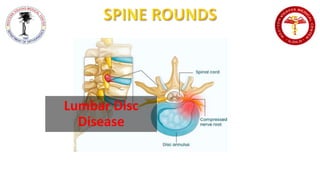
- A herniated disc occurs when the intervertebral disc's outer fibers (the annulus) are damaged and the soft inner material of the nucleus pulposus, under extreme pressure, bulges out through the annulus fibrosus ring. If the annulus tears near the spinal canal, the nucleus pulposus material can push into the spinal canal.