
Recommended
More Related Content
What's hot
What's hot (20)
Similar to OBSTRUCTIVE SLEEP APNEA.ppt
Similar to OBSTRUCTIVE SLEEP APNEA.ppt (20)
Recently uploaded
Recently uploaded (20)
OBSTRUCTIVE SLEEP APNEA.ppt
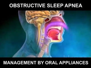
- 1. OBSTRUCTIVE SLEEP APNEA MANAGEMENT BY ORAL APPLIANCES
- 2. • APNEA : Cessation of air flow > 10seconds • HYPOPNEA : Decreased airflow > 30% from baseline lasting > 10seconds associated with > 4% oxyhemoglobin saturation • SLEEP : A normal, reversible, recurring behavioural state of disengagement and unresponsiveness to the environment that is characterized by typical changes in the electroencephelogram.
- 3. • SNORING : A respiratory sound generated in the upper airway during sleep, particularly deep (slow- wave) sleep and REM sleep. • OBSTRUCTIVE SLEEP APNEA (OSA) SYNDROME : A sleep-associated disorder of breathing with a reduction or complete airflow obstruction despite an ongoing effort by patient for breathing.
- 4. MUSCLES HOLD AIRWAY OPEN NORMAL BREATHING AIRWAY NARROWS WHEN MUSCLES RELAX SNORING AIRWAY COLLAPSES OR BLOCKED WHEN MUSCLES OVER RELAX OBSTRUCTIVE SLEEP APNEA
- 11. DIAGNOSIS
- 12. DIAGNOSTIC METHODS • APNEA-HYPOPNEA INDEX (AHI) : Number of apneas and hypopneas per hour of sleep • RESPIRATORY DISTURBANCE INDEX (RDI) : Number of apneas + hypopneas + RERAs per hour of sleep
- 13. Forms of OSAHS on basis of AHI Score S.No. Forms of OSAHS AHI Score 1 Mild AHI 5-14/hr 2 Moderate AHI 15-30/hr 3 Severe AHI >30/hr 4 Very severe AHI >40/hr
- 14. Subjective assessment by Epworth Sleepiness Scale (ESS) S.No. Types of Sleepiness ESS Score 1 Normal range ESS <11 2 mild subjective daytime sleepiness ESS =11 3 moderate subjective daytime sleepiness ESS =16 4 Severe subjective daytime sleepiness ESS >18
- 15. Objective assessment by Multiple Sleep Latency Test (MSLT) • Used to measure the time to fall asleep (using EEG criteria). This is performed in a dark room on at least four separate occasions across the day. This period of time is called as sleep latency.
- 16. Overnight Polysomnography • Electro-encephalography (EEG) - brain wave monitoring • Electromyography (EMG) - muscle tone monitoring • Recording thoracic-abdominal movements - chest and abdomen movements • Recording oro-nasal airflow - mouth and nose airflow • Pulse oximetry - heart rate and blood oxygen level monitoring • Electrocardiography (ECG) - heart monitoring • Sound and video recording
- 18. OXIMETRY • Cheap recording pulse oximeters are readily available; therefore oximetry is used as the first screening tool for OSAHS. • These are spectrophometric devices that are used for the detection and calculation of the differential absorption of light by presence of oxygenated and deoxygenated haemoglobin in blood. • This is a method for detection of the blood oxygen saturation
- 19. The techniques to visualize pharynx • Cineradiography, • Fiberoptic bronchoscopy, • Acoustic reflectance, • Forced expiratory maneuvers, • CT scanning and • lateral cephalometry ( more commonly used.)
- 20. CEPHALOMETRIC DIAGNOSIS • Adjunctive procedure for assessing craniofacial patterns associatedwith OSAS • Evaluation of nasopharyngeal obstruction, position of base of tongue, and pharyngeal relationship through specific airway parameters done. • Helps in :- • measuring posterior airway space • volume of pharyngeal airway • small size of nasopharyngeal airway with snoring. • depth of soft tissue of the posterior wall with nasal respiratory resistance • size of adenoidal mass and itsdistance from the posterior wall of antrum
- 22. TREATMENT • Behavioural interventions • Non-surgical interventions • Surgical interventions
- 23. Behavioural Interventions Lose weight Eliminate the use of alcohol, tobacco, and sedatives Sleep on one side Regularize sleep hours
- 24. Non-surgical Interventions Continuous positive airway pressure (CPAP): – Pneumatic splint to maintain upper airway patency throughout all phases of sleep – Treatment of choice – Improves subjective and objective sleepiness, cognitive function, vigilance, mood and quality of life measures. – Best results are obtained in those with an AHI of >15 – Side effects: epistaxis, sinusitis, rhinitis, dryness of the nasal passages, nasal bridge sores, claustrophobia, abdominal bloating, mouth leaks and noise
- 27. Oral appliance therapy • adjustable and nonadjustable – Anterior tongue repositioners – Mandibular posturing devices – Soft palate or uvula lifting devices
- 28. INDICATIONS :- 1) Patients with snoring or mild OSA who do not respond for treatment with behavioral measures. 2) Patients with moderate to severe OSA who refuse treatment with nasal CPAP. 3) Patients who are not appropriate for tonsillectomy, adenoidectomy, and tracheostomy. ADVANTAGES :- • Significant reduction in breathing pauses • Improvement of airflow for some patient with apnea • Reduction in the snoring and • High compliance level as compared to CPAP DISADVANTAGES : • Reciprocal forces are generated on the teeth and jaw by mandibular advancement splints which results in dry mouth, gum soreness, salivation, tooth pain, headaches, and TMJ problems
- 30. Anterior Tongue Repositioners • advances the tongue • tongue & mandible together with adjacent soft tissue • increases the posterior airway space • increases the activity of the genioglossal & lateral pterygoid muscles • effects a stretch induction of the pharyngeal motor system
- 32. Mandibular Posturing Devices Nocturnal Airway- Patency Appliance (NAPA) Thornton Adjustable Positioner (TAP) Klearway Appliance Silicone Positioner Appliance OASYS Oral/Nasal Airway System HERBST APPLIANCE
- 33. SOFT PALATE OR UVULA LIFTING DEVICE
- 34. RETRUDED JAW NARROW AIRWAY Mandibular Repositioning Device Lower jaw moves forward Constricted palate Closed airway Palatal expansion screw Arch expands
- 35. Macroglossia Obstructed airway Tongue retaining devices Pulls tongue forward Thick soft plate Closed airway Soft palate lifter Lifts palate upwards & open airways Retruded jaw Closed airway Mandible advancement surgery Jaw moves forward and airways enlarge
- 36. Surgical interventions • Uvulopalatopharyngoplasty (UPPP) – poor and unpredictable • Tonsillectomy • Tracheostomy • Mandibular advancement • Bariatric surgery • Nasal surgery
- 37. CONCLUSION • Orthodontic diagnosis may discover anatomic conditions that could cause respiratory obstructive sleep apnea • Enlarged tonsils or adenoids in a lateral cephalometric radiograph, or maxillary width deficiency and narrow nasal cavity in a P-A radiograph, are indications for questioning the patient about other symptoms.
- 38. • The clinician should be aware of the role of orthodontics in prevention and treatment of sleep disorders. • Four-bicuspid extraction therapy in an already hypoplastic skeletal pattern could further reduce tongue space and increase the possibility of sleep disorders later in life.
- 39. • Prevention of obstructive sleep apnea may be possible in young children through dentofacial orthopedics to maximize the development of the glossopharyngeal space, the nasomaxillary complex, and other dentofacial components.