
Recommended
More Related Content
What's hot
What's hot (20)
Similar to Internal anatomy of pulp space
Similar to Internal anatomy of pulp space (20)
More from MrinaliniDr
More from MrinaliniDr (12)
Recently uploaded
Recently uploaded (20)
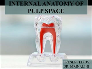
Internal anatomy of pulp space
- 2. + Introduction + History + Development of pulp + Pulp cavity Laws Of Pulp Cavity Coronal pulp(Pulp chamber) Roof and Floor Pulp horns Canal orifices Radicular pulp(Root canal) Classification of root canals Weine’s classification(1969,1982) Vertucci’s classification(1974) Grossman’s classification
- 3. Gulabiwala and Coworkers(2001) Seit and Bayirili(2001) Yoshioka and Villegas(2004) Other classifications o Classification by H M AAhmed(2017) o Classification by Rashmi Bansal et al.(2018) Isthmus o Identification o Classification o Clinical Significance Root canal ramification Terminologies Classification of root canal ramification Accessory Canal o Incidence
- 4. o Formation o Classification of accessory canal o Clinical significance Concept of radius of curvature and angle of curvature Classification of root canal curvature Ingle and Taintor(1980) and Pucci and Reig(1986) Zeidle’s classification of root canal system(1986) Schneider’s classification(1986) Wein’s classification Csaba Dobo Negi et al(1995) Relationship between degree of curvature and ledge formation Management of apical curvature Management of curvature in middle third + Regressive changes in anatomy of root canal
- 5. + Apical root anatomy Minor Constriction o Introduction o Topography o Position Major constriction o Introduction o Location o Associated studies Cementodentinal Junction Radiographic apex Significance of apical third Optimal working length Apical tissue
- 6. + Variations in the pulpal anatomy of teeth Variations in development C-shaped oIntroduction oIncidence oClassification(Melton’s and Fan’s classification for C-shaped canal) oExternal root anatomy of C shaped canal configuration molar oSignificance oManagement of C shaped canal Access cavity preparation Biomechanical preparation Obturation Post endodontic restoration
- 7. Gemination Fusion Concrescence Taurodontism Talon’s cusp Dilaceration Dentinogenesis imperfecta Dentin Dysplasia Extra root canal Missing root Dens evaginatus Dens invaginatus
- 8. Variations in shape of pulp cavity Gradual curve Apical curve C shaped canal Bayonet shaped Dilaceration Sickle shaped Variations in pulp cavity due to pathology Pulp stones Calcifications Internal resorption External resorption Variations in apical third Different locations of apex Accessory and lateral canals Open apex
- 9. + Methods of determining pulp anatomy Clinical methods Diagnostic method Anatomic studies Radiographs Radiovisiography Cone beam computed tomography Dental operating microscope Fiberoptic endoscope Magnetic resonance imaging Visualisation endogram In vitro methods Sectioning of teeth Use of dyes Filling and Clearing of teeth
- 10. Contrasting media Radiography Scanning electron microscopic analysis + Factors affecting internal anatomy – Age – Irritant – Calcification – Resorption + Pulp space anatomy of permanent teeth Maxillary central Maxillary lateral incisor Maxillary canine Maxillary first premolar Maxillary second premolar Maxillary first molar
- 11. Maxillary second molar Maxillary third molar Mandibular central Mandibular lateral incisor Mandibular canine Mandibular premolars Mandibular first molar Mandibular second molar Mandibular third molar + Difference from primary teeth + Conclusion + Previously asked questions
- 12. Attempting to treat the root-canal system without detailed anatomic description would be equivalent of a physician looking for an appendix without ever having read Gray’s Anatomy. -Paul Krasner
- 13. INTRODUCTION + Of all the phases of anatomic study in the human system, one of the most complex is the pulpal morphology. + For succcess of endodontic therapy, knowledge of pulp anatomy cannot be ruled out. + It is essential to have the knowledge of normal and usual configuration of the pulp cavity along with variations.
- 14. 1842:Investigation of tooth anatomy CARABELLI: published drawings of sectioned teeth detailing the root canal system 1870: MUHLREITER -first one to investigate root canal anatomy, sectioned teeth in all planes & described the internal anatomy with details 1890: G. V. BLACK-contributed with the study of the root canal anatomy in the 1st edition of his book 1892: ALFRED GYSI- presented pictures of histological sections of the tooth showing the complexity of the internal anatomy. 1901:PREISWERCK -injected molten metal within the pulp followed by complete decalcification of tooth and obtained a metal model of internal anatomy Diwan A, Sreedevi CR, Nagaraj T, Raghava V, Sinha P, Moushmi CB. Determination of internal anatomy of a permanent dentition: A review. Int J Contemp Dent Med Rev. 2015;2015. HISTORY
- 15. 1907: FISCHER- used celluloid instead of metal Better results, small ramifications of the replicas broke easily as celluloid was fragile. 1916: DEWEY –injected paraffin to study the root canal anatomy. 1917: HESS- injected root canals with vulcanized rubber, removed hard tissue by decalcification. Material:still valuable to the study of the root canal anatomy. 1918-1926: OKUMARA- studied internal anatomy of teeth using injection of dye & diaphonization 1923: CLYDE DAVIS- studied the anatomy of the apical third using ground sections of the tooth. Diwan A, Sreedevi CR, Nagaraj T, Raghava V, Sinha P, Moushmi CB. Determination of internal anatomy of a permanent dentition: A review. Int J Contemp Dent Med Rev. 2015;2015.
- 16. 1960: DE DEUS- first author to study systematically root canal anatomy of all dental groups using clearing technique (diaphonization). 1925: BARRET-studied the dental anatomy using serial histological sections 1955:MEYER & SCHEELE- using wax models demonstrated numerous lateral canals in the apical third of the root 1974: VERTUCCI & WILLIAMS- found a complex root canal system and identified eight configurations of the pulp space Diwan A, Sreedevi CR, Nagaraj T, Raghava V, Sinha P, Moushmi CB. Determination of internal anatomy of a permanent dentition: A review. Int J Contemp Dent Med Rev. 2015;2015. 1969:WEIN- first to categorize root canal configurations within a single root 2017:H M A AHMED- A new system for classification of root & root canal morphology
- 17. + Begins at 8th week of intrauterine life CEMENTUM
- 18. + Lies within the tooth + Enclosed by dentin all around except apical foramen Endodontic Science, Carlos Estrela, Volume 1 , 2nd edition,Page-531 Coronal pulp: Pulp chamber Radicular pulp: Root canal
- 19. PULP CAVITY PULP CHAMBER ROOT CANAL Roof Floor Pulp horn Canal Orifice Accessory and Lateral Canals Accessory Foramina Apical delta Apical foramen Krasner P, Rankow HJ. Anatomy of the pulp-chamber floor. Journal of endodontics. 2004 Jan 1;30(1):5-16.
- 20. Krasner and Rankow: studies pulp chamber of 500 extracted teeth Krasner P, Rankow HJ. Anatomy of the pulp-chamber floor. Journal of endodontics. 2004 Jan 1;30(1):5-16. Anatomic Laws 1. Relationship of pulp chamber to crown 2. Relationship of root canal orifice to pulp chamber floor
- 21. floor of the pulp chamber is always located in center of the tooth at the level of the CEJ Law of centrality walls of the pulp chamber are always concentric to external surface of the tooth at the level of CEJ Law of concentri city Relationship of pulp chamber to crown CEJ is the most consistent, repeatable landmark for locating the position of the pulp chamber Law of CEJ Krasner P, Rankow HJ. Anatomy of the pulp-chamber floor. Journal of endodontics. 2004 Jan 1;30(1):5-16.
- 22. Law of symmetry 1: except for maxillary molars, orifices of canals are equidistant from a line drawn in a mesial distal direction through the pulp-chamber floor. Law of symmetry 2: except for the maxillary molars, orifices of canals lie on a line perpendicular to a line drawn in a mesial-distal direction across the center of the floor of the pulp chamber Krasner P, Rankow HJ. Anatomy of the pulp-chamber floor. Journal of endodontics. 2004 Jan 1;30(1):5-16. Relationship of root canal orifice to pulp chamber floor
- 23. the color of the pulp-chamber floor is always darker than the walls Law of Color Change: orifices of root canals are always located at the junction of the walls and the floor Law of orifice location 1: Krasner P, Rankow HJ. Anatomy of the pulp-chamber floor. Journal of endodontics. 2004 Jan 1;30(1):5-16.
- 24. the orifices of the root canals are located at the angles in the floor-wall junction Law of orifice location 2: orifices of root canals are located at the terminus of the root developmental fusion lines Law of orifice location 3 Krasner P, Rankow HJ. Anatomy of the pulp-chamber floor. Journal of endodontics. 2004 Jan 1;30(1):5-16.
- 25. • Acquires shape and size of crown of the tooth + Roof : Dentin covering the pulp chamber occlusally or incisally. + Floor : Dentin bounding the pulp chamber near the cervix of the tooth particularly that forming the furcation area Parallel to roof Endodontic Science, Carlos Estrela, Volume 1 , 2nd edition,Page-531 Roof Floor
- 26. + Walls and angles : Walls: correspond to respective walls of the tooth surface. Angles correspond to the respective angles formed from the walls of pulp chamber. + Pulp horns: Between occlusal and pulp chamber Accentuation of roof of pulp chamber directly under a cusp or developmental lobe + Canal orifices: Openings in the floor of pulp chamber leading to root canals Continuous with pulp chamber and root canal Endodontic Science, Carlos Estrela, Volume 1 , 2nd edition,Page-531
- 27. + From canal orifices to apical foramen + Anterior teeth: Pulp chamber merges into root canal + Posterior teeth: Division becomes quite obvious Endodontic Science, Carlos Estrela, Volume 1 , 2nd edition,Page-531
- 28. Bansal R, Hegde S, Astekar M. Classification of Root Canal Configurations: A Review and a New Proposal of Nomenclature System for Root Canal Configuration.Journal of Clinical and Diagnostic Research,2018. •Weine et al. (1969) : first to categorize root canal configurations within a single root •Weine (1982): Type IV
- 29. Bansal R, Hegde S, Astekar M. Classification of Root Canal Configurations: A Review and a New Proposal of Nomenclature System for Root Canal Configuration.Journal of Clinical and Diagnostic Research,2018.
- 30. Vertucci et al. (1974): based on evaluation of 200 cleared maxillary 2nd premolars in which the pulp cavities were stained with dye Bansal R, Hegde S, Astekar M. Classification of Root Canal Configurations: A Review and a New Proposal of Nomenclature System for Root Canal Configuration.Journal of Clinical and Diagnostic Research,2018.
- 31. Bansal R, Hegde S, Astekar M. Classification of Root Canal Configurations: A Review and a New Proposal of Nomenclature System for Root Canal Configuration.Journal of Clinical and Diagnostic Research,2018.
- 32. + Seit and Bayirili in 2001 reported: 14 new root canal configuration Bansal R, Hegde S, Astekar M. Classification of Root Canal Configurations: A Review and a New Proposal of Nomenclature System for Root Canal Configuration.Journal of Clinical and Diagnostic Research,2018. Ahmed HM, Versiani MA, De‐Deus G, Dummer PM. A new system for classifying root and root canal morphology. International endodontic journal. 2017 Aug;50(8):761-70.
- 33. + Yoshioka and Villegas in 2004: Type V to Wein’s classification + Type V: A root canal configuration having more than 2 canals that branched off from the main canal more than 3mm from the apex defined as another main canal Bansal R, Hegde S, Astekar M. Classification of Root Canal Configurations: A Review and a New Proposal of Nomenclature System for Root Canal Configuration.Journal of Clinical and Diagnostic Research,2018.
- 34. + Christie wt al(1991), Carlsen & Alexandersen (2000), Baratto‐Filho et al.( 2002), Versiani et al (2012): Maxillary molars with four roots + Carlsen & Alexandersen (2000), Baratto‐Filho et al. (2002), Versiani et al. (2012): maxillary premolars with three canals + Belizzi & Hartwell (1981), Ahmed & Cheung (2012): the middle mesial canal + Pomeranz et al. (1981): distolingual root in mandibular molars + Kottoor et al. (2012) and Albuquerque et al. (2012) suggested a new nomenclature to classify root canal anatomy in maxillary and mandibular molars, respectively. Ahmed HM, Versiani MA, De‐Deus G, Dummer PM. A new system for classifying root and root canal morphology. International endodontic
- 35. + Simple, accurate and useful :information on root and root canal anatomy. + Does not address the degree of root and root canal curvature, degree of root/canal separation, exact level of bifurcation of canals/roots, accessory canals + Codes for three separate components: the tooth number, the number of roots and their configuration, and the root canal configuration Ahmed HM, Versiani MA, De‐Deus G, Dummer PM. A new system for classifying root and root canal morphology. International endodontic
- 36. Ahmed HM, Versiani MA, De‐Deus G, Dummer PM. A new system for classifying root and root canal morphology. International endodontic
- 37. Ahmed HM, Versiani MA, De‐Deus G, Dummer PM. A new system for classifying root and root canal morphology. International endodontic
- 38. Tooth number:FDI Root nomenclature: right side Course of canal: bracket Foramen through which canal is exiting at the apex: after slash Anatomic Variations: Left eg. C shaped canal- C,Taurodont-T Single root as R in the right side, Bansal R, Hegde S, Astekar M. Classification of Root Canal Configurations: A Review and a New Proposal of Nomenclature System for Root Canal Configuration.Journal of Clinical and Diagnostic Research,2018.
- 39. + Narrow ribbon shaped communication between the root canals containing pulp or pulpally derived tissues is called isthmus Endodontic Science, Carlos Estrela, Volume 1 , 2nd edition,Page-531
- 40. Endodontic Science, Carlos Estrela, Volume 1 , 2nd edition,Page-531
- 41. + Nidus for recurrent infection + Highest incidence: Mesial root of mandibular 1st molar + Cambruzzi & Marshall: Use of methylene blue dye for visualisation + Microscope: for identification + Ultrasonic :tips for preparation and filling Endodontic Science, Carlos Estrela, Volume 1 , 2nd edition,Page-531
- 42. + Main canal: Present in longitudinal axis, passes from roof of pulp chamber to apical foramen + Collateral canal: Located parallel to main canal, either capable of being reached or not by isolating the apical foramen, smaller in volume than main canal Endodontic Science, Carlos Estrela, Volume 1 , 2nd edition,Page-531
- 43. + Lateral canal: In cervical third and beginning of middle third, either perpendicular or not + Secondary canal: Apical third, either perpendicular to main canal or not + Accessory canal: Ramification of secondary canal which goes in direction of periodontium + Intercanal: Ramification between main and collateral or secondary canal + Recurring canal: Part of main canal not going through a discrete passage and returning to main canal Endodontic Science, Carlos Estrela, Volume 1 , 2nd edition,Page-531
- 44. + Reticular canal: Represents the mixture of three or more canals, ramification of the intercanal + Apical delta: Triangular area of root surrounded by main canal, accessory canal and periradicular tissues Endodontic Science, Carlos Estrela, Volume 1 , 2nd edition,Page-531
- 46. + Mitchell(1965): auxiliary, reticular and recurrent canals + De-Deus(1975): lateral canal, secondary canal and the accessory canal + AAE 2016: Accessory canal: branch of the main pulp canal or chamber that communicates with the external root surface. Lateral canal: type of accessory canal, located in the coronal or middle third of the root, extending horizontally from the main canal space Furcation canal: an accessory canal located in the furcation Endodontic Science, Carlos Estrela, Volume 1 , 2nd edition,Page-531
- 47. + Accessory canal: Fibrous tissue and connective tissue same as that of pulp but closely resembles connective tissue of periodontal ligament + Incidence: 2 to 3- 72% in posterior teeth 35% in anterior teeth(Seltzer,1966) 73.5% : apical third 11.4% : middle third 15.1%: cervical third Formation: Entrapment of PDL vessel in HERS during mineralisation Alothmani OS, Chandler NP, Friedlander LT. The anatomy of the root apex: A review and clinical considerations in endodontics. Saudi Endodontic
- 48. Detection of accessory canal: + Thickening of PDL or lesion in lateral wall of root + Usually becomes noticeble post obturation + Bulbous root: more ramification + Tortuous root canal or sharp bend in root: more chances Clinical significance: + Interchange of irritants + Deep periodontal pocket: Channel for toxic products into pulp + Inflammatory pulp tissue: Effect on periodontal tissue Alothmani OS, Chandler NP, Friedlander LT. The anatomy of the root apex: A review and clinical considerations in endodontics. Saudi Endodontic
- 49. + Yoshiuchi et al. (1972): staining and clearing method + Based on the region of the root: Kasahara et al. (1990), Miyashita et al. (1997), Adorno et al. (2010) Accessory canal at 5/10–9/10, 4/10–2/10, 1/10 or less of the root length: cervical, middle or apical location, respectively Ahmed HM, Neelakantan P, Dummer PM. A new system for classifying accessory canal morphology. International endodontic journal. 2018 Feb
- 50. Ahmed HM, Neelakantan P, Dummer PM. A new system for classifying accessory canal morphology. International endodontic journal. 2018 Feb
- 51. Lesser radius of curvature Less fatigue of instruments
- 52. Ingle and Taintor(1980) and Pucci and Reig(1986) + Apical curve + Gradual curve + Sickle shaped + Dilaceration + Bayonet Balani P, Niazi F, Rashid H. A brief review of the methods used to determine the curvature of root canals. Journal of Restorative Dentistry. 2015 Sep 1;3(3):57.
- 53. Zeidle’s classification of root canal system(1986) + Severe curve + Dilacerated curve + Bayonet curve + Apical bifurcation + Apical curve + Additional canals + Lateral and Accessory canals Balani P, Niazi F, Rashid H. A brief review of the methods used to determine the curvature of root canals. Journal of Restorative Dentistry. 2015 Sep 1;3(3):57.
- 54. •a mid-point marked on the file at the level of canal orifice •straight line drawn parallel to the image and that point is labeled as point A •second point is marked where the flare starts to deviate that is labeled point B •third point is marked at the apical foramen and is termed point C and the angle formed by the intersection of these lines is measured Easy: straight and curved less than 5 degree Average: curved more than 10 less than 25 Difficult: curved more than 25 Schneider’s classification(1986): Based on degree of curvature in root canal, measured using protactor Balani P, Niazi F, Rashid H. A brief review of the methods used to determine the curvature of root canals. Journal of Restorative Dentistry. 2015 Sep 1;3(3):57.
- 55. Balani P, Niazi F, Rashid H. A brief review of the methods used to determine the curvature of root canals. Journal of Restorative Dentistry. 2015 Sep 1;3(3):57. Point A: at the center of the canal orifices Point B: 2 mm below the orifices in the long axis of the canal Primary line: Point A and Point B Point C: 1 mm coronal to the apical foramen Point D: At apical foramen Secondary line: Point C and Point D
- 56. Weine’s classification: + Curvature of 30 to 45 degree + Curvature of 45 to 60 degree + Curvature of 60 to 90 degree + Curvature more than 90 degree + Bayonet shaped curve + Backman et al(1976) and Southard et al(1990) : Based on radius quotient(angle divided by radius) + Dabo Negi et al: Schnieder’s angle and radius of circle superimposed on curved part of root canal Balani P, Niazi F, Rashid H. A brief review of the methods used to determine the curvature of root canals. Journal of Restorative Dentistry. 2015 Sep 1;3(3):57.
- 57. Csaba Dobo Negi et al(1995) + Straight or ‘I form’ + Apical curve or ‘J form’ + Curved canal along its entire length or ‘C form’ + Multicurved or ‘S form’ Balani P, Niazi F, Rashid H. A brief review of the methods used to determine the curvature of root canals. Journal of Restorative Dentistry. 2015 Sep 1;3(3):57.
- 58. Balani P, Niazi F, Rashid H. A brief review of the methods used to determine the curvature of root canals. Journal of Restorative Dentistry. 2015 Sep 1;3(3):57. (More than 20O
- 59. Apical curvature: + Straight line access + Start : smaller diameter K file such as #08 or #10(precurved) + Chelating agent (EDTA) ,irrigation with sodium hypochlorite + Segal: reamer instead of K-file, more flexible . – Once removed, describes the degree, type, location, and direction of the curvature, – Due to its flexibility may lead to canal transportation. + Stainless steel files of smaller diameter with light passive movement ,diameter of glide path is then increased with nickel- titanium (NiTi) hand files before the preparation of the canal with rotary NiTi file Balani P, Niazi F, Rashid H. A brief review of the methods used to determine the curvature of root canals. Journal of Restorative Dentistry. 2015 Sep 1;3(3):57.
- 60. Managing middle curvature + Adequate access and good coronal third preparation + Coronal third preparation followed by the mid-portion preparation using precurved files + Precurved file: negotiating the canal and makes a glide path before rotary NiTi files are introduced for cleaning and shaping Balani P, Niazi F, Rashid H. A brief review of the methods used to determine the curvature of root canals. Journal of Restorative Dentistry. 2015 Sep 1;3(3):57.
- 61. + Receded pulp horns + Shorter and smaller pulp chamber + Narrower root canals( due to secondary or reparative dentin deposition) + Narrower minor diameter, wider major diameter + Reduced no. of accessory foramina(due to calcification of contained soft tissue) + Narrower or obliterated dentinal tubules Receded pulp horns Shorter and smaller pulp chamber Narrower root canals( due to secondary or reparative dentin deposition) Narrower minor diameter and wider major diameter Reduced no. of accessory foramina(due to calcification of contained soft tissue) Narrower or obliterated dentinal tubules
- 62. + Apical constriction(minor diameter/physiological foramen): Apical part of root canal having narrowest diameter short of apical foramina or radiographic apex May or may not coincide with CDJ Histologically: at the junction between pulpal connective tissue and interstitial loose connective tissue of periodontal ligament Alothmani OS, Chandler NP, Friedlander LT. The anatomy of the root apex: A review and clinical considerations in endodontics. Saudi Endodontic Journal. 2013 Jan 1;3(1):1.
- 63. Dummer et al: + Type A: Single constriction + Type B: Tapering constriction with narrowest portion of canal very near to actual apex + Type C: Number of constrictions present + Type D: Constriction followed by narrow, parallel portion of canal + 5th type: canal completely blocked with secondary dentin or cementum Alothmani OS, Chandler NP, Friedlander LT. The anatomy of the root apex: A review and clinical considerations in endodontics. Saudi Endodontic Journal. 2013 Jan 1;3(1):1.
- 64. The distance between the AC and AF ranged between 0.4-1.2 mm, while its reported location in relation to the root apex ranged between 0.5-1.01 mm Alothmani OS, Chandler NP, Friedlander LT. The anatomy of the root apex: A review and clinical considerations in endodontics. Saudi Endodontic Journal. 2013 Jan 1;3(1):1.
- 65. + Apical foramen(major diameter): Main apical opening on surface of root canal through which blood vessels enter Diameter: almost double the apical constriction, funnel shaped described as morning glory or hyperbolic + Alothmani OS, Chandler NP, Friedlander LT. The anatomy of the root apex: A review and clinical considerations in endodontics. Saudi Endodontic Journal. 2013 Jan 1;3(1):1.
- 66. + Changes as a result of functional influence on the teeth + Mesial migration or tipping: apex tilt to opposite side + Tissues entering pulp exert pressure on one wall of foramen : resorption and cementum deposition on opposing wall + Shifts with: Aging, mesial migration, occlusal drift and continuous cementum deposition Alothmani OS, Chandler NP, Friedlander LT. The anatomy of the root apex: A review and clinical considerations in endodontics. Saudi Endodontic Journal. 2013 Jan 1;3(1):1.
- 67. Deviation of the AF from the root apex is common, with a reported frequency ranging from 17-100% Alothmani OS, Chandler NP, Friedlander LT. The anatomy of the root apex: A review and clinical considerations in endodontics. Saudi Endodontic Journal. 2013 Jan 1;3(1):1.
- 68. Green’s study(1955,1956 and 1960): Major apical foramen situated directly at apex more frequently in: + Maxillary first premolar and mandibular second premolar + Maxillary central and lateral incisor + Maxillary molars and all mandibular teeth with exception of 2nd premolar: main apical foramina coincides with apices less frequently Green D.A stereo-binocular microscopic study of the root apices and surrounding areas of 100 mandibular molars.Oral Surg Oral Med Oral Pathol 1955;8:1298–1304. Green D.A stereomicroscopic study of the root apices of 400 maxillary and mandibular anterior teeth. Oral Surg Oral Med Oral Pathol 1956;9:1224– 32. Green D. Stereomicroscopic study of 700 root apices of maxillary and mandibular posterior teeth. Oral Surg Oral Med Oral Pathol 1960;13:728–33.
- 69. + Mean distance between major and minor diameter + Increased length in older individual: increased cementum + Cementodentinal junction: Usually lies 0.1mm from the apical foramen + Tooth apex: Radiographic apex Young person: 0.5mm Older person: 0.7mm Alothmani OS, Chandler NP, Friedlander LT. The anatomy of the root apex: A review and clinical considerations in endodontics. Saudi Endodontic Journal. 2013 Jan 1;3(1):1.
- 71. SIGNIFICANCE OF APICAL THIRD + Great degree of variation in shape and size: problem during endodontic procedure + Presence of accessory canal, pulp stones, areas of resorption, irregular secondary dentin: alter root canal therapy + Most of the curvature occurs in this area + Obturation should end at apical constriction + Apical 3mm is resected during endodontic surgery to eliminate canal abberations Alothmani OS, Chandler NP, Friedlander LT. The anatomy of the root apex: A review and clinical considerations in endodontics. Saudi Endodontic Journal. 2013 Jan 1;3(1):1.
- 72. Several apical reference points CDJ: •Prevent microbial escape into periapical tissues & block entry of tissue fluids into canal space (theoretically) •Histological point: cannot be located clinically and its appearance varies from tooth to tooth •Few teeth: located inside the root canal Apical foramen: •Cleaning and shaping short of AF: entire procedure is performed within root canal regardless of the position or existence of AC •Accurate location of the AF is only possible histologically Alothmani OS, Chandler NP, Friedlander LT. The anatomy of the root apex: A review and clinical considerations in endodontics. Saudi Endodontic Journal. 2013 Jan 1;3(1):1.
- 73. Apical constriction: •Result in least amount of tissue damage •Quality guidelines of European Society of Endodontology (2006) :working length determination should be as close as possible to the AC. •Divergent shape of canal apical to AC: difficult to adequately clean. •Most favorable histological response at the periapical region: instrumentation and filling ended at the level of the AC •Method of identifying AC not clear, teeth prepared 1 mm short of radiographic apex if the AF could not be identified radiographically Alothmani OS, Chandler NP, Friedlander LT. The anatomy of the root apex: A review and clinical considerations in endodontics. Saudi Endodontic Journal. 2013 Jan 1;3(1):1.
- 74. Apical constriction: •Instrumentation at level of AC: better treatment outcomes. •Kuttler: all root canal procedures should terminate 0.5 mm short of AF(nearest to AC) •Risks: leaving diseased tissue apical to AC. •Histologically not identified in many teeth. •Clinically: setting WL 1 mm short of radiographic apex may position the file exactly at AC in 22%, 35% and 11% of anteriors, premolars & molars respectively •Cementum deposition: alters relation of radiographic apex to AC Alothmani OS, Chandler NP, Friedlander LT. The anatomy of the root apex: A review and clinical considerations in endodontics. Saudi Endodontic Journal. 2013 Jan 1;3(1):1.
- 75. Radiographic apex •Include all apical ramifications in the disinfection and root filling procedures •Simon: suggested instrumentation to the radiographic apex and then stepping back to create an apical stop for the root filling •Results in under- or over-instrumentation as AF is usually not located at the radiographic apex. •in vitro:50% of the teeth had files extending beyond the AF when inserted till radiographic apex. •in vivo:extended beyond the AF in most cases Alothmani OS, Chandler NP, Friedlander LT. The anatomy of the root apex: A review and clinical considerations in endodontics. Saudi Endodontic Journal. 2013 Jan 1;3(1):1.
- 76. Alothmani OS, Chandler NP, Friedlander LT. The anatomy of the root apex: A review and clinical considerations in endodontics. Saudi Endodontic Journal. 2013 Jan 1;3(1):1. Normal periapical tissue: Working length 1mm short of radiographic apex Bone resorption: 1.5 mm short of apex Bone and apex resorption: 2mm short of apex
- 77. Alothmani OS, Chandler NP, Friedlander LT. The anatomy of the root apex: A review and clinical considerations in endodontics. Saudi Endodontic Journal. 2013 Jan 1;3(1):1. More fibrous, fewer cells Histologically (Yamashi et al,1986): larger concentration of glycogen, a condition compatible for presence of anaerobic environment Gross appearance: Collagenous tissue white in colour Fibrous tissue: acts as barrier against apical progression of pulpal inflammation
- 78. DEVELOPMENT
- 79. + Root and their root canals with their cross-sectional morphology C- shaped are called C-shaped canals + First documented in endodontic literature : Cooke and Cox in 1979 + Fusion of mesial and distal roots on either buccal or lingual root surface or due to failure of HERS to fuse on buccal or lingual root surface + Most common: Mandibular 2nd molars + May also be seen in: Mandibular 1st molar, Maxillary 1st and 2nd molar + Common in Asians and Caucasians Cooke HG, Cox FL. C-shaped canal configurations in mandibular molars. The Journal of the American Dental Association. 1979 Nov 1;99(5):836-9.
- 81. •High prevalence in mandibular second molars (2.7%-45.5%). •Incidence studies in mandibular premolars have been reported in Chinese, Indian and Iranian population, with the highest frequency being reported in the Chinese population (29.7%). •Bilateral occurrence of C-shaped canals: 70%-81%. Fernandes M, De Ataide I, Wagle R. C-shaped root canal configuration: A review of literature. Journal of conservative dentistry: JCD. 2014 Jul;17(4):312.
- 82. Fernandes M, De Ataide I, Wagle R. C-shaped root canal configuration: A review of literature. Journal of conservative dentistry: JCD. 2014 Jul;17(4):312.
- 83. Fernandes M, De Ataide I, Wagle R. C-shaped root canal configuration: A review of literature. Journal of conservative dentistry: JCD. 2014 Jul;17(4):312.
- 84. Fernandes M, De Ataide I, Wagle R. C-shaped root canal configuration: A review of literature. Journal of conservative dentistry: JCD. 2014 Jul;17(4):312. Type I: Canals merge into one before exit Type II: 2 Canals- separate exit Type III:1 canal curved and superimposed to radiolucent line
- 85. + A conical or square configuration of roots + Roots: occluso-apical groove on the buccal or lingual surface, (line of fusion between mesial and distal roots) + pulp chambers :greater apico-occlusal width with a low bifurcation + root canal system: broad, fan-shaped communications from the coronal to the apical third of the canal Fernandes M, De Ataide I, Wagle R. C-shaped root canal configuration: A review of literature. Journal of conservative dentistry: JCD. 2014 Jul;17(4):312.
- 86. + four radiographic characteristics that can allow prediction of the existence of this anatomical condition: radicular fusion radicular proximity a large distal canal blurred image of a third canal in between. + Crown morphology: does not present with any special features that can aid in the diagnosis. + A longitudinal groove on lingual or buccal surface of the root with a C-shaped anatomy may be present. Fernandes M, De Ataide I, Wagle R. C-shaped root canal configuration: A review of literature. Journal of conservative dentistry: JCD. 2014 Jul;17(4):312.
- 87. + Continuous C-shape or arc like Mesiobuccal-Distal (MB-D) + Number of canals: one to three + Oval or flat orifice: one or two canal + Round orifice: usually only one canal + Continuous C-shape orifice: 3 initial files are inserted, one at either end and one in the middle. + Oval orifice: two files inserted, one file at each end of the orifice + Exploration: small size endodontic files,(no. 8, 10, 15 K-file) with a small, abrupt apically placed curve, to ensure that irregularities are not missed. Fernandes M, De Ataide I, Wagle R. C-shaped root canal configuration: A review of literature. Journal of conservative dentistry: JCD. 2014 Jul;17(4):312. ACCESS CAVITY PREPARATION
- 88. • Cleaning and shaping • Orifice : widened with Gates Glidden drills. + C1 (continuous C type) & C2 (semicolon type) configurations :always have a narrow isthmus, avoid perforation during their preparation. + Narrow isthmus areas: GGdrills should not be used, cleaning should be carried out using a size 25 instrument or smaller. + High risk of root perforation at the thinner lingual walls of C- shaped canals during cleaning and shaping. Fernandes M, De Ataide I, Wagle R. C-shaped root canal configuration: A review of literature. Journal of conservative dentistry: JCD. 2014 Jul;17(4):312.
- 89. + Nickel-titanium rotary instruments safe + Enlargement to an apical dimension greater than size 30 (0.06 taper): not recommended. + Self-adjusting file (SAF) system: more efficacious than the protaper system for shaping of C-shaped canals. Fernandes M, De Ataide I, Wagle R. C-shaped root canal configuration: A review of literature. Journal of conservative dentistry: JCD. 2014 Jul;17(4):312.
- 90. + Large canal space: intracanal instruments reaching and debriding the entire portion is doubtful, irrigation procedures more significant. + Cleaning of the C-shaped canal system with rotary instruments: assisted by ultrasonic irrigation. + Use of chemical agents for disinfection: calcium hydroxide as an intracanal medicament for a period of 7-10 days. Fernandes M, De Ataide I, Wagle R. C-shaped root canal configuration: A review of literature. Journal of conservative dentistry: JCD. 2014 Jul;17(4):312.
- 91. Obturation Barnett technique: Placing a large diameter file in the most distal portion of the canal Seating the master cone in the mesial canal File is withdrawn and the master cone of the distal canal is seated Placement of accessory cones in the middle portion of the C- shaped canal. Fernandes M, De Ataide I, Wagle R. C-shaped root canal configuration: A review of literature. Journal of conservative dentistry: JCD. 2014 Jul;17(4):312.
- 92. + Following cleaning and shaping: RDT around canals usually 0.2 to 0.3 mm. + Resultant forces of compaction during obturation can exceed the dentin canal resistance resulting in root fracture and perforation of the root. + Thermoplasticized gutta-percha technique may prove to be more beneficial. + Aim of this technique: move gutta-percha and sealer into root canal system under hydraulic force. Fernandes M, De Ataide I, Wagle R. C-shaped root canal configuration: A review of literature. Journal of conservative dentistry: JCD. 2014 Jul;17(4):312.
- 93. + C-shaped canals : hydraulic forces can dramatically decrease and can seriously compromise the obturation quality due to: + (a) there are divergent areas that are frequently unshaped, which may offer resistance to obturating material flow + (b) communications exist between the main canals of the C- shape through which the entrapped filling materials that should be captured between the apical tug back area and the level of condensation may pass from one canal to another. Fernandes M, De Ataide I, Wagle R. C-shaped root canal configuration: A review of literature. Journal of conservative dentistry: JCD. 2014 Jul;17(4):312.
- 94. To overcome these: Walid's technique + Placing the master points simultaneously in the C-shaped canal + Large plugger is placed on one of the seared master points while the other master point is down packed with a smaller plugger. + This increases the resistance towards the passage of obturating material from one canal to another. + The smaller plugger is then held in place while the other point is down packed. + This offers backpressure on entrapped filling materials and enhances the seal. Fernandes M, De Ataide I, Wagle R. C-shaped root canal configuration: A review of literature. Journal of conservative dentistry: JCD. 2014 Jul;17(4):312.
- 95. Post endodontic restorations Prefabricated or cast posts increase the risk of creating a strip perforation. No prefabricated post (circular or conical i.e. of a circular cross section) would fit the C-shaped canals. Since the floor of the pulp chamber is deep: provide ample retention from the available undercuts. Chamber-retained, bonded amalgam or composite: better choice as the core or as the final restoration in these teeth. Fernandes M, De Ataide I, Wagle R. C-shaped root canal configuration: A review of literature. Journal of conservative dentistry: JCD. 2014 Jul;17(4):312.
- 96. GEMINATION Attempt at division of a single tooth resulting in incomplete formation of two teeth Mahendra L, Govindarajan S, Jayanandan M, Shamsudeen SM, Kumar N, Madasamy R. Complete bilateral gemination of maxillary incisors with
- 97. Mahendra L, Govindarajan S, Jayanandan M, Shamsudeen SM, Kumar N, Madasamy R. Complete bilateral gemination of maxillary incisors with Before treatment. Study Cast Intraoral periapical radiographs showing pre- and postendodontic treatment. Clinical photograph of split crowns
- 98. FUSION Union of two normally separated tooth germ Separate or fused pulp space Chipashvili N, Vadachkoria D, Beshkenadze E. Gemination or fusion?-challenge for dental practitioners (case study). Georgian Med News. 2011 May;194:28-33.
- 99. + Localization and access to the canals might pose additional difficulties. + Internal morphology varies and pulp chambers may be together or separated. + Communication between pulp chambers of fused teeth: common. Chipashvili N, Vadachkoria D, Beshkenadze E. Gemination or fusion?-challenge for dental practitioners (case study). Georgian Med News. 2011 May;194:28-33.
- 100. Clinical view of the fused teeth before treatment Separated pulp chamber and two root canals. Palatal view of endodontic access cavity. Radiographic view of teeth after treatment. Clinical view of resin composite veneer restoration. Radiographic view of teeth at the end of one month.
- 101. CONCRESCENCE Fusion after root formation Joined by cementum only Law L, Fishelberg G, Skribner JE, Lin LM. Endodontic treatment of mandibular molars with concrescence. Journal of Endodontics. 1994 Nov
- 102. Law L, Fishelberg G, Skribner JE, Lin LM. Endodontic treatment of mandibular molars with concrescence. Journal of Endodontics. 1994 Nov
- 103. TAURODONTISM Body of tooth enlarged at expense of root(Bull like teeth) Pulp chamber: extremely large(greater apicoocclusally) Pulp: Lacks normal constriction at cervical region Conditions: Klienfelter’s and Down’s syndrome Jafarzadeh H, Azarpazhooh A, Mayhall JT. Taurodontism: a review of the condition and endodontic treatment challenges. International endodontic journal. 2008 May;41(5):375-88.
- 104. + Wide variation in size and shape of pulp chamber + Varying degrees of obliteration and canal configuration + Apically positioned canal orifices and potential for additional root canal systems + Shifman & Buchner (1976): access to root canal orifices can easily obtained as floor of pulp chamber not affected by the formation of reactional dentine as in normal teeth. Jafarzadeh H, Azarpazhooh A, Mayhall JT. Taurodontism: a review of the condition and endodontic treatment challenges. International endodontic journal. 2008 May;41(5):375-88.
- 105. + Durr et al. (1980): morphology could hamper the location of the orifices, thus difficulty in instrumentation and filling + Exploration of grooves between all orifices, with magnification (Tsesis et al. 2003): additional orifices and canals + Complete removal of necrotic pulp : 2.5% sodium hypochlorite initially as an irrigant to digest pulp tissue(Prakash et al. 2005). + Application of final ultrasonic irrigation: ensure no pulp remains (Prakash et al. 2005). Jafarzadeh H, Azarpazhooh A, Mayhall JT. Taurodontism: a review of the condition and endodontic treatment challenges. International endodontic journal. 2008 May;41(5):375-88.
- 106. + Modified filling technique: combined lateral compaction in apical region with vertical compaction of elongated pulp chamber (Tsesis et al. 2003). + Hypertaurodont: vital pulpotomy instead of pulpectomy-treatment of choice (Shifman & Buchner 1976, Neville et al. 2002). + PRosthetic treatment: post-placement avoided for tooth reconstruction less surface area of the tooth is embedded in the alveolus (Tsesis et al. 2003). Jafarzadeh H, Azarpazhooh A, Mayhall JT. Taurodontism: a review of the condition and endodontic treatment challenges. International endodontic journal. 2008 May;41(5):375-88.
- 107. Chowdappa NS, Hegde MN, Shetty S, Bhat GT. " Management of taurodont right mandibular second molar tooth": A case
- 108. TALON’S CUSP Resembles eagle’s talon Projects lingually from cingulum area of maxillary or mandibular incisor varying extensions of pulp tissue, or maybe devoid of pulp tissue Shafer’s Oral Pathology, 7th edition
- 109. + DILACERATION + Extraordinary curving of root Etiology: Trauma during root development Jafarzadeh H, Abbott PV. Dilaceration: review of an endodontic challenge. Journal of endodontics. 2007 Sep 1;33(9):1025-30.
- 110. + “Scout file” : provide critical information regarding extent and direction of root canal dilaceration + Greater incidence : blocking, ledging, apical cavitation like transportation or zipping, perforation & instrument breakage + Precurvature of files: depends on curvature of the canal, size of the instrument and depth at which instrument is to be used Jafarzadeh H, Abbott PV. Dilaceration: review of an endodontic challenge. Journal of endodontics. 2007 Sep 1;33(9):1025-30.
- 111. + Severely curved canals:Instruments discarded after use(“single use instruments”) + Multi-visit approach : interappointment intracanal medicaments + Calcium hydroxide with glycerin rather than with sterile water. + Glycerin : significantly superior to water in regards to the length of filling and density in the apical third of curved canals Jafarzadeh H, Abbott PV. Dilaceration: review of an endodontic challenge. Journal of endodontics. 2007 Sep 1;33(9):1025-30.
- 112. + DENTINOGENESIS IMPERFECTA + Defective formation of dentin + Partial or total obliteration of pulp chamber or root canal due to continued formation of dentin Yeh PY, Pai SF, Lee YY, Yang SF. Dentinogenesis imperfecta: a challenge for root canal treatment-case report. Journal of Dental Sciences. 2008
- 113. + Multiple-purpose probes , Surgical-length contra-angle burs & Chelating agents: help gain access and improve the possibility of negotiating calcified canals + Perforations and ledges are common + Chelating agents not advised: further softening of original defective dentin + Periapical surgery: for a tooth with persistent apical pathosis + Rotary instruments : gentle force and as few times as required Yeh PY, Pai SF, Lee YY, Yang SF. Dentinogenesis imperfecta: a challenge for root canal treatment-case report. Journal of Dental Sciences. 2008
- 114. + DENTIN DYSPLASIA + Characterized by formation of normal enamel, atypical dentin and abnormal pulpal morphology, Obliterated canals Diwan A, Sreedevi CR, Nagaraj T, Raghava V, Sinha P, Moushmi CB. Determination of internal anatomy of a permanent dentition: A review. Int J Contemp Dent Med Rev. 2015;2015
- 115. + DENS INVAGINATUS(DENS IN DENTE) + Exaggeration of lingual pit Invagination of enamel organ into the dental papilla before calcification has occur Most commonly: max lateral incisor Tendency of plaque accumulation: predisposes to decay Gallacher A, Ali R, Bhakta S. Dens invaginatus: diagnosis and management strategies. British dental journal. 2016 Oct;221(7):383.
- 116. Gallacher A, Ali R, Bhakta S. Dens invaginatus: diagnosis and management strategies. British dental journal. 2016 Oct;221(7):383.
- 117. + Two canal orifices: one regular and one invagination opening + May present with wide open or ‘blunderbuss’ open apices + Class II lesions(close proximity with pulp): the invagination dressed with mineral trioxide aggregate (MTA), remaining defect restored with composite resin. Gallacher A, Ali R, Bhakta S. Dens invaginatus: diagnosis and management strategies. British dental journal. 2016 Oct;221(7):383.
- 118. + Ultrasonic alloy tips: debride the lesions + Irrigants :ultrasonically activated to maximise their efficacy and ensure that they reach all parts of the anomaly. + Pulpal portion of the tooth: treated with endodontic files, thorough irrigation of sodium hypochlorite + Thermoplastic gutta percha (to ensure that the complex anatomy has been completely sealed) Gallacher A, Ali R, Bhakta S. Dens invaginatus: diagnosis and management strategies. British dental journal. 2016 Oct;221(7):383.
- 119. Gallacher A, Ali R, Bhakta S. Dens invaginatus: diagnosis and management strategies. British dental journal. 2016 Oct;221(7):383.
- 120. + DENS EVAGINATUS + Anamolous tubercle or cusp on occlusal surface Tubercle wears off fast: Early exposure of accessory pulp horn that extend into tubercle May result in periradicular pathology in otherwise caries free teeth Common: Premolars Ayer A, Vikram M, Suwal P. Dens evaginatus: a problem-based approach. Case reports in dentistry. 2015;2015.
- 121. + Usually contains pulp tissue + Trauma during mastication fracture of the tubercle necrosis of pulp and periapical infection + Vital pulp: selective reduction of opposing occluding teeth + Fractured tubercle: it can be sealed with resin. + Pulp exposure(early phase of root development): mineral trioxide aggregate (MTA) pulpotomy. + Necrotic pulp: MTA root end barrier(immature apex) and conventional root canal treatment(mature tooth) Ayer A, Vikram M, Suwal P. Dens evaginatus: a problem-based approach. Case reports in dentistry. 2015;2015.
- 122. + Gradual curve: Most common + Apical curve: Commonly seen in maxillary lateral incisor and mesiobuccal root of maxillary molar + C-shaped canal: Common in mandibular molars Diwan A, Sreedevi CR, Nagaraj T, Raghava V, Sinha P, Moushmi CB. Determination of internal anatomy of a permanent dentition: A review. Int J
- 123. + Bayonet shaped canal: Common in premolars + Sickle shaped canal: Common in mandibular molars, Canal: Ribbon shaped Sakkir N, Thaha KA, Nair MG, Joseph S, Christalin R. Management of Dilacerated and S-shaped Root Canals-An Endodontist’s Challenge. Journal of clinical and diagnostic research: JCDR. 2014 Jun;8(6):ZD22.
- 124. + Strip perforation: very high. + Guttman: preflaring the coronal 1/3rd of the canal(reduce the angle of curvature). + Precurving the file: A precurved file traverses the curve better than a straight file. + Precurving is done in two ways: – Placing a gradual curve for the entire length of the file – Placing a sharp curve of nearly 45° near the apical end of the instrument Sakkir N, Thaha KA, Nair MG, Joseph S, Christalin R. Management of Dilacerated and S-shaped Root Canals-An Endodontist’s Challenge. Journal of clinical and diagnostic research: JCDR. 2014 Jun;8(6):ZD22.
- 125. + Smaller number files :follow canal curvature(flexibility). + Intermediate size files: allows smoother transition of instrument sizes to cause smoother cutting in curved canals (cutting 1 mm of No. 15 file makes it No. 17 file as there is an increase of 0.02 mm of diameter per mm of length). + Flexible files (NiTi files, Flex R files): maintain shape of curve & avoid procedural errors (ledge, zipping). Sakkir N, Thaha KA, Nair MG, Joseph S, Christalin R. Management of Dilacerated and S-shaped Root Canals-An Endodontist’s Challenge. Journal of clinical and diagnostic research: JCDR. 2014 Jun;8(6):ZD22.
- 126. + Coronal pre-flaring and crown down technique. + Balanced force technique: less prone to cause iatrogenic damage, decreases the extrusion of debris apically and maintains the instruments centrally within the root canal Sakkir N, Thaha KA, Nair MG, Joseph S, Christalin R. Management of Dilacerated and S-shaped Root Canals-An Endodontist’s Challenge. Journal of clinical and diagnostic research: JCDR. 2014 Jun;8(6):ZD22.
- 127. + Pulp stones and calcification: Calcified masses present in either coronal or radicular pulp or both 50% of teeth Due to injury or normal phenomenon Calcification sometimes obliterate the pulp Shafer’s Oral Pathology, 7th edition
- 129. + For locating calcified canals: LN bur (Caulk/ Denstply), the Mueller bur (Brasseler, Savannah) and thin ultrasonic tips. + Orifice location: DG-16 explorer. + Small files(No. 8 or No. 10 K – file):to negotiate the canal. + Alternative option: Canal Pathfinder(reduced flute), Pathfinder CS-greater shaft strength ( Kerr Manufacturing Co.) Kothari H. Calcified Canals–A Review.IOSR Journal of Dental and Medical Sciences.2014;13(5):38-43 LN BUR MUELLER BUR ORIFICE LOCATION
- 130. + Orifice not negotiated with a fine instrument: drill 1-2 mm into the center of the orifice with a No.2 round bur on slow speed & use the explorer to re-establish the canal orifice + Slow speed bur: remove whitish chips that accumulate in the orifice. + Light stream of air blown into the chamber: chips appear as white spots on dark floor of chamber and serve as markers for exploration or further troughing Kothari H. Calcified Canals–A Review.IOSR Journal of Dental and Medical Sciences.2014;13(5):38-43
- 131. + Grinding of floor: dark- colored dentin visible + Locating canals and initial penetration under the microscope is also aided by fine instruments like the Micro- Orifice Opener Kothari H. Calcified Canals–A Review.IOSR Journal of Dental and Medical Sciences.2014;13(5):38-43
- 132. Biomechanical Preparation + Coronal flaring: crown- down fashion . + Incremental instrumentation: new increments between established widths by cutting off a portion of the file tip(wider in diameter). + Extremely sclerotic canals: 0.5 mm segments trimmed (width increases by 0.01mm ) + size 10 into a size 11 (cutting shaft-flat tip, a metal nail file used to smooth the end and reestablish a bevel) Kothari H. Calcified Canals–A Review.IOSR Journal of Dental and Medical Sciences.2014;13(5):38-43
- 133. + Chelator preparations: adjuncts for root canal preparation, especially in narrow and calcified root canals. + Apical dentin: more frequently sclerosed and more mineralized. + EDTA solution into the pulp chamber (pipette, cotton pellet): to identify the entrance to calcified canals. + EDTA: not used initially , may lead to transportation due to increased dentin permeability Kothari H. Calcified Canals–A Review.IOSR Journal of Dental and Medical Sciences.2014;13(5):38-43
- 134. + Internal resorption: Resorption begins centrally within the tooth Mostly initiated by: Inflammation of pulp Oval shaped enlargement of root canal space Common: Maxillary central incisor R/F: Smooth widening of root canal wall Mittal S, Kumar T, Mittal S, Sharma J. “Internal root resorption: An endodontic challenge”: A case series. Journal of conservative dentistry: JCD. 2014 Nov;17(6):590.
- 135. + Materials : • MTA • Glass ionomer cement • Super EBA • Hydrophilic plastic polymer (2-hydroxyethyl methacrylate with barium salts) • Zinc oxide eugenol • Zinc acetate cement • Amalgam alloy(not used) • Composite resin(not used) • Thermoplasticized gutta-percha(injection or condensation techniques) Mittal S, Kumar T, Mittal S, Sharma J. “Internal root resorption: An endodontic challenge”: A case series. Journal of conservative dentistry: JCD. 2014 Nov;17(6):590.
- 136. + Nonsurgical pulp space therapy with a calcium hydroxide dressing: Andreasen. + MTA: repair material due to superior sealing ability, biocompatibility and fibroblastic stimulation. + Obturating material cold filling gutta-percha system (GuttaFlow®2) combines two products in one: Gutta-percha in powder form with a particle size of less than 30 μm and sealer. + Good flow properties, low solubility and tight seal of the root canal due to its slight expansion, hence, no forces exerted on the weakened tooth structure as in comparison to thermomechanical or cold lateral compaction Hegde N, Hegde MN. Internal and external root resorption management: a report of two cases. International journal of clinical pediatric dentistry. 2013 Jan;6(1):44.
- 137. + Different location of apical foramen + Accessory or lateral canals + Open apex(Blunderbass canal): Due to periapical pathology before completion of root development or as a result of trauma or injury causing pulpal exposure
- 138. 1. Diagnostic Measures: Exploration: Analysing anatomy by an experienced clinician Troughing Grooves - with ultrasonic tips Champagne Bubble Test: with sodium hypochlorite, bubbles at canal orifice due to liberation of free oxygen Diwan A, Sreedevi CR, Nagaraj T, Raghava V, Sinha P, Moushmi CB. Determination of internal anatomy of a permanent
- 139. 2.ANATOMIC STUDIES: Basic anatomy and frequent variations can be studied before endodontic procedure 3.RADIOGRAPHY: One of the most common methods of analyzing pulp space by a clinician Disadvantage: 2dimensional Overlying canals: Clark rule or SLOB rule Diwan A, Sreedevi CR, Nagaraj T, Raghava V, Sinha P, Moushmi CB. Determination of internal anatomy of a permanent dentition: A review. Int J
- 140. If canal suddenly stops in radicular region: bifurcated or trifurcated. To confirm this 2nd radiograph with 10-30 degree mesial angulation should be taken (Fast break appearance). Diwan A, Sreedevi CR, Nagaraj T, Raghava V, Sinha P, Moushmi CB. Determination of internal anatomy of a permanent dentition: A review. Int J
- 141. Lateral radiolucency: Lateral canals Knob like image: Apex that curves towards or away from the beam of the X-ray machine Multiple vertical lines: Possibility of thin roots Diwan A, Sreedevi CR, Nagaraj T, Raghava V, Sinha P, Moushmi CB. Determination of internal anatomy of a permanent dentition: A review. Int J
- 142. 4.RADIOVISIOGRAPHY: Mouyen et al(1989) + Provides additional visual information more easily because of mapping effect of radiopaque measuring instruments + Advantages: Less exposure to radiation Elimination of chemical processing Diwan A, Sreedevi CR, Nagaraj T, Raghava V, Sinha P, Moushmi CB. Determination of internal anatomy of a permanent dentition: A review. Int J
- 143. 5. CBCT + 3D imaging of root canals + Computer image processing. + Obtains up to 600 distinct images by rotating around patients head. + Model can be rotated in any plane in space and analyzed internally and externally, can be sectioned transversally and longitudinally. + Canal volume can also be evaluated. Diwan A, Sreedevi CR, Nagaraj T, Raghava V, Sinha P, Moushmi CB. Determination of internal anatomy of a permanent dentition: A review. Int J
- 144. 6. DENTAL OPERATING MICROSCOPE + Nylen(1922): first to develop a monocular microscope. + Apotheker(1978): developed the dentiscope, commercially available for dental surgery and other procedures. + Enables to take photos of high quality and magnification for documentation Diwan A, Sreedevi CR, Nagaraj T, Raghava V, Sinha P, Moushmi CB. Determination of internal anatomy of a permanent dentition: A review. Int J
- 145. 7. FIBEROPTIC ENDOSCOPE(ORASCOPE) Magnified intracanal visualisation 0.7mm flexible fiberoptic endoscope: canal morphology Difference between an orascope and an endoscope: orascope made of fiber optics and an endoscope made of glass rods Infection control: placing disposable, optical-grade, plastic sheaths over the distal end of the probe Diwan A, Sreedevi CR, Nagaraj T, Raghava V, Sinha P, Moushmi CB. Determination of internal anatomy of a permanent dentition: A review. Int J
- 146. 8. HIGH RESOLUTION COMPUTED TOMOGRAPHY 3D imaging of root canals, area, perimeter of cross-section and volume can be evaluated Diwan A, Sreedevi CR, Nagaraj T, Raghava V, Sinha P, Moushmi CB. Determination of internal anatomy of a permanent dentition: A review. Int J
- 147. 9. MAGNETIC RESONANCE IMAGING: Permits creation of two and three-dimensional reconstructions that can be rotated and sectioned. Diwan A, Sreedevi CR, Nagaraj T, Raghava V, Sinha P, Moushmi CB. Determination of internal anatomy of a permanent dentition: A review. Int J
- 148. + Non-destructive method + Tutton et al.(2002): determine the roots of multi rooted teeth, smaller branches of the neurovascular bundle could be clearly identified entering apical foramina. Diwan A, Sreedevi CR, Nagaraj T, Raghava V, Sinha P, Moushmi CB. Determination of internal anatomy of a permanent dentition: A review. Int J
- 149. 10.VISUALISATION ENDOGRAM: Utilises Ruddle’s solution. + Solution is passively injected in canal and radiograph is taken. + Advantage: Irrigation as well as visualization COMPONENTS Sodium hypochlorite: Dissolves organic tissues 17% EDTA: dissolves inorganic part Hypque: Iodine containing radiopaque contrast medium Diwan A, Sreedevi CR, Nagaraj T, Raghava V, Sinha P, Moushmi CB. Determination of internal anatomy of a permanent dentition: A review. Int J
- 150. Mechanism of action + Hypaque :visualize root canal system anatomy (water soluble and radiopaque contrast solution). + Sodium hypochlorite : solvent. + EDTA: improved penetration access cavity preparation Injection of ruddle’s solution Sodium hypochlorite dissolves the pulp and eliminates the bacteria within the root canal system. Iodine portion of the Ruddle’s solution flows into vacated spaces which are cleared by the solvent action of the solution. Diwan A, Sreedevi CR, Nagaraj T, Raghava V, Sinha P, Moushmi CB. Determination of internal anatomy of a permanent dentition: A review. Int J
- 151. IN-VITRO METHODS 1. TOOTH SECTIONING Diwan A, Sreedevi CR, Nagaraj T, Raghava V, Sinha P, Moushmi CB. Determination of internal anatomy of a permanent dentition: A review. Int J
- 152. 2. DYES: Methylene blue, Fluorescein sodium Stains vital or dystrophic pulp tissue Diwan A, Sreedevi CR, Nagaraj T, Raghava V, Sinha P, Moushmi CB. Determination of internal anatomy of a permanent dentition: A review. Int J
- 153. 3. FILLING AND CLEARING Tooth decalcified under 5% nitric acid or 10% hydrochloric acid dehydrated with varying concentration of alcohol immersed in clearing agents(xylene, benzene, methyl salicylate etc.) Tooth becomes transparent and pulp space can be visualised 4. RADIOGRAPHY Diwan A, Sreedevi CR, Nagaraj T, Raghava V, Sinha P, Moushmi CB. Determination of internal anatomy of a permanent dentition: A review. Int J
- 154. 5. CONTRASTING MEDIA: Iodine containing radiopaque contrast media could be ionic(Hypaque, Ruddle’s solution) or non-ionic(Saigram, Iopamido) Endogram: Radiographic appearance of pulp space in the tooth after receiving radiopaque contrasting media Diwan A, Sreedevi CR, Nagaraj T, Raghava V, Sinha P, Moushmi CB. Determination of internal anatomy of a permanent dentition: A review. Int J
- 155. 6. SEM ANALYSIS: Advanced and sophisticated method Determine number and size of apical foramen, accesory foramen and their distance from anatomic apex Diwan A, Sreedevi CR, Nagaraj T, Raghava V, Sinha P, Moushmi CB. Determination of internal anatomy of a permanent dentition: A review. Int J
- 156. + Uses focused beam of electrons for scanning a sample to produce an image. + Its a microscope that uses electron instead of light to form an image. + Advantages: – Large depth of field – Achieve resolution better than 1 nm. – Specimens can be observed in high vacuum, in low vacuum, in wet conditions, and at a wide range of cryogenic or elevated temperatures. Diwan A, Sreedevi CR, Nagaraj T, Raghava V, Sinha P, Moushmi CB. Determination of internal anatomy of a permanent dentition: A review. Int J
- 157. PULP SPACE ANATOMY OF TEETH + 52 pulp organs:32(permanent)+ 20(primary) + Total pulp volume in permanent teeth: 0.38cc + Mean pulp volume: 0.02cc + Largest average pulp volume: Maxillary molar + Lowest pulp volume: Mandibular incisor
- 158. MAXILLARY CENTRAL INCISOR MAXILLARY LATERAL INCISOR Average tooth length 22.5 22 mm Pulp chamber Equidistant from wall 3 pulp horns Wider & ovoid mesiodistally Similar to maxillary CI but smaller 2 or no pulp horn Broader mesiodistally Root canal Usually 1 root 1canal (99.4%), 2 canal(0.6%) Centrally located, conical in shape Lateral canal: 20% Usually 1 root 1canal De Deus(1992)- 3% with 2 root canal, Walvekar (1997): 3 root canal Lateral canal: 24% Cross-section Cervical: triangular(young), oval(old),Middle: ovoid Apical : round Cervical: ovoid(labiopalatally) Middle: ovoid Apical : round
- 159. MAXILLARY CENTRAL INCISOR MAXILLARY LATERAL INCISOR Others Root curvature: 75%- straight Distal -8%, Mesial -4% Labial -9%, Lingual-4% Root: deflected palatal & distal(more than 50%) Clinical significance Access cavity: Triangular to slightly oval Removal of lingual shoulder Access outline: more oval as tooth matures Labial Perforation: common Access cavity too far palatally: Straight line access difficult Access cavity: Smaller Gemination, fusion concrescence, dens invaginatus, talon’s cusp, microdontia : common. Removal of cervical constriction Lateral canals: more common Labial perforation: Most common
- 161. Average tooth length(mm) Pulp chamber Root canal Cross- section Others 26.5 Largest of all single rooted teeth Wider labiolingually No pulp horn Labiopalatally : triangular & mesiodistally: narrow & resembles flame Usually 1 root 1canal:96.5%, 2 or more canal: 3.5%(wider labiopalatally) One pulp horn Lateral canal: 30% Apical foramen centrally at anatomic apex in 14% cases Cervical: oval Middle: oval Apical : constricted Root: distally, Bifidity(rare) 39% straight root,32% distal,7% bayonet, 2% dilacerated, 13% labial MAXILLARY CANINE
- 162. + Access cavity: Oval( greater diameter labiopalatally) + Fenestration common, apical curettage difficult + Surgical access difficult: long root + 30% root: distal curve + Abscess in maxillary canine: perforates labial cortical plate below insertion of levator muscles of upper lip and drains in buccal vestibule + Perforation above insertion of levator muscles of lip: drainage of abscess in canine space resulting in cellulitis Cohen’s Pathways of pulp, 8th edition
- 163. MAXILLARY 1ST PREMOLAR MAXILLARY 2ND PREMOLAR Average tooth length 20.6 21.5 Pulp chamber Wider B-P(ovoid) 2 pulp horns Roof: coronal to cervical line & floor: below cervical line More wider B-P 2 pulp horn Roof: Deeper than 1st Pulp chamber deeper if 2 canals present Root canal 2 roots: 54.6% 21.9%-separated &32.7%-partially fused 2 canals at apex-29% Palatal canal: larger below palatal cusp & buccal canal below buccal cusp Lateral canal:49.5% Single root: 90.3% Partially fused: 7.7% 1 canal at apex: 75% 2 canals: separate or converge at apex Majority of canals: curved only 9% straight Lateral canal: 59.5%
- 164. MAXILLARY 1ST PREMOLAR MAXILLARY 2ND PREMOLAR Others Root: Straight: 38%, Distal:36%, Buccal:14%, Palatal:2%, Double rooted: Buccal-Palatal & straight common Palatal-Straight & buccal curve common Apical foramen on lateral root surface:78% (Guttman) Distal curve of root: 27%, Bayonet curve: 20.6% Clinical significance 2 canal: A/O-oval or slot 3 canal: triangular base towards buccal Maxillary sinus floor: consider Surgical procedure: Palatal root difficult to reach Access cavity design: 1 canal- B-L width corresponds to buccal & palatal pulp horn, 2 canal-identical to 1st PM, 3 canal- triangular Narrow ribbon likecanal: Difficult to obturate 1 canal: Orifice indistinct
- 166. Average tooth length: 20.8 mm Largest pulp chamber 4 pulp horns: MB,DB,MP,DP Pulpal roof: rhomboidal, floor: traingular Palatal orifice: Largest, oval MB orifice: Below MB cusp, long buccopalatally DB orifice: Distal & Palatal to MB orifice, accessible from mesial Cohen’s Pathways of pulp, 8th edition
- 167. MB root: broad buccopalatally, distal curve-78% DB: small, almost round, 54% straight, 17% mesial, 19% distal & 10% S or bayonet shaped Palatal: largest & longest, flat ribbon like wider mesiodistally, 40% straight, 55% buccally, lateral canal in 40% Oswald(1979)- Curvature in palatal canal is so common that it should be assumed that the curve is present unless proved otherwise Cohen’s Pathways of pulp, 8th edition
- 168. Ingle(2002): 3 canal in 41.1%, 4 canal in 56.5% and 2 canal in 2.4% MB: narrowest MB2-51.5-95.2% in vitro studies & 18.6-77.2% in vivo studies On avg. MB2: 1.8 mm away from MB canal in Palatomesial direction DB: single narrow taperiang, round at apical third Palatal canal: Ovoid mesiodistally, at apex round Buccal curvature: 85%, Curve to buccal and to palatal: 13% Cohen’s Pathways of pulp, 8th edition
- 169. Access cavity: Traingular, MB2 canal- Cloverleaf appearance or Shamrock preparation(Luebke) MB2 canal(troughing & countersinking by ultrasonic tips): from distopalatal angle as initial canal curvature is mesial Buccal curvature of palatal canal: may not be visible on radiograph Isthmus: Between MB canals (sometimes) Close to sinus floor Cohen’s Pathways of pulp, 8th edition
- 171. NON- NEGOTIABLE MB2 CANAL: Narrow, Diffuse calcification, Pulp stones,Tortuous pathway IDENTIFICATION + Piezoelectric ultrasonics + Dyes: Methylene blue, Chinese red + Bubble test + Fiberoptics + Explorer + Red Line Test + Magnification + Radiograph: If in WL film, file not centered in dimensions of root, presence of another canal Shetty K, Yadav A, Babu VM. Endodontic management of maxillary first molar having five root canals with the aid of spiral computed tomography. Saudi Endodontic Journal. 2014 Sep 1;4(3):149.
- 172. Shetty H, Sontakke S, Karjodkar F, Gupta P, Mandwe A, Banga KS. A Cone Beam Computed Tomography (CBCT) evaluation of MB2 canals in endodontically treated permanent maxillary molars. A retrospective study in Indian population. Journal of clinical and experimental dentistry. 2017 Jan;9(1):e51.
- 173. Case reports of number of root canals in permanent maxillary first molar and method used to identify canals
- 174. Tooth length: 20mm Pulp chamber: similar to 1st molar, narrower MD Roof: rhomboidal, floor: obtuse traingular Sometimes all 3 canals in straight line Greater incidence: root fusion and C shaped canal Root canals: Less divergent, fewer lateral canals Cohen’s Pathways of pulp, 8th edition
- 175. Palatal root: straight and 37% buccal curve MB root: distal curve, 22% straight Distal root: straight, 17% mesial curve Usually 3 canal Fused buccal root: 2 canal 1 conical root: 1 canal 16% apical foramen centrally located MB orifice: more mesial and buccal than first molar Cohen’s Pathways of pulp, 8th edition
- 176. Access cavity 4: rhomboidal, 3: traingular, 2: ovoid wider buccopalatally To enhance radiographic visibility: A more perpendicular and distoangular radiograph Closer to maxillary sinus than 1st molar Cohen’s Pathways of pulp, 8th edition
- 177. Tooth length: 17mm Pulp chamber: similar to 2nd molar Cases of 4 to 5 root canal orifice or conical chamber with 1 root canal 3 well developed roots, may be fused, 1-4 or more roots Root canal: 1 to 4 or in rare cases 5, C shpaed canal Close to maxillary sinus and tuberosity Cohen’s Pathways of pulp, 8th edition
- 178. MANDIBULAR CENTRAL INCISOR MANDIBULAR LATERAL INCISOR Average tooth length 20.7 20.7 Pulp chamber Small, flat mesiodistally 3 pulp horns(young tooth) Wide labiolingually larger dimension Root canal 1 flat root, narrow mesiodistally wide labiolingually 1 canal 1 foramina:70% 2 canal, 1 foramen:5%, 1-2-1: 22%, 2 canal 2 foramen: 3% Root: Larger than central incisor Straight, distally or labially curved 1 canal,1 foramen: 56.9%, 2-1: 14.7%, 2 canal 2 foramen: 13.9%
- 179. MANDIBULAR CENTRAL INCISOR MANDIBULAR LATERAL INCISOR Others Cervical: ovoid Middle: ribbon shaped(labiolingual) Apical : round Lateral canals: 20% Apical foramen at center of root: 25% Isthmus: 20% of teeth at 1mm level, 30% at 2mm,55% at 3 mm (Mauger & Schindler,1998) Lateral canals: 20% Apical foramen at center of root: 20%
- 181. + Access cavity: Long oval + 2 canals: may not be appreciated on radiograph + 2nd canal: Usually lingual to main canal + Surgical access: Difficult + Removal of lingual shoulder, + Gemination & fusion common Cohen’s Pathways of pulp, 8th edition
- 182. Average tooth length Pulp chamber Root canal Cross-section Others 23mm Similar to maxillary canine but small in dimension Narrow mesiodistally Only 1 pulp horn in adults Cervical constriction Single root but 2.3% cases: 2 roots and 2 canal 1 canal: 78%, 2-1: 5%, 1-2-1: 18%, 2 canal 2 foramen:2% 1 root canal: broad in middle third followed by constriction Cervical: ovoid, middle: ovoid, apical: round 68%: straight root, 20% distal curve Lateral canals: 30% Apical foramen at center: 30% C/S:Removal of lingual shoulder: to gain access to 2nd canal MANDIBULAR CANINE
- 184. + Access cavity: Oval + Old patient: secondary dentin deposition- Incorporation of incisal edge for straight line access Cohen’s Pathways of pulp, 8th edition
- 185. MANDIBULAR 1ST PREMOLAR MANDIBULAR 2ND PREMOLAR Average tooth length 21.6 22.3 Pulp chamber MD width: narrow Prominent buccal horn Small lingual pulp horn Crown tilt: 300 Similar to 1st premolar, Lingual pulp horn more prominent under well developed lingual cusp Root canal Short conical root, apical third may divide into 2 or 3 roots 1 canal 1 foramen: 70%, 1-2-1: 4%, 1-2: 24%, 2 canal 2 foramen: 1.5%, 3-2: 2% 1 root, very rarely 2 or 3 roots 1canal 1 foramen:97.5%, 1-2: 2.5% Root curvature: distal- 40%, straight-39%
- 186. MANDIBULAR 1ST PREMOLAR MANDIBULAR 2ND PREMOLAR Others Cervical: ovoid, middle: ovoid, apical: round 48% root: straight, 35% distal Lateral canal: 44.3% Apical foramen at apex: 15%
- 187. MANDIBULAR 1ST PREMOLAR MANDIBULAR 2ND PREMOLAR
- 188. + Acess cavity: oval(wider mesiodistally), extends on cusp tip to gain straight line access + Close to mental nerve + Distal tilt: Angulation of bur Cohen’s Pathways of pulp, 8th edition
- 189. Tooth length: 21mm Pulp chamber: roof-rectangular, floor-rhomboidal 4 pulp horns Roof: cervical third just above cervix Floor: Cervical third of root 3 orifice: MB, ML, D Cohen’s Pathways of pulp, 8th edition
- 190. MB orifice: Below MB cusp ML orifice: Depression formed by mesial and lingual wall, A groove usually connects MB and ML orifice Distal: oval, widest dia buccolingually, distal to buccal groove 2 roots: Wide and flat buccolingually 3 roots: few cases, either mesial or distal, known as RADIX ENTOMOLARIS in Eurasian and Indian population(less than 5% cases) Cohen’s Pathways of pulp, 8th edition
- 191. Mesial root: 2 canal 2 foramen: 41%, 2-1 : 28%, 2-1-2 : 10%, 1 canal 1 foramen: 12%, 1-2 : 8% Distal root: 1 canal 1 foramen: 70%, 1-2 : 8%, 2-1: 15%, 2 canal 2 foramen: 5% Mesial root: distally curve in 84% cases Distal root: straight Lateral canal: 23% in furcation area, 45% in mesial root & 30% in distal root Cohen’s Pathways of pulp, 8th edition
- 193. + Access cavity: Trapezoidal or rhomboidal + C shaped canal + Overenlargement of mesial canals: avoided Cohen’s Pathways of pulp, 8th edition
- 194. Bansal R, Hegde S, Astekar M. Morphology and prevalence of middle canals in the mandibular molars: A
- 199. + Below dentinal projection in the groove between 2 main canals + Layer of dentin in groove: lighter + Average length of groove: 1.07-2.81mm + Average depth: 1.05 + Sherwani et al(2016): 67% cases in indian population,middle mesial canal in the center, 20% closer to ML and 12% to MB Chavda SM, Garg SA. Advanced methods for identification of middle mesial canal in mandibular molars: An in vitro study. Endodontology. 2016 Jul 1;28(2):92.
- 200. Tooth length: 19.8mm Pulp chamber: smaller Root canal orifices: smaller & closer together Roots: 2 in 71% cases, 1 in 27% and 3 in 2% cases Lateral canals: Mesial root-45% and distal root-34%, Furcation area-11% Cohen’s Pathways of pulp, 8th edition
- 201. 3 root canals, most frequent variation: 2 canals All 3 canals are small and ovoid in cervical and middle third and round in apical third Cohen’s Pathways of pulp, 8th edition CLINICAL SIGNIFICANCE C shaped canal May be only one mesial canal
- 202. + Tooth length: 18.5mm + Pulp chamber: similar to 1st and 2nd molar, Large and possess many anamolous configuration + 2 roots 2 canal, occasionally 1 root 1 canal or 3 root 3 canal Cohen’s Pathways of pulp, 8th edition CLINICAL SIGNIFICANCE Anatomy: unpredictable Varying access preparation shape Alveolar socket: may project onto lingual plate of the mandible
- 203. STRUCTURE OF DECIDUOUS AND PERMANENT PULP PULP CHAMBER PULPAL OUTLINE PULP HORNS Larger in comparison to crown Follows DEJ more closely Closer to outer surface Smaller in comparison to crown Follows DEJ less closely Away from outer surface DECIDUOUS PERMANENT
- 204. STRUCTURE OF DECIDUOUS AND PERMANENT PULP PULP CHAMBER ROOT CANAL BLOOD SUPPLY Porous, presence of accessory canal Ribbon like Enlarged apical foramen, thus abundant blood supply Less accessory canal Well defined, less branching Foramens are restricted, reduced blood supply favours calcific response DECIDUOUS PERMANENT
- 205. + The cause of most endodontic failure is inadequate biomechanical preparation of root canal system. + This can be due to inadequate knowledge of root canal anatomy. + Therefore, the only way to provide the best environment for success is to have thorough knowledge about the root canal system along with its variations.
- 206. + The cause of most endodontic failure is inadequate biomechanical preparation of root canal system + This can be due to inadequate knowledge of root canal anatomy + A systemic knowledge of pulp chamber floor anatomy can provide greater certainty about the total number of root canal in a particular tooth. + Therefore, the only way to provide the best environment for success is to have thorough knowledge about the root canal system along with its variations
- 207. + Describe in detail internal anatomy of maxillary 1st and 2nd molar + Describe in detail internal anatomy of mandibular 1st and 2nd molar + Internal anatomy of permanent teeth and its clinical significance in restorative dentistry and endodontics + Describe structure of root apex and clinical significance + Management of curved canal + Management of calcified canal
- 208. 1. Diwan A, Sreedevi CR, Nagaraj T, Raghava V, Sinha P, Moushmi CB. Determination of internal anatomy of a permanent dentition: A review. Int J Contemp Dent Med Rev. 2015;2015. 2. Endodontic Science, Carlos Estrela, Volume 1 , 2nd edition.Page-531-563 3. Krasner P, Rankow HJ. Anatomy of the pulp-chamber floor. Journal of endodontics. 2004 Jan 1;30(1):5-16. 4. Goto G, Zhang Y. Study of cervical pulp horns in human primary molars. The Journal of clinical pediatric dentistry. 1995;20(1):41-4. 5. Bansal R, Hegde S, Astekar M. Classification of Root Canal Configurations: A Review and a New Proposal of Nomenclature System for Root Canal Configuration.Journal of Clinical and Diagnostic Research,2018. 6. Ahmed HM, Versiani MA, De‐Deus G, Dummer PM. A new system for classifying root and root canal morphology. International endodontic journal. 2017 Aug;50(8):761-70. 7. Alothmani OS, Chandler NP, Friedlander LT. The anatomy of the root apex: A review and clinical considerations in endodontics. Saudi Endodontic Journal. 2013 Jan 1;3(1):1.
- 209. 8. Ahmed HM, Neelakantan P, Dummer PM. A new system for classifying accessory canal morphology. International endodontic journal. 2018 Feb 1;51(2):164-76. 9. Balani P, Niazi F, Rashid H. A brief review of the methods used to determine the curvature of root canals. Journal of Restorative Dentistry. 2015 Sep 1;3(3):57. 10. Green D.A stereo-binocular microscopic study of the root apices and surrounding areas of 100 mandibular molars.Oral Surg Oral Med Oral Pathol 1955;8:1298–1304. 11. Green D.A stereomicroscopic study of the root apices of 400 maxillary and mandibular anterior teeth. Oral Surg Oral Med Oral Pathol 1956;9:1224–32. 12. Green D. Stereomicroscopic study of 700 root apices of maxillary and mandibular posterior teeth. Oral Surg Oral Med Oral Pathol 1960;13:728–33. 13. Cooke HG, Cox FL. C-shaped canal configurations in mandibular molars. The Journal of the American Dental Association. 1979 Nov 1;99(5):836-9. 14. Fernandes M, De Ataide I, Wagle R. C-shaped root canal configuration: A review of literature. Journal of conservative dentistry: JCD. 2014 Jul;17(4):312. 15. Mahendra L, Govindarajan S, Jayanandan M, Shamsudeen SM, Kumar N, Madasamy R. Complete bilateral gemination of maxillary incisors with separate root canals. Case reports in dentistry. 2014;2014
- 210. 16. Chipashvili N, Vadachkoria D, Beshkenadze E. Gemination or fusion?- challenge for dental practitioners (case study). Georgian Med News. 2011 May;194:28-33. 17. Law L, Fishelberg G, Skribner JE, Lin LM. Endodontic treatment of mandibular molars with concrescence. Journal of Endodontics. 1994 Nov 1;20(11):562-4. 18. Jafarzadeh H, Azarpazhooh A, Mayhall JT. Taurodontism: a review of the condition and endodontic treatment challenges. International endodontic journal. 2008 May;41(5):375-88. 19. Chowdappa NS, Hegde MN, Shetty S, Bhat GT. " Management of taurodont right mandibular second molar tooth": A case report. Journal of Indian Academy of Dental Specialist Researchers. 2014 Jul 1;1(2):80. 20. Jafarzadeh H, Abbott PV. Dilaceration: review of an endodontic challenge. Journal of endodontics. 2007 Sep 1;33(9):1025-30. 21. Yeh PY, Pai SF, Lee YY, Yang SF. Dentinogenesis imperfecta: a challenge for root canal treatment-case report. Journal of Dental Sciences. 2008 Jun 1;3(2):117-22. 22. Diwan A, Sreedevi CR, Nagaraj T, Raghava V, Sinha P, Moushmi CB. Determination of internal anatomy of a permanent dentition: A review. Int J Contemp Dent Med Rev. 2015;2015
- 211. 23. Gallacher A, Ali R, Bhakta S. Dens invaginatus: diagnosis and management strategies. British dental journal. 2016 Oct;221(7):383 24. Ayer A, Vikram M, Suwal P. Dens evaginatus: a problem-based approach. Case reports in dentistry. 2015;2015. 25. Sakkir N, Thaha KA, Nair MG, Joseph S, Christalin R. Management of Dilacerated and S-shaped Root Canals-An Endodontist’s Challenge. Journal of clinical and diagnostic research: JCDR. 2014 Jun;8(6):ZD22. 26. Kothari H. Calcified Canals–A Review.IOSR Journal of Dental and Medical Sciences.2014;13(5):38-43. 27. Mittal S, Kumar T, Mittal S, Sharma J. “Internal root resorption: An endodontic challenge”: A case series. Journal of conservative dentistry: JCD. 2014 Nov;17(6):590. 28. Hegde N, Hegde MN. Internal and external root resorption management: a report of two cases. International journal of clinical pediatric dentistry. 2013 Jan;6(1):44. 29. Diwan A, Sreedevi CR, Nagaraj T, Raghava V, Sinha P, Moushmi CB. Determination of internal anatomy of a permanent dentition: A review. Int J Contemp Dent Med Rev. 2015;2015
Editor's Notes
- Biological consideration on root canal filling: Castellucei A and Becciani R
- Carabelli:1st comprehensive and systemic description of root canal anatomy Marco A. Versiani Jesus D. Pécora Manoel D. Sousa-Neto A Brief History of the First Studies on the Root Canal Anatomy
- diaphanization. (biology) A technique in which a biological specimen is treated to make the skin and tissue transparent whilst retaining body shape and staining ...
- diaphanization. (biology) A technique in which a biological specimen is treated to make the skin and tissue transparent whilst retaining body shape and staining ...
- Give textbook reference
- Law of CEJ: Distance from external surface of crown to walls of pulp chamber is same throughout the surfaces of tooth. Law of centrality prevents unnecessary undermining and weakening of marginal ridges as a centered preparation is indicated. Though the crown is angulated with the root, CEJ as a landmark is reliable This is law of concentricity, which helps in extending the access properly. When a bulge of the CEJ is seen it is expected that the pulp chamber also extend in that direction In this study, CEJ was the North Star for locating the pulp chamber and orifices. A common error of over enlargement internally is by starting an access too far mesially leading to perforations.3 This is prevented by using CEJ as the guide line.
- Textbook
- Due to partial fusion of root canal
- Textbook
- accessory orifice(s) (aO), through the canal (C) to the accessory foramen (foramina) C: CORONAL, M: MIDDLE,A: APICAL , D:APICAL DELTA
- What is radius of curvature
- such as ethylenediaminetetraacetic acid
- According to Kuttler (1955), the narrowest diameter of the canal is definitely not at the site of exit of the canal from the tooth but usually occurs within the dentin, just prior to the initial layers of cementum
- According to Kuttler (1955), the narrowest diameter of the canal is definitely not at the site of exit of the canal from the tooth but usually occurs within the dentin, just prior to the initial layers of cementum
- Textbook
- Apex locator determines which apical root structure
- Textbook
- C-shaped root canal configuration: A review of literature Marina Fernandes1, Ida de Ataide1, Rahul Wagle2 1 Department of Conservative Dentistry and Endodontics, Goa Dental College and Hospital, Bambolim, India 2 Consulting Endodontist, Goa, Indi
- C-shaped root canal configuration: A review of literature Marina Fernandes1, Ida de Ataide1, Rahul Wagle2 1 Department of Conservative Dentistry and Endodontics, Goa Dental College and Hospital, Bambolim, India 2 Consulting Endodontist, Goa, Indi
- Conical or square root with a vague, radiolucent longitudinal line separating the root into mesial and distal parts
- Compressed Ni Ti files, with hollow center means no metal in center and via these hollow areas irrigant is provided
- Gemination or fusion? - challenge for dental practitioners (case study). Chipashvili N1, Vadachkoria D, Beshkenadze E.
- A 14-yr-old female presented to the Emergency Clinic at the University of Medicine and Dentistry of New Jersey Dental School with a chief compliant of intermittent pain in the mandibular left quadrant for l-wk duration. Her medical history was noncontributory. Clinical examination showed that the crowns of teeth 18 and 19 seemed fused. The mesial interproximal area of the crown of tooth 18 and the distal interproximal area of the crown of tooth 19 were cariously involved. Neither tooth was sensitive to palpation or percussion. Both were responsive to pulp testing. No probable pockets were present. A periapical radiograph of teeth 18 and 19 revealed that the mesial root of tooth 18 and the distal root of tooth 19 seemed to join together (Fig. 1). The carious lesion of tooth 18 seemed very close to the mesial pulp horn, and the carious lesion of tooth 19 seemed incipient. No obvious periapical rarefaction was associated with either tooth. A clinical diagnosis ofpulpitis of tooth 18 caused by caries was made. It was decided to excavate the decay to determine the extent of carious involvement. FIG 1. Mesial root of tooth 18 and distal root of tooth 19 seem joined. Tooth 18 has a mesial interproximal caries, and tooth 19 has a distal interproximal incipient caries. No obvious periapical rarefaction is associated with both teeth. 562 The patient was anesthetized with local anesthetic, and the teeth were isolated with rubber dam. During caries removal of tooth 18, a carious pulp exposure occurred. A pulpotomy was performed, and the chamber closed with a sterile cotton pellet and Cavit. The incipient caries on tooth 19 was excavated without evidence of pulp exposure and a temporary restoration with IRM placed. At the subsequent visit, careful exploration of the pulp chamber of tooth 18 revealed only two canals: one mesial and one distal. Working lengths were determined, and the canals were chemomechanically debrided, irrigated with 1% sodium hypochlorite, and dried with paper points. The tooth was medicated with a Calasept dressing and closed with Cavit. Three wk later, a periapical radiolucent area suddenly developed around the apices of both teeth 18 and 19. Tooth 18 was obturated with Grossman's root canal sealer and guttapercha points using lateral condensation, and a temporary restoration with Cavit placed (Fig. 2). Postoperative radiographs of tooth 18 at 3 and 6 months revealed that the size of the periapical lesion remained essentially the same (Fig. 3). Seven months later, tooth 19 became sensitive to percussion and not responsive to vitality testing. The pulp was completely necrotic when the pulp chamber DISCUSSION This case presents several interesting aspects in endodontic treatment: unusual root anatomy, difficulty in diagnosis, and possible irreversible pulp injury of a vital healthy tooth caused by extension of the inflammatory periapical pathosis of the adjacent tooth. Radiographically, the mesial root of tooth 18 and the distal root of tooth 19 appear to be fused. Neverthe- FIG 5. One-yr postoperative radiograph of tooth 19. Note that the size of the periapical rarefaction had substantially decreased. Both teeth 18 and 19 remained symptom free. less, several radiographs seem to indicate that the teeth have separate root canals. Most likely, teeth 18 and 19 are joined by cementum, thus designated as a "concrescence" (2). Neither radiographic nor clinical examination revealed any communication between the two pulp cavities. Pulp testing presented a complex problem, because it was not known whether the vitality testing truly represented the response of tooth 18 or 19. There are two possible reasons why tooth 19 became nonvital. First, there may have been a radiographicaUy undetectable canal connecting the root canal system of teeth 18 and 19. The microorganisms in tooth 18 invaded tooth 19. Therefore, the periapical pathosis around both teeth continued to persist after completion of root canal treatment of tooth 18. However, it is doubtful from histomorphogenesis of the root that a canal can be formed to connect two teeth joined by concrescence. Second, perhaps the pulp of tooth 19 was devitalized by the periapical inflammatory process of tooth 18, and the microorganisms then invaded the necrotic pulp
- Taurodontism: a review of the condition and endodontic treatment challenges H. Jafarzadeh1, A. Azarpazhooh2 & J. T. Mayhall3
- Obturation: GP cones in two canal( distal and MB), ML filled with thermoplasticised GP Nagesh Satyappa Chowdappa1, Mithra Nidarsh Hegde1, Shishir Shetty1, Ganesh Tulsidas Bhat
- NiTI, SAFETY TIP,
- Dentinogenesis imperfecta: a challenge for root canal treatment -case report PEI-YING YEH1 SHENG-FANG PAI2 YA-YUN LEE3 SHUE-FEN YANG3,4
- Dens invaginatus - A review & case report SURUCHI SISODIA * RAHUL MARIA ** ANISHA MARIA ***
- Dens invaginatus: diagnosis and management strategies A. Gallacher,*1 R. Ali2 and S. Bhakta3
- Case Report Dens Evaginatus: A Problem-Based Approach A. Ayer,1 M. Vikram,1 and P. Suwal2
- The balanced force movements of the file are [14]:–clockwise 60°, so that it binds against the wall and advances apically – anticlockwise 120° with apical pressure, so as to crush and break off the engaged dentinal wall. -clockwise 60° without apical advancement, allows flutes to be loaded with debris and removed from the canal. The balanced force technique is less prone to cause iatrogenic damage, decreases the extrusion of debris apically and maintains the instruments centrally within the root canal Management of Dilacerated and S-shaped Root Canals - An Endodontist’s Challenge Nasil Sakkir,1 Khaleel Ahamed Thaha,2 Mali G Nair,3 Sam Joseph,4 and R Christalin5
- LN(Long necked) bur: half round bur, in broken instruments
- Apexogenesis:allow vital pulp vital and complete development of root, pulpotomy Apexification:necrottic pulp, material placed, formation of osteocementum or bone like tissue
- Radiographs are the “eyes” of the dentists when performing many procedures. In 1895, Wilhelm Konard Roentgen discovered the cathode rays, which have contributed greatly to improve the dental health. Radiography is one of the most common methods of analyzing the pulp space by a clinician, but one must remember that this is only a two-dimensional image of a 3D object hence the clinician should analyze the pulp space, three-dimensionally,and this comes through experience
- Radiographs are the “eyes” of the dentists when performing many procedures. In 1895, Wilhelm Konard Roentgen discovered the cathode rays, which have contributed greatly to improve the dental health. Radiography is one of the most common methods of analyzing the pulp space by a clinician, but one must remember that this is only a two-dimensional image of a 3D object hence the clinician should analyze the pulp space, three-dimensionally,and this comes through experience
- the “SLOB” rule
- Monocular microscope: one lens
- Hypaque acts as a radiopaque medium, and it is as radiopaque as gutta percha
- A CBCT evaluation of MB2 canals in endodontically treated maxillary molars Heeresh shetty
- DYE METHODS, PRE OP RADIOGRAPH, POST OP RADIOGRAPH, CBCT, MAGNIFICATION