
Recommended
Recommended
More Related Content
Similar to Cardiovascular assessment.pptx
Similar to Cardiovascular assessment.pptx (20)
Recently uploaded
Recently uploaded (20)
Cardiovascular assessment.pptx
- 2. DEMONSTRATION ON CARDIOVASCULAR ASSESSMENT PRESENTED BY- MONU KUMAR YADAV B.SC (N) 2ND YEAR BHAARATH COLLEGE OF NURSING CHENNAI -73
- 3. INTRODUCTIION Cardiovascular system consists of heart and blood vessels . The heart acts as a pump and the blood vessels are channels carrying blood . Cardiovascular assessment will help to identify significant factors that can influence cardiovascular health such as high blood cholesterol , smoking use , diabetes or hypertension
- 4. CENTRAL VENOUS PRESSURE - It is the venous pressure as measured at the right atrium done by means of a catheter introduced through the median cubital vein to the superior vena cava BLOOD PRESSURE Blood pressure is the amount of force ( pressure ) that blood flow on the walls of the blood vessels as it passes through them
- 5. Systolic blood pressure systolic blood pressure (SBP) is the maximum pressure exerted during ventricular systole. It ranges between 100 to 140 mmhg Diastolic blood pressure Diastolic blood pressure (DBP ) is the minimum pressure during ventricular diastole It ranges from 60 to 90 mmhg with an average of 80 mmhg It is the measure of peripheral resistance .
- 6. ANATOMY AND PHYSIOLOGY OF HEART The heart is covered by an outer fibrous pericardium and inner serous pericardium. The space between two layers is termed the pericardial cavity The fluid present in the pericardial cavity ensure smooth movement of the heart by acting as lubricant The human heart consist of four chambers , two atria and two ventricles Two atria or two ventricles contract simultaneously as a single unit The two atria are separated by the interatrial septum Two ventricles are the separated by the interventricular septum The opening between the right atrium is guarded by the tricuspid valve The opening between the left atrium and left ventricle is guarded by the bicuspid valve
- 8. The wall of the heart has three layers- 1 .epicardium 2. myocardium 3. endocardium Epicardium – It is a serous layer covering the heart . It is the outer layer of the heart Myocardium – it is the middle layer of the heart . It is madeup of cardiac muscle cells which are involuntary Endocardium – it is a single layer of endothelial cells lining the inner surface of the heart
- 9. CONDUCTING SYSTEM OF THE HEART :- This is special system is a combination of specialized excitable tissue and pathway which are collectively termed the conducting system . It is also known as junctional tissue 1. sinoatrial node ( SA node ) 2. atrioventricular node ( av node ) 3. bundle of hits 4. right and left bundle branches 5. purkinje fibers
- 11. ASSESSMENT – HISTORY The purpose of the cardiovascular health assessment is to provide information about your cardiovascular symptoms and how they developed. A complete cardiovascular history will give you indications to potential or underlying cardiovascular illness or diseases states Past health history • it is important to ask questions about your patients past health history . • The past health history should collect information related to the cardiovascular system .
- 12. Lifestyle and psychosocial status – • Nutrition • Smoking • Alcohol • Exercise • Drugs • Family hiostry Assessment article- • Stethoscope • Sphygmomanometer • Pen light • Pen • Measure tap • Wrist watch
- 13. Inspection – Eye- the presence of yellowish plaques of the eyelids could indicate hyper lipo proteinemia a risk factor for hypertension Chest- • observe the chest for overall torso contour • See pectus excavatum and pectus carinatum ( pigeon chest ) Skin- • Clubbing- the presence of clubbing indicates chronic poor oxygen perfusion to the distal tissue of the hand and feet .
- 14. cyanosis— The presence of cyanosis also denotes chronic poor oxygen delivery to the peripheral tissue of the hands and feet. Xanthomas— the presence of yellow plaques under the skin excorialted through the skin could indicate hyper lipo protein emia a risk factor for hypertension. Edema— The presence of edema can be caused by several factor although most commonly is associated with decreased cardiac function leading to decreased capillary flow
- 15. Palpation – use the palm of hand to feel the chest wall for the point of maximal impulse (PMI) , which is usually found at the apex of the heart . The apical pulse is generally located in the 5th intercostal space about 7-9 cm to the left of the midline . Palpate the peripheral arteries include the branchial , radial, femoral , popliteal , dorsalis pedis and posterior tibial . These should feel similar bilaterally
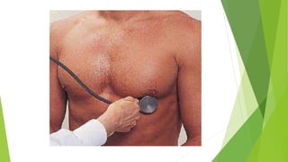
- 16. Chest percussion – • Normally only the left border of heart can be detected by percussion . • It extends from the sternum to mid clavicular line in the 3rd to 4th intercostal space . Ausculation- • Ausculation is listening to the sounds of the body during a physical examination . • There are four heart sounds S1 , S2 ,S3 and S4 These sounds are produced due to the closure of valves or due to turbulence to the flow of blood during different phages of the cardiac cycle . The heart sounds are produced due to vibrations set up by the closure of valves and the vibrations of walls of the heart. blood vessel and column of blood in the vessel .
- 17. The first sound ( S1 ) and second sound (S2) heart sounds are heard by using the stethoscope Abnormal sounds – Heart murmurs are sounds such as whooshing or swishing made by rapid , choppy , blood flow through the heart . The sounds can be heard with a device called stethoscope . Heart murmurs can be present at birth or develop later in life.
- 19. Symptoms – • chest pain • dizziness • cough • swollen liver • fating • blue or gray lips and fingers • shortness of breath laboratory tests- Blood tests Electrocardiogram ( ECG ) Stress test Echocardiogram Chest x-ray Cardia MRI
- 20. Conclusion – Integrating the cardiovascular health history and physicial exam takes practice. It is not enough to simply ask the right questions and perform the physical exam . As the patients nurse you must critically analyze all of the data you are obtaining. Summary- Till now we discussed about cardiovascular assessment , its define , central venous pressure , blood pressure, about heart and conducting system of heart , assessment , article, inspection, palpation , percussion ,auscultation , abnormally sounds of the heart ,laboratory tests. Hope you all under stood well
- 21. Journal abstract – Artificial intelligence in cardiovascular prevention Ciccarelli, Michele , gialluria Published – may 2023 Abstract Prevention and effective treatment of cardiovascular disease are progressive issues that grow in tandem with the average age of the world population. Over recent decades , the potential role of artificial intelligence in cardiovascular medicine has been increasingly reconized because of the incredible amount of real . World data regarding patient health status and health care delivery that can be collected from a variety of sources wherein patients information is routinely collected . Including patient registries , clinical case reports . Reimbursement claims and billing reports , medical device and electric health like any other health data , RWD can be analyzed in accordance with high quality research methods and its analysis can deliver valuable patient centric insights complementing the information obtained from conventional clinical trials.
- 22. Artificial intelligence application on RWD has the potential to detect a patients health trajectory leading to personalized medicine and tailored treatment this article reviews the benefits of artificial intelligence in cardiovascular prevention and management . Focusing on diagnostic and therapeutic improvements without neglecting the limitations of this new scientific approach. Bibliography d. Venkatesh, HH Sudhakar (2019) basics of medical physiology for nursing students , Wolters Kluwer publication , page no 82-117