
Recommended
More Related Content
What's hot
What's hot (20)
Similar to PHYSICAL ASSESSMENT OF THORAX AND LUNGS..pptx
Similar to PHYSICAL ASSESSMENT OF THORAX AND LUNGS..pptx (20)
Recently uploaded
Recently uploaded (20)
PHYSICAL ASSESSMENT OF THORAX AND LUNGS..pptx
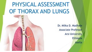
- 1. PHYSICAL ASSESSMENT OF THORAX AND LUNGS Dr. Milka D. Madhale Associate Professor Arsi University Ethiopia Asella
- 2. OBJECTIVES Discuss brief anatomy and physiology of lung and thorax. Demonstrate assessment of lung and thorax. Identify normal assessment of lung and thorax. Identify abnormal assessment of thorax and lung. Apply the caring value during assessment of thorax and lung.
- 3. BRIEF ANATOMY AND PHYSIOLOGY OF LUNGS AND THORAX THORACIC CAVITY It is situated between the neck and the diaphragm. Thoracic cavity has lungs, heart, oesophagus, trachea and thymus.
- 5. IMPORTANT FUNCTIONS OF RESPIRATORY SYSTEM It brings oxygen into our bodies, which we need for our cells to live and function properly. It helps to get rid of carbon dioxide, which is a waste of product of cellular function. ( Respiration is the movement of oxygen from the outside environment to the cells within tissues, and the transport of carbon dioxide in the opposite direction.)
- 9. MECHANISM OF RESPIRATION (BRIEF OVERVIEW) Air enters the nostrils--------Nose filters the incoming air— ----Air passes in the pharynx to the larynx ( protected by epiglottis, which prevents food entering in the passage of lungs)------Air goes into trachea(windpipe)-------Trachea divides into two bronchi-------Each bronchus divides bronchioles----Bronchioles enters into alveolar duct ( the actual exchange of gases occurs in the alveoli) ( Oxygen from air in the alveoli goes into the blood and carbon dioxide in the blood goes out)
- 10. INSPECTION Patient is asked to sit upright on the examination table with arms aside. Ensure adequate lighting. Examine the anterior and posterior chest. Before examination , wash the hands with soap and water.
- 11. General observations: Fast respiratory rate. Cyanosis(blue discoloration on skin) Unusual posture. Using accessary muscle for breathing. Inward movement of intercostal muscles. Note if patient is coughing. Note if patients voice is hoarse. Note wheezing. Abnormal breathing pattern.
- 12. Peripheral examination: Hand examination: Assess the flapping tremor (asterisks): It is the tremor of hand when the wrist is extended. It resembles like bird flapping its wing.( Caused by CARBON DIOXIDE retention, if they have received bronchodilator therapy.)
- 13. Nicotine staining on the fingers:(sign of identifying smokers) Both thumbnails side by side (Diamond shape is normal) Abnormalities are seen in pulmonary fibrosis, cystic fibrosis, bronchogenic carcinoma)
- 14. Erythema Nodosum:( Red, painful, tender lumps) May be the first sign of a systemic disease such as Tuberculosis.
- 15. Facial flushing (Indication of carbon dioxide retention- Dyspnea) Nasal polyps and epistaxis
- 16. Pink and healthy tongue Bluish tongue-Central cyanosis
- 17. Horner's syndrome Observe for Horners syndrome: ( constricted pupil, ptosis, decreased sweating on the face shows brachial plexus compression—tumor in the apex of the lungs)
- 18. CHEST INSPECTION: Chest Scars: Chest scars can be from previous thoracotomy
- 19. SHAPES OF THE CHEST:
- 20. Normal Chest: The chest cavity is outlined on each side by the white bony structures that represent the ribs of the chest wall. On the top portion of the chest is the neck and the collar bones (clavicles). On the bottom, the chest cavity is bordered by the diaphragm under which is the abdominal cavity. No deformity is seen on the chest.
- 21. Contd…… Barrel chest: It refers, to an increase in the anterior posterior diameter of the chest wall resembling the shape of a barrel, most often associated with emphysema. Pectus excavatum: It is a congenital deformity of the anterior thoracic wall in which the sternum and rib cage grow abnormally. This produces a caved-in or sunken appearance of the chest. It can either be present at birth or not develop until puberty.
- 22. Pectus carinatum (L carīnātus, equiv. to carīn(a) keel), also called pigeon chest, is a malformation of the chest characterized by a protrusion of the sternum and ribs. It is distinct from the related malformation Pectus excavatum.
- 23. Contd…..
- 24. PALPATION: 1. Examination with the hands, feeling for organs, masses, or infiltration of a part of the body, feeling the heart or pulse beat, or vibrations in the chest. 2. Touching, feeling, or perceiving by the sense of touch.
- 25. Assessing lymphadenopathy Palpate the cervical lymph nodes with both hands, one on each side of the patient's face. Start at the preauricular glands and then work down, palpating with the ends of your fingers: jugulodigastric, submandibular, submental, anterior cervical, supraclavicular, posterior cervical, posterior auricular, occipital lymph nodes. Assess for axillary lymphadenopathy by holding the patient's arm near the elbow with one hand and palpating in the axilla with your other hand.
- 26. Palpate the trachea Palpate the trachea Position yourself in front of the patient. Place your right index finger in the sternal notch. Palpate the lateral borders of the trachea to determine if it is in normal (midline) position. A deviated trachea can indicate lung pathology The trachea will be deviated away from the side of an effusion or lung mass, and towards the side of a pneumothorax, collapsed lung, or atelectasis.
- 27. Palpate the chest wall. • Use the palm of your right or left hand to assess for any obvious point tenderness, masses, or rib deformities. • Palpate the chest anteriorly and posteriorly. Any differences between right and left can indicate abnormal underlying lung tissue. • Note any evidence of subcutaneous emphysema.It is observed when air gets into the subcutaneous tissues, and is associated with lung collapse secondary to trauma or a ruptured bronchial tube. Assess chest expansion • Place your hands, with thumbs touching, in the midline and extend your fingers to make contact with the lateral edges of the chest anteriorly, just below the level of the nipples. • Ask the patient to take a deep breath. The thumbs should separate by approximately 5 cm or more in normal chest expansion (this technique can also be utilized posteriorly).
- 28. Place your hands at the lower part of the chest. Ask the patient to say 99 or 1-2-1 The vibration felt against your hand should be the same in each hand.
- 29. PERCUSSION The act or technique of tapping the surface of a body part to learn the condition of the parts beneath by the resultant sound
- 30. PERCUSSION Posterior Chest Anterior Chest
- 31. PROCEDURE: Make sure the patient is undressed till the waist. Position the patient on examination table at 30-40 degree angle and approach from the right side. Percuss both posteriorly and anteriorly , starting on the back. Place non-dominant hand with middle finger pressed and hyperextended firmly. The firmer the finger is pressed to the chest wall, the harder the percussion note. Make sure other fingers and palm are not pressed against the chest wall. It is advisable to keep finger nail short.
- 32. Contd….. Percuss on the chest in the intercostal spaces. Resonant percussion note ------ Heard over a normal air filled lung. Dull Percussion note ------ The sound over solid tissues--- Indicates the consolidation. Tympanic percussion note ---- ( a drum like sound when percussing hollow organ) ----- Pleural effusion Stony Duller sound than dull sound ---- COPD, Emphysema or pneumothorax.
- 33. AUSCULTATION: ( Actions of listening sounds from the heart, lungs, and other organs ,typically with stethoscope.)
- 34. Points for auscultation of the lungs:
- 35. Position the patient: ask the patient to lean forward or sit upright in order to examine posteriorly. Asking the patient to fold arms or place hands on opposing shoulders also helps to get maximal exposure to the lung fields. Place the diaphragm of the stethoscope on the patient's chest, and ask the patient to take deep breaths in and out through the mouth. Auscultate at five levels posteriorly and anteriorly, comparing side by side. Normal breath sounds are called vesicular breath sounds, which are low-pitched sounds louder on inspiration and softer on expiration. They should be symmetrical posteriorly. Note the presence and location of abnormal (adventitious) extra breath sounds, such as crackles, wheezing, rhonchi, stridor, or pleural friction rub. Procedure:
- 36. Note the characteristics of any abnormal breath sounds (if present): loudness, quality, duration, and whether they occur during inspiration or expiration (i.e., timing in the respiratory cycle). Many abnormal breath sounds are best heard after asking the patient to cough. Assess for Broncho phony, an increased sound transmission over the consolidated lung, when asking the patient to say "99" or "1-2-1 Assess for whispering sound, While auscultating with the stethoscope, ask the patient to whisper "99" or "1-2-1." In the consolidated lung, the sound will actually be heard better and more clearly with the stethoscope. Contd……..
- 37. Breath Sounds Bronchial Sound These are hollow sound, tubular sounds that are lower pitched. They can be auscultated over trachea, where they are considered normal. If it is heard over the peripheral lung field, considered as abnormal, which indicate pneumonia, atelectasis, pleural effusion.
- 38. Contd…. Broncho-vesicular sounds Crackles/Crepitations/ Rales These are normal sounds on the mid chest area in between the scapula, where inspiration and expiration periods are equal. These are abnormal sounds made in respiratory disease during inhalation in CCF, Pulmonary edema
- 39. Contd…….. Wheezing Ronchi It is a high pitched , whistling sound made while breathing often associated with difficulty in breathing.Heard in Bronchial Asthma It is continuous low pitched, rattling sounds, often resembles snoring. Heard in COPD, Chronic Bronchitis, Cystic fibrosis.
- 40. Stridor Rub It is high pitched crowing breath sound heard during respiration. Heard in upper airway narrowing or obstruction It is the audible medical sign( a friction sound) Heard in pleurisy, viral infection and influenza Contd…..
- 41. Application of caring value Patient value is a key element in patient care. Respect his opinion and decision. Allow him/her to communicate. Guide him/her in all the steps he has to follow. Nurse must have professional value to provide safe and high quality care.