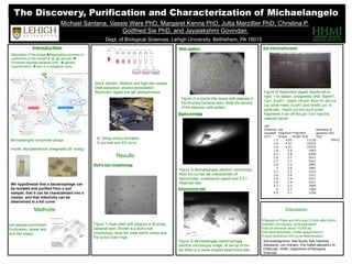
1. Introduction
Wild-type
Methods
•Plaques on Plate are bull’s eye, 2-3mm after 24hrs
•Electron microscopy- pure population
•Size of Genome: about 70,000 bp
•Gel electrophoresis: cluster assignment H
•Future Directions: Kill curve determination
Results
Discussion
Adsorption of the phagedegradative enzymes or
contraction of the sheath & tail @ cell wall
Enzymes degrade bacterial DNA, genetic
recombination lytic or a lysogenic cycle
Michaelangelo temperate phage
model: Mycobacterium smegmatis (M. smeg)
We hypothesize that a bacteriophage can
be isolated and purified from a soil
sample; that it can be characterized into a
cluster; and that infectivity can be
determined in a kill curve
soil sample enrichment
Purification: streak test,
and titer assay.
Acknowledgments: Sea faculty Sea Teaching
Assistants, Lee Graham, The Hatfull laboratory At
Pittsburgh, HHMI, Department of Biological
Sciences
Figure 1) Agar plate with plaques in M.smeg
bacterial lawn. Shown is a Bull’s eye
morphology. Note the clear sterile center and
the turbid outer rings
Figure 2) a typical titer assay with plaques in
the M.smeg bacterial lawn. Note the density
of the plaques- web pattern
Figure 4) Michaelangelo bacteriophage
electron microscopy image. At the tip of the
tail there is a clover shaped attachment site.
Figure 3) Michaelangelo electron microscopy.
Note the curved tail characteristic of
siphoviridae, icosahedral capsid and 2.5:1
Head:tail ratio
Figure 5) Restriction digest. Bands left to
right: 1-kb ladder, undigested DNA, BamH1,
Cla1, EcoR1, HaeIII, HindIII. Bam H1 did not
cut, while HaeII, EcoR1 and HindIII cut. In
particular, HaeIII cut into such small
fragments it ran off the gel. Cla1 had the
clearest bands
Stock solution: Medium and high titer lysates
DNA extraction: alcohol precipitation
Restriction digest and gel eletrophoresis
M. Smeg colony formation
% survival and Kill curve
claI
Distance
traveled
(cm)
log
fragment
length
Fragment
length (bd)
estimate of
genome size
(bp)
1.5 4.05 11220 79452
1.6 4.01 10232
1.6 4.01 10232
1.8 3.9 7943
2.2 3.8 6309
2.5 3.7 5011
2.7 3.7 5011
2.9 3.6 3981
3 3.6 3981
3.3 3.5 3162
3.4 3.4 2511
3.5 3.4 2511
3.6 3.4 2511
3.7 3.3 1995
4 3.2 1584
4.5 3.1 1258
Bull’s eye morphology
Web pattern Gel electrophoresis
Siphoviridae
Attachment site