SlideShare a Scribd company logo
1 of 1
Download to read offline
PerkinElmer, Inc., 940 Winter Street, Waltham, MA USA (800) 762-4000
or (+1) 203 925-4602 www.perkinelmer.com
Efficient AlphaLISA immunoassays for the detection and
characterization of monoclonal antibody biotherapeutics
Geneviève Pinard, Claire Normand, Valérie Paquet, Wolfgang Reintsch, Lenka Rihakova,
Mireille Caron, Michael Lomotan, Julie Bédard, Véronique Brechler and Francesco Lipari
PerkinElmer BioSignal, Inc., 1744 William St., Montreal, QC, H3J 1R4, Canada
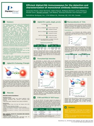
Immunoassays are commonly used in the development
and characterization of therapeutic monoclonal
antibodies (TAbs). There is an increasing need for
efficient and sensitive assays to detect and
characterize TAbs during various stages of
development. The most common technology for
performing immunoassays is the enzyme-linked
immunosorbent assay (ELISA), which is a robust
method but requires multiple and time-consuming
steps that can lead to variable data. The AlphaLISA®
assay format is a chemiluminescent homogeneous
bead-based technology which does not require washing
or separation steps, and as such provides significant
advantages over ELISA. AlphaLISA assays are typically
performed in 96- or 384-well plates and can be run
with sample volumes as low as 5 µL, conserving
valuable test materials. The total assay time is less
than 3 hours, which is half the time of a normal ELISA.
Several new AlphaLISA assays have been developed to
characterize monoclonal antibody biotherapeutics:
• Detection of fucosylated IgG by use of an anti-IgG
antibody and a core fucose-specific lectin.
• Detection of bioprocess contaminants such as host
cell proteins (CHO and PER.C6® HCP).
• Quantitation of therapeutic antibodies measuring total
human IgG1 or IgG4.
Data provided here demonstrate that AlphaLISA
technology provides a versatile assay platform for
improving various analytical assays in the modern
biotherapeutics development laboratory.
The biotinylated anti-analyte antibody binds to the Streptavidin-
coated Alpha Donor beads while another anti-analyte antibody is
conjugated to AlphaLISA Acceptor beads. In the presence of the
analyte, the beads come into close proximity. The 680 nm laser
excitation of the Donor beads provokes the release of singlet
oxygen molecules that triggers a cascade of energy transfer in the
Acceptor beads resulting in a sharp peak of light emission at 615
nm.
AlphaLISA products (PerkinElmer):
Standalone products:
• AlphaLISA Acceptor beads: Lens culinaris agglutinin (LCA)
(#AL140), Anti-Human IgG1 (#AL141), Anti-Human IgG4
(#AL142).
• Alpha Donor beads: Streptavidin Donor beads (#6760002S)
AlphaLISA kits:
• Chinese Hamster Ovary Cell Host Cell Proteins (CHO HCP)
(#AL210)
• CHO HCP (broad reactivity) (#AL301)
• PER.C6® Cell Host Cell Proteins (PER.C6® HCP) (#AL302)
• Human Immunoglobulin G subclass 1 (IgG1) (pharmacokinetic)
(#AL303)
• Human Immunoglobulin G subclass 4 (IgG4) (pharmacokinetic)
(#AL304).
Other materials required for fucosylated IgG4 detection
assay (section 5):
• Human IgG4, Fitzgerald (#31-AI20)
• AffiniPure F(ab')2 Fragment Goat Anti-Human IgG, Fcγ
Fragment Specific, Bethyl Laboratories (#A80-248A)
5 mL Standard or Sample
Add 10 mL of AlphaLISA Anti-Analyte Acceptor beads
Incubate 30 minutes
Add 25 mL of Streptavidin Alpha Donor beads
Incubate 30 minutes
Read at 615 nm using EnVision® or EnSpire® Multimode
Plate Reader and analyze data
Add 10 mL of Biotinylated Anti-Analyte Antibody
Incubate 60 minutes
Precision of CHO HCP assay (Cat. no. AL210). Assay precision
data were calculated from a total of 18 assays. Two operators
performed three independent assays using three different kit lots.
Each assay consisted of one standard curve and three control
samples of high (A), medium (B) and low (C) concentrations,
assayed in triplicate.
Catalogue
number
Kit name
Lower detection
limit * (ng/mL)
Upper limit **
(mg/mL)
AL210 CHO HCP 0.18 0.3
AL301
CHO HCP (broad
reactivity)
0.45 1
AL302 PER.C6® HCP 0.78 3
The carbohydrates or glycans linked on the biotherapeutic
antibody may play a major role in the mechanism of action as well
as biodistribution of the drug. Detection of core fucose on
monoclonal antibodies using Alpha technology can be used for
antibody screening and characterization. The fucosylated IgG
assay is based on the detection of core fucose by lens culinaris
agglutinin (LCA), which has a high specificity for 1,6-linked
fucose 1.
Detection of fucosylated IgG4 in serum-free cell culture medium.
Human IgG4 from myeloma plasma was diluted in cell culture
medium (Hybridoma-SFM Gibco #12045-084). Fucosylated IgG4
was then detected in a typical AlphaLISA protocol using
Streptavidin Alpha Donor beads, biotinylated anti-human IgG and
AlphaLISA LCA Acceptor beads.
The CHO and PER.C6® HCP assays measure the presence of
contaminating proteins in purification samples. Two CHO HCP kits
have been developed (AL210 and AL301). The second product is
identified as “broad reactivity”, because the antibodies have been
shown to detect more CHO proteins than the previous kit. Ideally,
both assays should be tested for a given process to determine the
most suitable. The lower detection limit and upper limit of
detection are provided in the following table.
During the exploratory and pre-clinical phases, pharmacokinetic
studies of TAbs are commonly performed in rodents or monkeys
(non-human primates). TAb is injected into the animal and then
measured in the serum at different time points. The TAb is
measured using a generic IgG assay or a ligand binding assay.
A combination of an anti-human IgG antibody, monkey-
adsorbed, and a subclass specific anti-IgG1 or 4 antibody are
used to obtain highly sensitive assays to measure IgG1 or IgG4
in animal serum.
Monoclonal antibodies specific to either IgG1 or IgG4 subclass
were conjugated to AlphaLISA Acceptor beads to be used for
AlphaLISA assays to measure TAb in serum via a ligand binding
assay. A human monoclonal antibody specific to TNF was used
as a model to demonstrate the application of the assay for
detection of a TAb in monkey serum.
Summary8
Summary
AlphaLISA Technology Principle
Materials
AlphaLISA: quick, simple, precise
Fucosylated IgG detection5
CHO and PER.C6® HCP assays6
Pharmacokinetics of TAbs71 4
2
3
Typical assay precision data
Sample
Mean
(pg/mL)
SD
(pg/mL)
%CV
(n=18)
A 92 819 6 955 7.5
B 10 765 641 6.0
C 1 049 92 8.8
Intra-assay precision
Sample
Mean
(pg/mL)
SD
(pg/mL)
%CV
(n=6)
A 92 819 10 650 11.5
B 10 765 1 137 10.6
C 1 049 180 17.2
Inter-assay precision
Generic IgG assay
Sensitivity and specificity of the human IgG1 assay. Human IgG1
purified from myeloma plasma or total IgG from cynomolgus
monkey were diluted in buffer and detected using a typical
AlphaLISA protocol. The human IgG1 is detected down to a level
of 20 pg/mL, whereas 300 ng/mL of monkey IgG gives only
background signal.
Ligand binding assay
Dilution factor % Recovery
1 100
2 89
4 83
8 83
16 84
Linearity
Spike (ng/mL) % Recovery
100 85
10 82
1 86
Spike recovery
Accuracy of the human IgG1 assay in monkey serum. 10%
monkey serum was used as analyte diluent. A pool of monkey
sera spiked with 1000 ng/mL of human IgG1 was initially diluted
10-fold with buffer. This sample containing 100 ng/mL IgG1 was
used for the linearity experiment. The recovery for linearity was
calculated using the undiluted sample (1X) as the 100% value.
For spike recovery, the recovery was calculated based on the
theoretical amount of IgG1 present.
Detection of anti-TNF antibody in monkey serum. A monoclonal
antibody (IgG1 subclass) against TNF was diluted in 10%
monkey serum and detected using anti-IgG1-conjugated
Acceptor beads with biotinylated TNF as ligand. The lower
detection limit was 0.3 ng/mL.
Sensitive and reproducible immunoassays were developed for
analyses of biotherapeutics. These assays can be applied in
different stages of TAb development, such as cloning and
expression, process development and pharmacokinetic studies.
References
1. Tateno, H. et al. Comparative analysis of core-fucose-binding lectins from
Lens culinaris and Pisum sativum using frontal affinity chromatography.
Glycobiology 19, 527–536 (2009).
* Calculated by interpolating the average background counts (12 wells without
analyte) + 3 x standard deviation value on the standard curve.
** Corresponds to analyte concentration on the standard curve giving maximum
signal.
Excitation
680 nm
Emission
615 nm
Streptavidin-coated
Alpha Donor Bead
LCA lectin-conjugated
AlphaLISA Acceptor Bead
Biotinylated
Anti-human IgG
Monoclonal
antibody
Excitation
680 nm
Emission
615 nm
Streptavidin-coated
Alpha Donor Bead
Anti-IgG1-conjugated
AlphaLISA Acceptor Bead
Biotinylated
TNF
Anti-
TNF
antibody

More Related Content

What's hot

Aai 2007-elis array-poster-070515
Aai 2007-elis array-poster-070515Aai 2007-elis array-poster-070515
Aai 2007-elis array-poster-070515Elsa von Licy
 
Host Cell Protein Analysis - Measuring the Forest, or Counting theTrees
Host Cell Protein Analysis - Measuring the Forest, or Counting theTreesHost Cell Protein Analysis - Measuring the Forest, or Counting theTrees
Host Cell Protein Analysis - Measuring the Forest, or Counting theTreesTed Kocot
 
Clinical Samples & Disease State Plasma Newsflash October 2016
Clinical Samples & Disease State Plasma Newsflash October 2016Clinical Samples & Disease State Plasma Newsflash October 2016
Clinical Samples & Disease State Plasma Newsflash October 2016BBISolutions
 
ELISA Kits for Cancer Research
ELISA Kits for Cancer ResearchELISA Kits for Cancer Research
ELISA Kits for Cancer ResearchElabscience
 
BBI Solutions - Human Proteins
BBI Solutions - Human ProteinsBBI Solutions - Human Proteins
BBI Solutions - Human ProteinsBBISolutions
 
Research Symposium Poster (Spring - April 2014)
Research Symposium Poster (Spring - April 2014)Research Symposium Poster (Spring - April 2014)
Research Symposium Poster (Spring - April 2014)William Parker
 
Disease State Plasma
Disease State PlasmaDisease State Plasma
Disease State PlasmaBBISolutions
 
07 elisa kits for vocal cord cancer research
07 elisa kits for vocal cord cancer research07 elisa kits for vocal cord cancer research
07 elisa kits for vocal cord cancer researchElabscience
 
Replacing animal-derived components in regulatory in vitro tests
Replacing animal-derived components in regulatory in vitro testsReplacing animal-derived components in regulatory in vitro tests
Replacing animal-derived components in regulatory in vitro testsDr Carol Barker-Treasure
 
SOJ Biochem 2016
SOJ Biochem 2016SOJ Biochem 2016
SOJ Biochem 2016Bob Matson
 
SAGE Student Research Conference Poster- The Effect of Purified Acetaminophen...
SAGE Student Research Conference Poster- The Effect of Purified Acetaminophen...SAGE Student Research Conference Poster- The Effect of Purified Acetaminophen...
SAGE Student Research Conference Poster- The Effect of Purified Acetaminophen...Melissa McCoy, MS, MBA
 
January 2015 - Antibodies Newsflash
January 2015 - Antibodies NewsflashJanuary 2015 - Antibodies Newsflash
January 2015 - Antibodies NewsflashBBISolutions
 
Antibodies - Key Marker Performance - July 2015
Antibodies - Key Marker Performance - July 2015Antibodies - Key Marker Performance - July 2015
Antibodies - Key Marker Performance - July 2015BBISolutions
 
Measure Target Cancer Cell Death in Co-Cultures
Measure Target Cancer Cell Death in Co-CulturesMeasure Target Cancer Cell Death in Co-Cultures
Measure Target Cancer Cell Death in Co-CulturesDiscoverX Corporation
 
Oncodesign aacr 2018 development of a high throughput in vitro screening pl...
Oncodesign aacr 2018   development of a high throughput in vitro screening pl...Oncodesign aacr 2018   development of a high throughput in vitro screening pl...
Oncodesign aacr 2018 development of a high throughput in vitro screening pl...Florence Fombertasse
 

What's hot (19)

Aai 2007-elis array-poster-070515
Aai 2007-elis array-poster-070515Aai 2007-elis array-poster-070515
Aai 2007-elis array-poster-070515
 
Host Cell Protein Analysis - Measuring the Forest, or Counting theTrees
Host Cell Protein Analysis - Measuring the Forest, or Counting theTreesHost Cell Protein Analysis - Measuring the Forest, or Counting theTrees
Host Cell Protein Analysis - Measuring the Forest, or Counting theTrees
 
Clinical Samples & Disease State Plasma Newsflash October 2016
Clinical Samples & Disease State Plasma Newsflash October 2016Clinical Samples & Disease State Plasma Newsflash October 2016
Clinical Samples & Disease State Plasma Newsflash October 2016
 
ELISA Kits for Cancer Research
ELISA Kits for Cancer ResearchELISA Kits for Cancer Research
ELISA Kits for Cancer Research
 
BBI Solutions - Human Proteins
BBI Solutions - Human ProteinsBBI Solutions - Human Proteins
BBI Solutions - Human Proteins
 
Research Symposium Poster (Spring - April 2014)
Research Symposium Poster (Spring - April 2014)Research Symposium Poster (Spring - April 2014)
Research Symposium Poster (Spring - April 2014)
 
Disease State Plasma
Disease State PlasmaDisease State Plasma
Disease State Plasma
 
EBposter_HeatStress
EBposter_HeatStressEBposter_HeatStress
EBposter_HeatStress
 
07 elisa kits for vocal cord cancer research
07 elisa kits for vocal cord cancer research07 elisa kits for vocal cord cancer research
07 elisa kits for vocal cord cancer research
 
Replacing animal-derived components in regulatory in vitro tests
Replacing animal-derived components in regulatory in vitro testsReplacing animal-derived components in regulatory in vitro tests
Replacing animal-derived components in regulatory in vitro tests
 
SOJ Biochem 2016
SOJ Biochem 2016SOJ Biochem 2016
SOJ Biochem 2016
 
SAGE Student Research Conference Poster- The Effect of Purified Acetaminophen...
SAGE Student Research Conference Poster- The Effect of Purified Acetaminophen...SAGE Student Research Conference Poster- The Effect of Purified Acetaminophen...
SAGE Student Research Conference Poster- The Effect of Purified Acetaminophen...
 
2015 PBL Assay Solutions
2015 PBL Assay Solutions2015 PBL Assay Solutions
2015 PBL Assay Solutions
 
January 2015 - Antibodies Newsflash
January 2015 - Antibodies NewsflashJanuary 2015 - Antibodies Newsflash
January 2015 - Antibodies Newsflash
 
Antibodies - Key Marker Performance - July 2015
Antibodies - Key Marker Performance - July 2015Antibodies - Key Marker Performance - July 2015
Antibodies - Key Marker Performance - July 2015
 
Measure Target Cancer Cell Death in Co-Cultures
Measure Target Cancer Cell Death in Co-CulturesMeasure Target Cancer Cell Death in Co-Cultures
Measure Target Cancer Cell Death in Co-Cultures
 
Oncodesign aacr 2018 development of a high throughput in vitro screening pl...
Oncodesign aacr 2018   development of a high throughput in vitro screening pl...Oncodesign aacr 2018   development of a high throughput in vitro screening pl...
Oncodesign aacr 2018 development of a high throughput in vitro screening pl...
 
Biopra activities
Biopra activitiesBiopra activities
Biopra activities
 
GLP-1 Poster_02JUN15
GLP-1 Poster_02JUN15GLP-1 Poster_02JUN15
GLP-1 Poster_02JUN15
 

Similar to 44-150724PST_Peptalk_2013_AlphaLISA_immunoassays

POLYMERASE CHAIN REACTION (PCR) AND ENZYME-LINKED.pptx
POLYMERASE CHAIN REACTION (PCR) AND ENZYME-LINKED.pptxPOLYMERASE CHAIN REACTION (PCR) AND ENZYME-LINKED.pptx
POLYMERASE CHAIN REACTION (PCR) AND ENZYME-LINKED.pptxAreebWaheed
 
Top-Selling ELISA Kits 2016 - Elabscience
Top-Selling ELISA Kits 2016 - ElabscienceTop-Selling ELISA Kits 2016 - Elabscience
Top-Selling ELISA Kits 2016 - ElabscienceElabscience
 
Ag Ab Reaction Ii Final
Ag Ab Reaction Ii FinalAg Ab Reaction Ii Final
Ag Ab Reaction Ii Finaldeepak deshkar
 
portrait poster for iFAAM 2016 Amsterdam RY 3
portrait poster for iFAAM 2016 Amsterdam RY 3portrait poster for iFAAM 2016 Amsterdam RY 3
portrait poster for iFAAM 2016 Amsterdam RY 3Ross Yarham
 
ria elisa biochem.pdf
ria elisa biochem.pdfria elisa biochem.pdf
ria elisa biochem.pdfRinaDas9
 
MDC Connects: Biomarker identification - Assessing Immune Function
MDC Connects: Biomarker identification - Assessing Immune FunctionMDC Connects: Biomarker identification - Assessing Immune Function
MDC Connects: Biomarker identification - Assessing Immune FunctionMedicines Discovery Catapult
 
AUTOMATION IN SEROLOGY.pptx educational
AUTOMATION IN SEROLOGY.pptx  educationalAUTOMATION IN SEROLOGY.pptx  educational
AUTOMATION IN SEROLOGY.pptx educationalaryajayakottarathil
 
Generation of Anti-Idiotypic Responses Against an 85KDa Breast Tumor Associat...
Generation of Anti-Idiotypic Responses Against an 85KDa Breast Tumor Associat...Generation of Anti-Idiotypic Responses Against an 85KDa Breast Tumor Associat...
Generation of Anti-Idiotypic Responses Against an 85KDa Breast Tumor Associat...lsijjournal456
 
Pyrogen and its various test
Pyrogen and its various testPyrogen and its various test
Pyrogen and its various testPrachi Barbhaiya
 
2015 PBL Assay Solutions
2015 PBL Assay Solutions2015 PBL Assay Solutions
2015 PBL Assay Solutionsstephenaparent
 
ELISA Powerpoint Presentation
ELISA Powerpoint PresentationELISA Powerpoint Presentation
ELISA Powerpoint PresentationAnita Singh
 

Similar to 44-150724PST_Peptalk_2013_AlphaLISA_immunoassays (20)

POLYMERASE CHAIN REACTION (PCR) AND ENZYME-LINKED.pptx
POLYMERASE CHAIN REACTION (PCR) AND ENZYME-LINKED.pptxPOLYMERASE CHAIN REACTION (PCR) AND ENZYME-LINKED.pptx
POLYMERASE CHAIN REACTION (PCR) AND ENZYME-LINKED.pptx
 
Top-Selling ELISA Kits 2016 - Elabscience
Top-Selling ELISA Kits 2016 - ElabscienceTop-Selling ELISA Kits 2016 - Elabscience
Top-Selling ELISA Kits 2016 - Elabscience
 
ratanna paper
ratanna paperratanna paper
ratanna paper
 
Ag Ab Reaction Ii Final
Ag Ab Reaction Ii FinalAg Ab Reaction Ii Final
Ag Ab Reaction Ii Final
 
portrait poster for iFAAM 2016 Amsterdam RY 3
portrait poster for iFAAM 2016 Amsterdam RY 3portrait poster for iFAAM 2016 Amsterdam RY 3
portrait poster for iFAAM 2016 Amsterdam RY 3
 
Khushbu_Simoa_06-16
Khushbu_Simoa_06-16Khushbu_Simoa_06-16
Khushbu_Simoa_06-16
 
ria elisa biochem.pdf
ria elisa biochem.pdfria elisa biochem.pdf
ria elisa biochem.pdf
 
Antibodies & Serums
Antibodies & SerumsAntibodies & Serums
Antibodies & Serums
 
MDC Connects: Biomarker identification - Assessing Immune Function
MDC Connects: Biomarker identification - Assessing Immune FunctionMDC Connects: Biomarker identification - Assessing Immune Function
MDC Connects: Biomarker identification - Assessing Immune Function
 
AUTOMATION IN SEROLOGY.pptx educational
AUTOMATION IN SEROLOGY.pptx  educationalAUTOMATION IN SEROLOGY.pptx  educational
AUTOMATION IN SEROLOGY.pptx educational
 
Generation of Anti-Idiotypic Responses Against an 85KDa Breast Tumor Associat...
Generation of Anti-Idiotypic Responses Against an 85KDa Breast Tumor Associat...Generation of Anti-Idiotypic Responses Against an 85KDa Breast Tumor Associat...
Generation of Anti-Idiotypic Responses Against an 85KDa Breast Tumor Associat...
 
ELISA Notes
ELISA NotesELISA Notes
ELISA Notes
 
Elisa
Elisa Elisa
Elisa
 
Elisa technique
Elisa techniqueElisa technique
Elisa technique
 
Pyrogen and its various test
Pyrogen and its various testPyrogen and its various test
Pyrogen and its various test
 
Elisa
ElisaElisa
Elisa
 
2015 PBL Assay Solutions
2015 PBL Assay Solutions2015 PBL Assay Solutions
2015 PBL Assay Solutions
 
Elisa
ElisaElisa
Elisa
 
Elisa ppt
Elisa pptElisa ppt
Elisa ppt
 
ELISA Powerpoint Presentation
ELISA Powerpoint PresentationELISA Powerpoint Presentation
ELISA Powerpoint Presentation
 

44-150724PST_Peptalk_2013_AlphaLISA_immunoassays

  • 1. PerkinElmer, Inc., 940 Winter Street, Waltham, MA USA (800) 762-4000 or (+1) 203 925-4602 www.perkinelmer.com Efficient AlphaLISA immunoassays for the detection and characterization of monoclonal antibody biotherapeutics Geneviève Pinard, Claire Normand, Valérie Paquet, Wolfgang Reintsch, Lenka Rihakova, Mireille Caron, Michael Lomotan, Julie Bédard, Véronique Brechler and Francesco Lipari PerkinElmer BioSignal, Inc., 1744 William St., Montreal, QC, H3J 1R4, Canada Immunoassays are commonly used in the development and characterization of therapeutic monoclonal antibodies (TAbs). There is an increasing need for efficient and sensitive assays to detect and characterize TAbs during various stages of development. The most common technology for performing immunoassays is the enzyme-linked immunosorbent assay (ELISA), which is a robust method but requires multiple and time-consuming steps that can lead to variable data. The AlphaLISA® assay format is a chemiluminescent homogeneous bead-based technology which does not require washing or separation steps, and as such provides significant advantages over ELISA. AlphaLISA assays are typically performed in 96- or 384-well plates and can be run with sample volumes as low as 5 µL, conserving valuable test materials. The total assay time is less than 3 hours, which is half the time of a normal ELISA. Several new AlphaLISA assays have been developed to characterize monoclonal antibody biotherapeutics: • Detection of fucosylated IgG by use of an anti-IgG antibody and a core fucose-specific lectin. • Detection of bioprocess contaminants such as host cell proteins (CHO and PER.C6® HCP). • Quantitation of therapeutic antibodies measuring total human IgG1 or IgG4. Data provided here demonstrate that AlphaLISA technology provides a versatile assay platform for improving various analytical assays in the modern biotherapeutics development laboratory. The biotinylated anti-analyte antibody binds to the Streptavidin- coated Alpha Donor beads while another anti-analyte antibody is conjugated to AlphaLISA Acceptor beads. In the presence of the analyte, the beads come into close proximity. The 680 nm laser excitation of the Donor beads provokes the release of singlet oxygen molecules that triggers a cascade of energy transfer in the Acceptor beads resulting in a sharp peak of light emission at 615 nm. AlphaLISA products (PerkinElmer): Standalone products: • AlphaLISA Acceptor beads: Lens culinaris agglutinin (LCA) (#AL140), Anti-Human IgG1 (#AL141), Anti-Human IgG4 (#AL142). • Alpha Donor beads: Streptavidin Donor beads (#6760002S) AlphaLISA kits: • Chinese Hamster Ovary Cell Host Cell Proteins (CHO HCP) (#AL210) • CHO HCP (broad reactivity) (#AL301) • PER.C6® Cell Host Cell Proteins (PER.C6® HCP) (#AL302) • Human Immunoglobulin G subclass 1 (IgG1) (pharmacokinetic) (#AL303) • Human Immunoglobulin G subclass 4 (IgG4) (pharmacokinetic) (#AL304). Other materials required for fucosylated IgG4 detection assay (section 5): • Human IgG4, Fitzgerald (#31-AI20) • AffiniPure F(ab')2 Fragment Goat Anti-Human IgG, Fcγ Fragment Specific, Bethyl Laboratories (#A80-248A) 5 mL Standard or Sample Add 10 mL of AlphaLISA Anti-Analyte Acceptor beads Incubate 30 minutes Add 25 mL of Streptavidin Alpha Donor beads Incubate 30 minutes Read at 615 nm using EnVision® or EnSpire® Multimode Plate Reader and analyze data Add 10 mL of Biotinylated Anti-Analyte Antibody Incubate 60 minutes Precision of CHO HCP assay (Cat. no. AL210). Assay precision data were calculated from a total of 18 assays. Two operators performed three independent assays using three different kit lots. Each assay consisted of one standard curve and three control samples of high (A), medium (B) and low (C) concentrations, assayed in triplicate. Catalogue number Kit name Lower detection limit * (ng/mL) Upper limit ** (mg/mL) AL210 CHO HCP 0.18 0.3 AL301 CHO HCP (broad reactivity) 0.45 1 AL302 PER.C6® HCP 0.78 3 The carbohydrates or glycans linked on the biotherapeutic antibody may play a major role in the mechanism of action as well as biodistribution of the drug. Detection of core fucose on monoclonal antibodies using Alpha technology can be used for antibody screening and characterization. The fucosylated IgG assay is based on the detection of core fucose by lens culinaris agglutinin (LCA), which has a high specificity for 1,6-linked fucose 1. Detection of fucosylated IgG4 in serum-free cell culture medium. Human IgG4 from myeloma plasma was diluted in cell culture medium (Hybridoma-SFM Gibco #12045-084). Fucosylated IgG4 was then detected in a typical AlphaLISA protocol using Streptavidin Alpha Donor beads, biotinylated anti-human IgG and AlphaLISA LCA Acceptor beads. The CHO and PER.C6® HCP assays measure the presence of contaminating proteins in purification samples. Two CHO HCP kits have been developed (AL210 and AL301). The second product is identified as “broad reactivity”, because the antibodies have been shown to detect more CHO proteins than the previous kit. Ideally, both assays should be tested for a given process to determine the most suitable. The lower detection limit and upper limit of detection are provided in the following table. During the exploratory and pre-clinical phases, pharmacokinetic studies of TAbs are commonly performed in rodents or monkeys (non-human primates). TAb is injected into the animal and then measured in the serum at different time points. The TAb is measured using a generic IgG assay or a ligand binding assay. A combination of an anti-human IgG antibody, monkey- adsorbed, and a subclass specific anti-IgG1 or 4 antibody are used to obtain highly sensitive assays to measure IgG1 or IgG4 in animal serum. Monoclonal antibodies specific to either IgG1 or IgG4 subclass were conjugated to AlphaLISA Acceptor beads to be used for AlphaLISA assays to measure TAb in serum via a ligand binding assay. A human monoclonal antibody specific to TNF was used as a model to demonstrate the application of the assay for detection of a TAb in monkey serum. Summary8 Summary AlphaLISA Technology Principle Materials AlphaLISA: quick, simple, precise Fucosylated IgG detection5 CHO and PER.C6® HCP assays6 Pharmacokinetics of TAbs71 4 2 3 Typical assay precision data Sample Mean (pg/mL) SD (pg/mL) %CV (n=18) A 92 819 6 955 7.5 B 10 765 641 6.0 C 1 049 92 8.8 Intra-assay precision Sample Mean (pg/mL) SD (pg/mL) %CV (n=6) A 92 819 10 650 11.5 B 10 765 1 137 10.6 C 1 049 180 17.2 Inter-assay precision Generic IgG assay Sensitivity and specificity of the human IgG1 assay. Human IgG1 purified from myeloma plasma or total IgG from cynomolgus monkey were diluted in buffer and detected using a typical AlphaLISA protocol. The human IgG1 is detected down to a level of 20 pg/mL, whereas 300 ng/mL of monkey IgG gives only background signal. Ligand binding assay Dilution factor % Recovery 1 100 2 89 4 83 8 83 16 84 Linearity Spike (ng/mL) % Recovery 100 85 10 82 1 86 Spike recovery Accuracy of the human IgG1 assay in monkey serum. 10% monkey serum was used as analyte diluent. A pool of monkey sera spiked with 1000 ng/mL of human IgG1 was initially diluted 10-fold with buffer. This sample containing 100 ng/mL IgG1 was used for the linearity experiment. The recovery for linearity was calculated using the undiluted sample (1X) as the 100% value. For spike recovery, the recovery was calculated based on the theoretical amount of IgG1 present. Detection of anti-TNF antibody in monkey serum. A monoclonal antibody (IgG1 subclass) against TNF was diluted in 10% monkey serum and detected using anti-IgG1-conjugated Acceptor beads with biotinylated TNF as ligand. The lower detection limit was 0.3 ng/mL. Sensitive and reproducible immunoassays were developed for analyses of biotherapeutics. These assays can be applied in different stages of TAb development, such as cloning and expression, process development and pharmacokinetic studies. References 1. Tateno, H. et al. Comparative analysis of core-fucose-binding lectins from Lens culinaris and Pisum sativum using frontal affinity chromatography. Glycobiology 19, 527–536 (2009). * Calculated by interpolating the average background counts (12 wells without analyte) + 3 x standard deviation value on the standard curve. ** Corresponds to analyte concentration on the standard curve giving maximum signal. Excitation 680 nm Emission 615 nm Streptavidin-coated Alpha Donor Bead LCA lectin-conjugated AlphaLISA Acceptor Bead Biotinylated Anti-human IgG Monoclonal antibody Excitation 680 nm Emission 615 nm Streptavidin-coated Alpha Donor Bead Anti-IgG1-conjugated AlphaLISA Acceptor Bead Biotinylated TNF Anti- TNF antibody