Chyme Composition and Intestinal Alkaline Conditions
•Download as PPT, PDF•
5 likes•1,083 views


Recommended
More Related Content
What's hot
What's hot (20)
Viewers also liked
Viewers also liked (20)
Similar to Chyme Composition and Intestinal Alkaline Conditions
Similar to Chyme Composition and Intestinal Alkaline Conditions (20)
Chyme Composition and Intestinal Alkaline Conditions
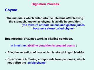
- 1. Chyme The materials which enter into the intestine after leaving the stomach, known as chyme, is acidic in condition. (the mixture of food, mucus and gastric juices became a slurry called chyme) But intestinal enzymes work in alkaline condition. In intestine, alkaline condition is created due to : - Bile, the secretion of liver which is stored in gall bladder - Bicarbonate buffering compounds from pancreas, which neutralize the acidic chyme Digestion Process
- 2. Digestion Process Intestine When acidic chyme enter into intestine, the secretin and cholecystokinin (CCK) hormones stimulate the secretion of pancreatic enzymes and bile from liver. The secretin and CCK secreted from intestinal epithelial cell wall and is transported by blood stream to pancreas, gall bladder and liver. Function of secretin: increases the secretion of pancreas and bile from liver Function of CCK: increases the pancreatic secretion and causes contraction of gall bladder to secrete bile In intestine the source of enzymes are as follows: - Pancreas (secrets zymogens of enzymes and bicarbonate) - Secretory cell or intestinal wall - Intestinal microflora
- 3. Digestion mechanism of different classes of food Food substances of fish i) Protein, carbohydrate and fat - digested first and then absorbed ii) Vitamins, minerals and water - directly absorbed
- 4. Protein digestion Enzymes involved in protein digestion are called proteolytic enzyme The proteolytic enzymes are two types - Endopeptidases - Exopeptidases • Endopeptidases: the enzymes which breakdown the central peptide bonds and form smaller peptide chians are called endopeptidases. eg. pepsin, trypsin, chymotripsin Pepsin- a major proteolytic enzyme in fish stomach. Active at – pH 1.5 - 3.0 • Exopeptidases: the enzymes which breakdown the terminal peptide bonds and separate amino acid are called exopeptidases. eg. carboxypeptidases.
- 5. Some intestinal proteolytic enzymes (Proteases) Intestinal proteases Sources Zymogen form End product of digestion Types of enzymes Trypsin Pancreas Trypsionogen Shorter chain peptide Endopeptidase Chymotripsin Pancreas Chymotripsinogen Shorter chain peptide Endopeptidase Carboxypeptidase Pancreas Procarboxypeptid ase Single amino acid Exopeptidase Aminopeptidase Epithelium cells of intestine Proaminopeptidase Splits off individual A.A. from amino terminal end of peptide chain Exopeptidease Dipeptidase Epithelium cells of intestine Breakdown dipeptides into constituent AA Exopeptidease
- 6. Zymogen: inactive form of enzyme is called zymogen (also called proenzyme) Enzymes Pepsin Trypsin Chymotriopsin Carboxypeptidase Aminopeptidase Dipeptidase Lipase Esterase Zymogen Pepsinogen Trypsinogen Chymotripsinogen Procarboxypeptidase Proaminopeptidase Prodipeptidase Prolipase Proesterase Zymogen form of some enzymes:
- 7. Carbohydrate digestion The enzyme associated with carbohydrate digestion are called carbohydrases The common carbohydrates in fish food are: i) Glycogen – storage carbohydrate of animals ii) Starch - storage carbohydrate of plants iii) Chitin – structural carbohydrate of animals iv) Cellulose - structural carbohydrate of plants
- 8. Starch and Glycogen Amylase – hydrolyze the starch and glycogen Amylase are 2 types: i) α amylase and ii) β amylase Digestive amylase of animal is generally α amylase which acts on α(1,4) glucosidic linkage of both starch and glycogen and converts into a mixture of glucose and maltose. Starch contain – amylose and amylopectin [contain both α (1,4) and α(1,6) linkage] Glycogen is structurally similar to amylopectin but their only difference is that glycogen contain more branch than amylopectin (i.e. contain more α(1,6) linkage)
- 9. Chitin Chitin is breakdown by the chitinase enzyme This enzyme is found particularly in fish species that eat crustaceans or zooplankton - secreted by stomach and pancreas. Additional source, bacteria (living within the intestine of fish) Chitin di and trimers of N-acetylglucosamine Monomer chitinase N-acetylglucosaminidaseN-acetylglucosaminidase (N-acetyl glucose amine)
- 10. Cellulose Cellulose is broken down by the enzyme cellulase. Fishes generally can not produce cellulase (probably derived from intestinal bacteria). Among cellulase producing bacteriaVibrio and Aeromonas are usually dominant in fish Cellulose Cellulase Glucose Cellulase breakdown the β(1,4) linkage of cellulose and produce glucose Other cabohydrases Source Substrate End product Maltase Intestinal epithelium Maltose Glucose Sucrase Intestinal epithelium Sucrose Glucose and Fructose Lactase Intestinal gland Lactose Galactose and Glucose Cellobiase Cellobiose Glucose
- 11. Lipid digestion At first large fat molecules are converted into smaller globules by the action of bile and then they can be hydrolyzed by lipolytic enzymes. This process known as emulsification Lipase and esterases (called lipolytic enzyme) are involved in fat digestion, secreted from pyloric caeca and intestinal mucosa and pancreas Lipid Fatty acids and glycerol Lipase No fat digestion- occurs in mouth Very little digestion – in stomach Most of the fat is digested in the intestine (at pH 6.8-7.6) Name of the enzymes Secretion place Function of enzyme Lipases Pancreas, pyloric caeca, intestinal mucosa Hydrolyze fat molecules into monoglycerides, glycerol and free fatty acid Esterases Intestine Hydrolyze simple ester of low molecular weight
- 12. Absorption - transfer of digested food materials from the digestive tract into blood stream and lymphatic system Types of absorption: i) Passive diffusion ii) Active transport iii) Pinocytosis Passive diffusion Active transport Pinocytosis It does not require expenditure of energy Occurs in response to concentration gradient Digested food materials are not transported by carrier system Relatively smaller food molecules are absorbed It requires expenditure of energy Occurs against concentration gradient Digested food materials are transported by carrier system. Carriers are highly specific for their substances Relatively smaller food molecules are absorbed Intake of food materials by fusion and constriction of the portion of the membrane of absorbing cell Fairly large molecules are absorbed Pinocytosis: A mechanism by which cells ingest extracellular fluid and its contents Mechanism of pinocytosis
- 13. Absorption site -occurs mainly in intestine also, stomach, rectum can be the absorption site The enterocytes are the absorptive cell of the intestinal epithelium. -The enterocytes form villi and microvilli which provide a large surface area for absorption Villi: are tiny, finger- like structures that protrude from the wall of the intestine and have additional extensions called microvilli (singular: microvillus) which protrude from epithelial cells lining villi. They increase the absorptive area of the intestinal wall. Digested nutrients (including sugars and amino acids) pass into the villi. Circulating blood then carries these nutrients away. Villi
- 14. Absorption of lipid: lipid is absorbed as – i) Free fatty acid ii) Glycerol iii) Monoglycerides iv) Large molecules – Pinocytosis Absorption of protein: protein is absorbed as – i) Amino acid ii) Di and tripeptides iii) Polypeptides - Pinocytosis Carrier system of AA i) Neutral amino acid carrier ii) Basic amino acid carrier iii) Acidic amino acid carrier Absorption of carbohydrates: carbohydrates are absorbed as – Monosaccharide – Active transport Passive transport Active transport
- 15. Digestibility Of the food ingested, only a part is absorbed and used for metabolic processes. The non absorbed portion of the diet leaves the body as faeces Digestibility = Food intake – faecal excretion Food intake X 100 Energy utilization N= F + E= Q + W Where, N = Energy content of food intake F = Energy content of faeces E = Energy content of nitrogen excretion (via gills, kidney, NH3, urea etc.) Q = Total metabolic energy consumption W = Energy gain of the organism
- 16. FCR (Food Conversion Ratio) = Weight gain Taken food PCR (Protein Conversion Rate): means how much protein is applied to produce more weight of fish is known by PCR Food Conversion Efficiency (FCE): Growth Food intake X 100 %
- 17. Summary of Digestion Location Source of Enzyme Digestive Process Mouth Stomach Small intestine Salivary glands Pancreas Intestine Polysaccharides Action continues until salivary amylase is inactivated by acidic pH Undigested polysaccharides Maltose & small polysac. Disaccharides hydrolyzed to monosaccharide as follows: Maltose Glucose + Glucose Sucrose Glucose + Fructose Lactose Glucose + Galactose Carbohydrate digestion Maltose + Small poly. Salivary amylase eg. starch Pancreatic amylase Maltase Sucrase Lactase
- 18. Protein digestion Location Source of Enzyme Digestive Process Stomach Small intestine Stomach (gastric gland) Pancreas Protein Short polypeptides Polypeptides Polypeptides Peptides + Dipeptides Free a. a. A - A - A A - A Pepsin A – A – A – A – A A – A – A – A – A Polypeptides + Dipeptides A – A – A A – A Trypsin Chymotripsin Polypeptides and free a. a. A – A – A A A Carboxy- peptidase A – A – A – A A A A Peptideses, Dipeptidases
- 19. Lipid digestion Location Source of Enzyme Digestive Process Small intestine Liver Pancreas Glob fat Emulsified fat (individual triacylglycerol) Triacylglycerol Fatty acid + Glycerol Bile salts Pancreatic lipase