SlideShare a Scribd company logo
1 of 40
PCL471 Laboratory
Receptor Pharmacology
Maurice Shen
maurice.shen@utoronto.ca
What is a Receptor?
• Paul Ehrlich (1800s) : Drug might act as a “Magic Bullet”
directed at a vulnerable “Receptor”
• A component of a cell or organism that interacts with a
specific ligand and initiates a chain of biochemical events
leading to the ligand’s observed effects.
– Ligands can be exogenous (drugs) or endogenous (hormones
and neurotransmitters etc.)
• 3 main characteristics of Ligand/Receptor Interactions:
– Saturable
– Reversible Binding
– Dose-Response Relationship
• Agonists: Drugs that elicit biological effects that are
comparable to endogenous ligands.
– Full: Maximal Efficacy
– Partial: Sub-maximal Efficacy
– Inverse: Produces opposite biological effects as the full
agonists
• Antagonists: Drugs that bind to the receptor but do not
elicit any biological effect; Zero Efficacy.
– Reversible & Competitive: transiently bound to the same site as
Agonists
– Non-competitive: bound to a different site as Agonists
– Irreversible: covalently bind to the same site as Agonists
Clinical Utilities?
Various Types of Drugs
• Ligand-Gated Ion Channels: Trans-membrane channels
that allows for the influx of ions such as Na+
, Ca2+
,Cl-
.
– Neurotransmitters systems: Glutamate, GABA, Acetylcholine
• Receptor Tyrosine Kinases: Trans-membrane
polypeptides that autophosphorylate at tyrosine residues
upon activation
– Growth Factors: Insulin, Epidermal Growth Factor (EGF), Platelet-
derived Growth Factor (PDGF).
• Nuclear Hormone Receptors: Cytosolic receptors that
regulates gene transcription within a cell.
– Hormones: Glucocorticoids, Thyroid Hormone, Sex Steroids etc.
Various Types of Receptors
• G Protein-Coupled Receptors: 7 trans-membrane
proteins that are coupled to specific G proteins; elicit
second messenger-mediated signaling cascade upon
activation.
– Largest family of cell surface receptors, more than 50% of drugs
on the market target GPCRs.
– Wide range of ligands including hormones, neurotransmitters,
light, and olfactory stimuli.
– Downstream signaling may lead to gene transcription, protein
synthesis, protein phosphorylations
Dopamine Receptors are GPCRs
Various Types of Receptors
• The use of varying concentrations of a specific
radiolabelled ligand to determine receptor affinity to
the ligand (Kd) and total receptor density (Bmax) in a
membrane preparation.
• The theory of radioligand binding experiments are
based on the Laws of Mass Action, where:
Ligand + Receptor [Ligand-Receptor]
Radioligand Binding Assays
Kon
Koff
1. Binding occurs when ligand and receptor collide due
to random diffusion and when the collision has the
correct orientation and sufficient energy.
The rate of association is defined by:
[ligand] x [receptor] x Kon
Steps of Ligand-Receptor Interactions
2. Once the bound complex is formed, the ligand and
receptor remain bound for a random amount of
time, determined by their relative affinity for one
another
The rate of dissociation is defined by:
[ligand-receptor] x Koff
Steps of Ligand-Receptor Interactions
3. After dissociation, the ligand and receptor remain
unchanged.
4. Equilibrium is reached when the rate of Ligand-Receptor
complex formation equals to the rate of dissociation.
Therefore, at equilibrium:
[ligand] x [receptor] x Kon = [ligand-receptor] x Koff
[ligand] x [receptor] Koff
[ligand-receptor] Kon
The Dissociation Constant (Kd) is defined as the concentration of
ligand that occupies half of the available receptors at equilibrium
= = Kd
Assumptions Made in the Model
1. All receptors are equally accessible to the ligand.
2. All receptors are either ligand-bound or ligand-free.
The model ignores partial binding
1. Neither the receptor nor the ligand is altered by
binding.
The model ignores changes in receptor conformation
1. All bindings are reversible.
Drug Bindings
1. Total Binding: Total radioligand bound to the cell
membrane.
2. Non-specific Binding: Determined by measuring the
radioligand binding in the presence of excess cold drug
with high affinity to the receptor of interest.
Assumes all receptors are bound by the cold drug
3. Specific Binding: The difference between Total and
Non-specific binding.
Specific binding curve is shown as an rectangular
hyperbolic function that yields Kd and Bmax
Non-specific Binding
Drug Bindings
1. Total Binding: Total radioligand bound to the
membrane.
2. Non-specific Binding: Determined by measuring the
radioligand binding in the presence of excess cold drug
with high affinity to the receptor of interest.
Assumes all receptors are bound by the cold drug
3. Specific Binding: The difference between Total and
Non-specific binding.
Specific binding curve is shown as a rectangular
hyperbolic function that yields Kd and Bmax
Saturation Binding Curves
Scatchard Plot
Advantages:
Requires very few data points
Easy to interpret visually (Kd & Bmax)
Disadvantage:
Inaccurate
Radioactivity Measurements
1. β particles emitted from the radioactive ligand transfer
energy to flours in the solvent.
2. Excited flours dissipate energy by emitting light, which is
detected by photomultiplier tube.
Today…
1. COS-7 cells transfected with either D1 or D5 cDNA;
membranes harvested by differential centrifugation
2. Two-point saturation binding of D1 and D5 dopamine
receptors using radiolabeled dopamine receptor
antagonist, [3
H] SCH-23390.
3. Radiation counts measured using scintillation counter.
Can you identify Dx and Dy based on the Kd derived
from the 2-point Scatchard plot?
Week 2
1. Incubation Period: 45 minutes
Unlabeled Competitive Drugs
Group 1: A
Group 2: B
Group 3: C
Group 4: D
Group 5: E
Both Dx and Dy
Can you identify Dx and Dy based on the
Kd derived from the 2-point Scatchard
plot?
No,
Why
?
Both D1 and D5 receptors exhibit similarly high
affinity for SCH23390. Cannot tell the difference
between Dx and Dy based on Kd.
Must employ another method to identify Dx and Dy…
Competition Binding
The use of competition curves:
Measures the binding of a single concentration of radioligand
in the presence of increasing concentrations of an unlabeled
drug
1. To screen for compounds that can selectively bind to a
specific receptor
2. To validate an assay:

Compete with a cold drug with known potency that
was derived from functional assays.

Confirms the cold drug’s binding affinity to the
receptor.
Competition Binding Curve
Total Radioligand
binding
Non-specific
binding
The cold drug compete with the
radioligand at the same site on
the receptor.
The drop of specific binding
from 90% to 10% represents an
81-fold increase in [cold drug],
which usually corresponds to 2
log units
Competition Binding Curve
IC50: the concentration of cold drug that displaces 50% of specific
binding of the radiolabeled drug from the receptor
Competition Binding Curve
Which drug has higher affinity to this receptor?
IC50 is determined by..
1. The concentration of the radioligand:
Higher [radioligand] would require higher [cold drug] to displace
radioligand binding.
2. The affinity of the radioligand to the receptor:
Higher affinity of the radioligand would require higher [cold
drug] to displace 50% of specific binding
Therefore..
IC50 may vary depending on the experimental setups;
A universal value is needed to describe the affinity of a
drug to the receptor.
Inhibition Constant, Ki
Defined as the concentration of unlabeled drug that will bind to half the
receptor binding sites at equilibrium in the absence of radioligand or other
competitors.
Ki is a property of the unlabeled drug and the receptor,
it can be obtained from the Cheng-Prusoff Equation:
Ki =
IC50
[D]
Kd
1+
[D] = concentration of radiolabeled drug
Kd = dissociation constant of the radiolabeled drug
IC50 is obtained from the competition curve of the cold drug
Assumptions made in the Cheng-Prusoff
Equation..
1. Only a small fraction of labeled and unlabeled ligands
has bound
2. Homogenous receptor population
3. No cooperativity
Binding to one binding site does not alter the drug’s affinity to
another site.
4. Reaction has reacted equilibrium
5. Reversible binding that follows the Laws of Mass
Action
6. Kd of the radioligand is known
GPCR Cycle
Ternary Complex Model of GPCR
G-protein coupling determines GPCR’s affinity to Agonist
binding.
G-protein coupled state: The G-protein is bound to GDP; GPCR is
in the inactive state and has high affinity to agonists.
G-protein uncoupled state: The G-protein is bound to GTP; GPCR
is activated and has low affinity to agonists.
G-protein coupling does not affect GPCR’s affinity to
Antagonist binding.
GPCR displays high affinity to antagonist binding despite the state of
G-protein coupling.
Competition Binding Curves of
Agonist vs. Antagonist
Agonist curve is biphasic due to the high/low affinity states, with
two IC50 and thus two Ki (Kihigh vs Kilow)
Antagonist curve is uniphasic due to single high affinity state
Biphasic Nature of Agonist Binding Curve
The two Ki values are approximately 2 log units apart
Today…
Perform competition binding experiments using the following
compounds with [3H]SCH23390 to elucidate the identity of Dx
and Dy through the rank order of potency of these compounds
Butaclamol
Dopamine
Noradrenaline
Spiperone
SCH23390
Other Binding Techniques
Affinity Chromatography
Separates proteins on the basis of reversible interaction between a
protein and a specific ligand that is coupled to a chromatography matrix

Used to purify receptor of interest with high selectivity

Requires a selective ligand covalently linked to chromatography matrix

Ligand-Receptor binding must be reversible

Receptor is eluted via changes in pH or ionic strength, or via competition
Affinity Chromatography
Other Binding Techniques
Photoaffinity Labeling
Direct probing of a target protein through a covalent bond which is
photochemically introduced between a ligand and its specific receptor

The ligand is linked to a photoreactive group sensitive to UV light

After binding to the receptor, UV light induces covalent linkage

The Ligand-Receptor complex can be identified by SDS-PAGE
Advantageous for Target Identification via quantification of the complex
formed; more complex will form if a ligand has high affinity vs low affinity
Photoaffinity Labeling
Other Binding Techniques
Receptor Autoradiography
Direct binding of a selective radioligand to its receptor in native tissue

Shows the distribution of the receptors in native tissue; ie brain slices.

Allows for quantitative analysis of the receptors

Ligand of choice determines selectivity
In theory similar to Immunohistochemistry, with the use of selective
ligands instead of antibodies
Receptor Autoradiography
Other Binding Techniques
Positron Emission Tomography (PET)
Most commonly used isotope is fluorodeoxyglucose (FDG), which is
glucose with radioactive flourine attached

Imaging technique which produces a three dimensional image
or map of functional processes in the body

System detects pairs of gamma rays emitted indirectly by a
positron-emitting radioisotope

Isotope is introduced in body on a metabolically active
molecule
Positron Emission Tomography (PET)
Lab Reports (double spaced)
1. Title Page
2. Body (Max 5 pages)

Introduction

Methods (can use tables)

Results (report data from your group)

Discussion (Dx, Dy, Drug A-E identification, summary, errors)
1. References
2. Graphs (must be hand-drawn)
A. 2-point Scatchard Plot (group)
B. 15-point Scatchard Plot (lab manual)
C. Competition Binding Curve (group)
Axis, Figure Legends at bottom
Lab Reports (double spaced)
1. Tables
A. Kd, Bmax from 2-point and 15-point Scatchard Plot for Dx and
Dy.
B. IC50 and Ki of all competitor drugs for Dx and Dy
Descriptions on top, Abbreviations
• Calculations
Give one example for each calculation, show step by step, be
specific

Specific Binding, Free, Bound/Free, Kd, Bmax, IC50, Ki
(shown as % of 10 -14
) etc.
Questions?
maurice.shen@utoronto.ca
Subject Title: PCL471

More Related Content

What's hot

Qsar and drug design ppt
Qsar and drug design pptQsar and drug design ppt
Qsar and drug design pptAbhik Seal
 
molecular docking its types and de novo drug design and application and softw...
molecular docking its types and de novo drug design and application and softw...molecular docking its types and de novo drug design and application and softw...
molecular docking its types and de novo drug design and application and softw...GAUTAM KHUNE
 
Protein microarray .pptx
Protein microarray .pptxProtein microarray .pptx
Protein microarray .pptxashharnomani
 
2D Gel Electrophoresis
2D Gel Electrophoresis2D Gel Electrophoresis
2D Gel Electrophoresisishwarpatil22
 
Structure based drug design
Structure based drug designStructure based drug design
Structure based drug designADAM S
 
PubChem and Its Applications for Drug Discovery
PubChem and Its Applications for Drug DiscoveryPubChem and Its Applications for Drug Discovery
PubChem and Its Applications for Drug DiscoverySunghwan Kim
 
NMR of protein
NMR of proteinNMR of protein
NMR of proteinJiya Ali
 
Virtual screening techniques
Virtual screening techniquesVirtual screening techniques
Virtual screening techniquesROHIT PAL
 
Structure based and ligand based drug designing
Structure based and ligand based drug designingStructure based and ligand based drug designing
Structure based and ligand based drug designingDr Vysakh Mohan M
 
Polyclonal and monoclonal antibody production
Polyclonal and monoclonal antibody productionPolyclonal and monoclonal antibody production
Polyclonal and monoclonal antibody productionN.H. Shankar Reddy
 
Molecular Docking Using Autodock
Molecular Docking Using AutodockMolecular Docking Using Autodock
Molecular Docking Using AutodockSapan Shah
 
Functional genomics
Functional genomicsFunctional genomics
Functional genomicsajay301
 
BACTERIAL RHODOPSIN.
BACTERIAL RHODOPSIN.BACTERIAL RHODOPSIN.
BACTERIAL RHODOPSIN.Amisav
 
co immunoprecipitation
co immunoprecipitationco immunoprecipitation
co immunoprecipitationssuser60e34a
 
X-ray Crystallography & Its Applications in Proteomics
X-ray Crystallography & Its Applications in Proteomics X-ray Crystallography & Its Applications in Proteomics
X-ray Crystallography & Its Applications in Proteomics Akash Arora
 

What's hot (20)

Qsar and drug design ppt
Qsar and drug design pptQsar and drug design ppt
Qsar and drug design ppt
 
molecular docking its types and de novo drug design and application and softw...
molecular docking its types and de novo drug design and application and softw...molecular docking its types and de novo drug design and application and softw...
molecular docking its types and de novo drug design and application and softw...
 
Protein microarray .pptx
Protein microarray .pptxProtein microarray .pptx
Protein microarray .pptx
 
SBDD.pptx
SBDD.pptxSBDD.pptx
SBDD.pptx
 
Biosensor
BiosensorBiosensor
Biosensor
 
Autodock review ppt
Autodock review pptAutodock review ppt
Autodock review ppt
 
2D Gel Electrophoresis
2D Gel Electrophoresis2D Gel Electrophoresis
2D Gel Electrophoresis
 
Structure based drug design
Structure based drug designStructure based drug design
Structure based drug design
 
Zinc finger protein
Zinc finger proteinZinc finger protein
Zinc finger protein
 
PubChem and Its Applications for Drug Discovery
PubChem and Its Applications for Drug DiscoveryPubChem and Its Applications for Drug Discovery
PubChem and Its Applications for Drug Discovery
 
Biosensors and Their Applications
Biosensors and Their Applications Biosensors and Their Applications
Biosensors and Their Applications
 
NMR of protein
NMR of proteinNMR of protein
NMR of protein
 
Virtual screening techniques
Virtual screening techniquesVirtual screening techniques
Virtual screening techniques
 
Structure based and ligand based drug designing
Structure based and ligand based drug designingStructure based and ligand based drug designing
Structure based and ligand based drug designing
 
Polyclonal and monoclonal antibody production
Polyclonal and monoclonal antibody productionPolyclonal and monoclonal antibody production
Polyclonal and monoclonal antibody production
 
Molecular Docking Using Autodock
Molecular Docking Using AutodockMolecular Docking Using Autodock
Molecular Docking Using Autodock
 
Functional genomics
Functional genomicsFunctional genomics
Functional genomics
 
BACTERIAL RHODOPSIN.
BACTERIAL RHODOPSIN.BACTERIAL RHODOPSIN.
BACTERIAL RHODOPSIN.
 
co immunoprecipitation
co immunoprecipitationco immunoprecipitation
co immunoprecipitation
 
X-ray Crystallography & Its Applications in Proteomics
X-ray Crystallography & Its Applications in Proteomics X-ray Crystallography & Its Applications in Proteomics
X-ray Crystallography & Its Applications in Proteomics
 

Viewers also liked

radioligand binding studies
radioligand binding studiesradioligand binding studies
radioligand binding studiesankit
 
Medicinal chemistry Basics: Receptor II
Medicinal chemistry Basics: Receptor IIMedicinal chemistry Basics: Receptor II
Medicinal chemistry Basics: Receptor IIRahul Patil PhD
 
section 4, chapter 9: energy for muscles
section 4, chapter 9: energy for musclessection 4, chapter 9: energy for muscles
section 4, chapter 9: energy for musclesMichael Walls
 
HIGH THROUGHPUT SCREENING Technology
HIGH THROUGHPUT SCREENING  TechnologyHIGH THROUGHPUT SCREENING  Technology
HIGH THROUGHPUT SCREENING TechnologyUniversity Of Swabi
 
High throughput screening (HTS) at iNovacia
High throughput screening (HTS) at iNovaciaHigh throughput screening (HTS) at iNovacia
High throughput screening (HTS) at iNovaciaiNovacia AB
 
High throughput screening
High throughput screeningHigh throughput screening
High throughput screeningajivengan
 
High throughput screening
High throughput screening High throughput screening
High throughput screening RewariBhavya
 
High throughput screening
High throughput screeningHigh throughput screening
High throughput screeningManish Kumar
 
Complexation and protein binding
Complexation and protein bindingComplexation and protein binding
Complexation and protein bindingeckotanglao
 
Receptor Pharmacology
Receptor PharmacologyReceptor Pharmacology
Receptor PharmacologyTulasi Raman
 
Drug receptor interactions
Drug receptor interactionsDrug receptor interactions
Drug receptor interactionsraj kumar
 
HPLC Principle,Instrumentation and Application
HPLC Principle,Instrumentation and ApplicationHPLC Principle,Instrumentation and Application
HPLC Principle,Instrumentation and ApplicationAlakesh Pradhan
 

Viewers also liked (13)

HTS
HTSHTS
HTS
 
radioligand binding studies
radioligand binding studiesradioligand binding studies
radioligand binding studies
 
Medicinal chemistry Basics: Receptor II
Medicinal chemistry Basics: Receptor IIMedicinal chemistry Basics: Receptor II
Medicinal chemistry Basics: Receptor II
 
section 4, chapter 9: energy for muscles
section 4, chapter 9: energy for musclessection 4, chapter 9: energy for muscles
section 4, chapter 9: energy for muscles
 
HIGH THROUGHPUT SCREENING Technology
HIGH THROUGHPUT SCREENING  TechnologyHIGH THROUGHPUT SCREENING  Technology
HIGH THROUGHPUT SCREENING Technology
 
High throughput screening (HTS) at iNovacia
High throughput screening (HTS) at iNovaciaHigh throughput screening (HTS) at iNovacia
High throughput screening (HTS) at iNovacia
 
High throughput screening
High throughput screeningHigh throughput screening
High throughput screening
 
High throughput screening
High throughput screening High throughput screening
High throughput screening
 
High throughput screening
High throughput screeningHigh throughput screening
High throughput screening
 
Complexation and protein binding
Complexation and protein bindingComplexation and protein binding
Complexation and protein binding
 
Receptor Pharmacology
Receptor PharmacologyReceptor Pharmacology
Receptor Pharmacology
 
Drug receptor interactions
Drug receptor interactionsDrug receptor interactions
Drug receptor interactions
 
HPLC Principle,Instrumentation and Application
HPLC Principle,Instrumentation and ApplicationHPLC Principle,Instrumentation and Application
HPLC Principle,Instrumentation and Application
 

Similar to PCL471Laboratory Slides

1. theories of d r intersctn presentn
1. theories of d r intersctn presentn1. theories of d r intersctn presentn
1. theories of d r intersctn presentnsuniu
 
Receptor theory for lecture .ppt
Receptor theory for lecture .pptReceptor theory for lecture .ppt
Receptor theory for lecture .pptsksarje2021
 
Advanced Medicinal Chemistry of GPCR Receptor
Advanced Medicinal Chemistry of GPCR ReceptorAdvanced Medicinal Chemistry of GPCR Receptor
Advanced Medicinal Chemistry of GPCR Receptorsaurabh gupta
 
THEORIES OF DRUG RECEPTOR INTERACTION
THEORIES OF DRUG RECEPTOR INTERACTIONTHEORIES OF DRUG RECEPTOR INTERACTION
THEORIES OF DRUG RECEPTOR INTERACTIONAbhishek Ghara
 
Role of receptors in drug design
Role of receptors in drug designRole of receptors in drug design
Role of receptors in drug designRoshni Ann
 
Molecular mechanisms of drugs
Molecular mechanisms of drugs Molecular mechanisms of drugs
Molecular mechanisms of drugs Moun Rafayel
 
Drugs: From Molecules to Man
Drugs: From Molecules to ManDrugs: From Molecules to Man
Drugs: From Molecules to Manmeducationdotnet
 
Biological & mathematical interpolation of receptors in tissue
Biological & mathematical interpolation of receptors in tissueBiological & mathematical interpolation of receptors in tissue
Biological & mathematical interpolation of receptors in tissuearcho24
 
In-Silico Identification of inhibitors for Controlling Rice Blast
In-Silico Identification of inhibitors for Controlling Rice BlastIn-Silico Identification of inhibitors for Controlling Rice Blast
In-Silico Identification of inhibitors for Controlling Rice BlastNisha Juyal
 
Drug receptor interactions
Drug receptor interactionsDrug receptor interactions
Drug receptor interactionsSaurabh Negi
 
Quantative Structure-Activity Relationships (QSAR)
Quantative Structure-Activity Relationships (QSAR)Quantative Structure-Activity Relationships (QSAR)
Quantative Structure-Activity Relationships (QSAR)Atai Rabby
 
QSAR : Activity Relationships Quantitative Structure
QSAR : Activity Relationships Quantitative StructureQSAR : Activity Relationships Quantitative Structure
QSAR : Activity Relationships Quantitative StructureSaramita De Chakravarti
 
Biological drug targets.pptx
Biological drug targets.pptxBiological drug targets.pptx
Biological drug targets.pptxPurushothamKN1
 

Similar to PCL471Laboratory Slides (20)

1. theories of d r intersctn presentn
1. theories of d r intersctn presentn1. theories of d r intersctn presentn
1. theories of d r intersctn presentn
 
Receptor theory for lecture .ppt
Receptor theory for lecture .pptReceptor theory for lecture .ppt
Receptor theory for lecture .ppt
 
Advanced Medicinal Chemistry of GPCR Receptor
Advanced Medicinal Chemistry of GPCR ReceptorAdvanced Medicinal Chemistry of GPCR Receptor
Advanced Medicinal Chemistry of GPCR Receptor
 
THEORIES OF DRUG RECEPTOR INTERACTION
THEORIES OF DRUG RECEPTOR INTERACTIONTHEORIES OF DRUG RECEPTOR INTERACTION
THEORIES OF DRUG RECEPTOR INTERACTION
 
Role of receptors in drug design
Role of receptors in drug designRole of receptors in drug design
Role of receptors in drug design
 
Molecular mechanisms of drugs
Molecular mechanisms of drugs Molecular mechanisms of drugs
Molecular mechanisms of drugs
 
Mechanism of drug action
Mechanism of drug actionMechanism of drug action
Mechanism of drug action
 
Drugs: From Molecules to Man
Drugs: From Molecules to ManDrugs: From Molecules to Man
Drugs: From Molecules to Man
 
Biological & mathematical interpolation of receptors in tissue
Biological & mathematical interpolation of receptors in tissueBiological & mathematical interpolation of receptors in tissue
Biological & mathematical interpolation of receptors in tissue
 
In-Silico Identification of inhibitors for Controlling Rice Blast
In-Silico Identification of inhibitors for Controlling Rice BlastIn-Silico Identification of inhibitors for Controlling Rice Blast
In-Silico Identification of inhibitors for Controlling Rice Blast
 
Radioreceptor assay
Radioreceptor assayRadioreceptor assay
Radioreceptor assay
 
Drug receptor interactions
Drug receptor interactionsDrug receptor interactions
Drug receptor interactions
 
Receptor ppt
Receptor pptReceptor ppt
Receptor ppt
 
Chapter 5 allosteric drug effects
Chapter 5  allosteric drug effectsChapter 5  allosteric drug effects
Chapter 5 allosteric drug effects
 
Quantative Structure-Activity Relationships (QSAR)
Quantative Structure-Activity Relationships (QSAR)Quantative Structure-Activity Relationships (QSAR)
Quantative Structure-Activity Relationships (QSAR)
 
QSAR : Activity Relationships Quantitative Structure
QSAR : Activity Relationships Quantitative StructureQSAR : Activity Relationships Quantitative Structure
QSAR : Activity Relationships Quantitative Structure
 
Receptor
Receptor Receptor
Receptor
 
Biological drug targets.pptx
Biological drug targets.pptxBiological drug targets.pptx
Biological drug targets.pptx
 
CADD
CADDCADD
CADD
 
Receptor
ReceptorReceptor
Receptor
 

PCL471Laboratory Slides

  • 1. PCL471 Laboratory Receptor Pharmacology Maurice Shen maurice.shen@utoronto.ca
  • 2. What is a Receptor? • Paul Ehrlich (1800s) : Drug might act as a “Magic Bullet” directed at a vulnerable “Receptor” • A component of a cell or organism that interacts with a specific ligand and initiates a chain of biochemical events leading to the ligand’s observed effects. – Ligands can be exogenous (drugs) or endogenous (hormones and neurotransmitters etc.) • 3 main characteristics of Ligand/Receptor Interactions: – Saturable – Reversible Binding – Dose-Response Relationship
  • 3. • Agonists: Drugs that elicit biological effects that are comparable to endogenous ligands. – Full: Maximal Efficacy – Partial: Sub-maximal Efficacy – Inverse: Produces opposite biological effects as the full agonists • Antagonists: Drugs that bind to the receptor but do not elicit any biological effect; Zero Efficacy. – Reversible & Competitive: transiently bound to the same site as Agonists – Non-competitive: bound to a different site as Agonists – Irreversible: covalently bind to the same site as Agonists Clinical Utilities? Various Types of Drugs
  • 4. • Ligand-Gated Ion Channels: Trans-membrane channels that allows for the influx of ions such as Na+ , Ca2+ ,Cl- . – Neurotransmitters systems: Glutamate, GABA, Acetylcholine • Receptor Tyrosine Kinases: Trans-membrane polypeptides that autophosphorylate at tyrosine residues upon activation – Growth Factors: Insulin, Epidermal Growth Factor (EGF), Platelet- derived Growth Factor (PDGF). • Nuclear Hormone Receptors: Cytosolic receptors that regulates gene transcription within a cell. – Hormones: Glucocorticoids, Thyroid Hormone, Sex Steroids etc. Various Types of Receptors
  • 5. • G Protein-Coupled Receptors: 7 trans-membrane proteins that are coupled to specific G proteins; elicit second messenger-mediated signaling cascade upon activation. – Largest family of cell surface receptors, more than 50% of drugs on the market target GPCRs. – Wide range of ligands including hormones, neurotransmitters, light, and olfactory stimuli. – Downstream signaling may lead to gene transcription, protein synthesis, protein phosphorylations Dopamine Receptors are GPCRs Various Types of Receptors
  • 6. • The use of varying concentrations of a specific radiolabelled ligand to determine receptor affinity to the ligand (Kd) and total receptor density (Bmax) in a membrane preparation. • The theory of radioligand binding experiments are based on the Laws of Mass Action, where: Ligand + Receptor [Ligand-Receptor] Radioligand Binding Assays Kon Koff
  • 7. 1. Binding occurs when ligand and receptor collide due to random diffusion and when the collision has the correct orientation and sufficient energy. The rate of association is defined by: [ligand] x [receptor] x Kon Steps of Ligand-Receptor Interactions 2. Once the bound complex is formed, the ligand and receptor remain bound for a random amount of time, determined by their relative affinity for one another The rate of dissociation is defined by: [ligand-receptor] x Koff
  • 8. Steps of Ligand-Receptor Interactions 3. After dissociation, the ligand and receptor remain unchanged. 4. Equilibrium is reached when the rate of Ligand-Receptor complex formation equals to the rate of dissociation. Therefore, at equilibrium: [ligand] x [receptor] x Kon = [ligand-receptor] x Koff [ligand] x [receptor] Koff [ligand-receptor] Kon The Dissociation Constant (Kd) is defined as the concentration of ligand that occupies half of the available receptors at equilibrium = = Kd
  • 9. Assumptions Made in the Model 1. All receptors are equally accessible to the ligand. 2. All receptors are either ligand-bound or ligand-free. The model ignores partial binding 1. Neither the receptor nor the ligand is altered by binding. The model ignores changes in receptor conformation 1. All bindings are reversible.
  • 10. Drug Bindings 1. Total Binding: Total radioligand bound to the cell membrane. 2. Non-specific Binding: Determined by measuring the radioligand binding in the presence of excess cold drug with high affinity to the receptor of interest. Assumes all receptors are bound by the cold drug 3. Specific Binding: The difference between Total and Non-specific binding. Specific binding curve is shown as an rectangular hyperbolic function that yields Kd and Bmax
  • 12. Drug Bindings 1. Total Binding: Total radioligand bound to the membrane. 2. Non-specific Binding: Determined by measuring the radioligand binding in the presence of excess cold drug with high affinity to the receptor of interest. Assumes all receptors are bound by the cold drug 3. Specific Binding: The difference between Total and Non-specific binding. Specific binding curve is shown as a rectangular hyperbolic function that yields Kd and Bmax
  • 14. Scatchard Plot Advantages: Requires very few data points Easy to interpret visually (Kd & Bmax) Disadvantage: Inaccurate
  • 15. Radioactivity Measurements 1. β particles emitted from the radioactive ligand transfer energy to flours in the solvent. 2. Excited flours dissipate energy by emitting light, which is detected by photomultiplier tube.
  • 16. Today… 1. COS-7 cells transfected with either D1 or D5 cDNA; membranes harvested by differential centrifugation 2. Two-point saturation binding of D1 and D5 dopamine receptors using radiolabeled dopamine receptor antagonist, [3 H] SCH-23390. 3. Radiation counts measured using scintillation counter. Can you identify Dx and Dy based on the Kd derived from the 2-point Scatchard plot?
  • 17. Week 2 1. Incubation Period: 45 minutes Unlabeled Competitive Drugs Group 1: A Group 2: B Group 3: C Group 4: D Group 5: E Both Dx and Dy
  • 18. Can you identify Dx and Dy based on the Kd derived from the 2-point Scatchard plot? No, Why ? Both D1 and D5 receptors exhibit similarly high affinity for SCH23390. Cannot tell the difference between Dx and Dy based on Kd. Must employ another method to identify Dx and Dy…
  • 19. Competition Binding The use of competition curves: Measures the binding of a single concentration of radioligand in the presence of increasing concentrations of an unlabeled drug 1. To screen for compounds that can selectively bind to a specific receptor 2. To validate an assay:  Compete with a cold drug with known potency that was derived from functional assays.  Confirms the cold drug’s binding affinity to the receptor.
  • 20. Competition Binding Curve Total Radioligand binding Non-specific binding The cold drug compete with the radioligand at the same site on the receptor. The drop of specific binding from 90% to 10% represents an 81-fold increase in [cold drug], which usually corresponds to 2 log units
  • 21. Competition Binding Curve IC50: the concentration of cold drug that displaces 50% of specific binding of the radiolabeled drug from the receptor
  • 22. Competition Binding Curve Which drug has higher affinity to this receptor?
  • 23. IC50 is determined by.. 1. The concentration of the radioligand: Higher [radioligand] would require higher [cold drug] to displace radioligand binding. 2. The affinity of the radioligand to the receptor: Higher affinity of the radioligand would require higher [cold drug] to displace 50% of specific binding Therefore.. IC50 may vary depending on the experimental setups; A universal value is needed to describe the affinity of a drug to the receptor.
  • 24. Inhibition Constant, Ki Defined as the concentration of unlabeled drug that will bind to half the receptor binding sites at equilibrium in the absence of radioligand or other competitors. Ki is a property of the unlabeled drug and the receptor, it can be obtained from the Cheng-Prusoff Equation: Ki = IC50 [D] Kd 1+ [D] = concentration of radiolabeled drug Kd = dissociation constant of the radiolabeled drug IC50 is obtained from the competition curve of the cold drug
  • 25. Assumptions made in the Cheng-Prusoff Equation.. 1. Only a small fraction of labeled and unlabeled ligands has bound 2. Homogenous receptor population 3. No cooperativity Binding to one binding site does not alter the drug’s affinity to another site. 4. Reaction has reacted equilibrium 5. Reversible binding that follows the Laws of Mass Action 6. Kd of the radioligand is known
  • 27. Ternary Complex Model of GPCR G-protein coupling determines GPCR’s affinity to Agonist binding. G-protein coupled state: The G-protein is bound to GDP; GPCR is in the inactive state and has high affinity to agonists. G-protein uncoupled state: The G-protein is bound to GTP; GPCR is activated and has low affinity to agonists. G-protein coupling does not affect GPCR’s affinity to Antagonist binding. GPCR displays high affinity to antagonist binding despite the state of G-protein coupling.
  • 28. Competition Binding Curves of Agonist vs. Antagonist Agonist curve is biphasic due to the high/low affinity states, with two IC50 and thus two Ki (Kihigh vs Kilow) Antagonist curve is uniphasic due to single high affinity state
  • 29. Biphasic Nature of Agonist Binding Curve The two Ki values are approximately 2 log units apart
  • 30. Today… Perform competition binding experiments using the following compounds with [3H]SCH23390 to elucidate the identity of Dx and Dy through the rank order of potency of these compounds Butaclamol Dopamine Noradrenaline Spiperone SCH23390
  • 31. Other Binding Techniques Affinity Chromatography Separates proteins on the basis of reversible interaction between a protein and a specific ligand that is coupled to a chromatography matrix  Used to purify receptor of interest with high selectivity  Requires a selective ligand covalently linked to chromatography matrix  Ligand-Receptor binding must be reversible  Receptor is eluted via changes in pH or ionic strength, or via competition
  • 33. Other Binding Techniques Photoaffinity Labeling Direct probing of a target protein through a covalent bond which is photochemically introduced between a ligand and its specific receptor  The ligand is linked to a photoreactive group sensitive to UV light  After binding to the receptor, UV light induces covalent linkage  The Ligand-Receptor complex can be identified by SDS-PAGE Advantageous for Target Identification via quantification of the complex formed; more complex will form if a ligand has high affinity vs low affinity
  • 35. Other Binding Techniques Receptor Autoradiography Direct binding of a selective radioligand to its receptor in native tissue  Shows the distribution of the receptors in native tissue; ie brain slices.  Allows for quantitative analysis of the receptors  Ligand of choice determines selectivity In theory similar to Immunohistochemistry, with the use of selective ligands instead of antibodies
  • 37. Other Binding Techniques Positron Emission Tomography (PET) Most commonly used isotope is fluorodeoxyglucose (FDG), which is glucose with radioactive flourine attached  Imaging technique which produces a three dimensional image or map of functional processes in the body  System detects pairs of gamma rays emitted indirectly by a positron-emitting radioisotope  Isotope is introduced in body on a metabolically active molecule
  • 39. Lab Reports (double spaced) 1. Title Page 2. Body (Max 5 pages)  Introduction  Methods (can use tables)  Results (report data from your group)  Discussion (Dx, Dy, Drug A-E identification, summary, errors) 1. References 2. Graphs (must be hand-drawn) A. 2-point Scatchard Plot (group) B. 15-point Scatchard Plot (lab manual) C. Competition Binding Curve (group) Axis, Figure Legends at bottom
  • 40. Lab Reports (double spaced) 1. Tables A. Kd, Bmax from 2-point and 15-point Scatchard Plot for Dx and Dy. B. IC50 and Ki of all competitor drugs for Dx and Dy Descriptions on top, Abbreviations • Calculations Give one example for each calculation, show step by step, be specific  Specific Binding, Free, Bound/Free, Kd, Bmax, IC50, Ki (shown as % of 10 -14 ) etc. Questions? maurice.shen@utoronto.ca Subject Title: PCL471

Editor's Notes

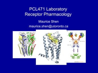
  1. I assume the majority of you guys are in the specialist program, so most of what I’m going to talk about today will probably just be a brief review of what you already know from PCL201 and PCL302. These are the crystal structures of 3 different types of receptors, which are GABAA receptor, dopamine D3 receptor, and insulin receptor. I put them there simply to make the slides look cool.
  2. So what is a receptor? The concept of a receptor exists all the way back in the 1800s, where this dude, Paul Ehrlich, proposed that a drug might act as a magic bullet that targets a specific receptor when he was trying to develop an anti-syphilis drug. I don’t know why he was studying syphilis but he did. Nowadays the existence of a receptor is well established, and as some of you probably know, this year’s Nobel price for chemistry was given to Robert Leftkowiz and Brian Kobilka who studied the G protein coupled receptors. A receptor is defined as a component of a cell or organism that interacts with a specific ligand and initiates a chain of biochemical events leading to the ligand’s observed effects. A ligand for a receptor can be endogenous or exogenous. For example, the endogenous ligand for the dopamine receptors would be dopamine, and an exogenous ligand would be L-DOPA. There are 3 main characteristics of ligand/receptor interactions. The binding of a ligand to its receptor is often saturable and reversible, and there exists a dose-response relationship where increasing concentration of a ligand will lead to enhanced biological effect until all the receptors are saturated.
  3. Since we are pharmacologists, we are more interested in exogenous ligands, or drugs, as opposed to endogenous ligands. We can classify drugs into different types based on their abilities to regulate known biological processes. Agonists are rugs that elicit biological effects that are comparable to endogenous ligands. For instance, L-DOPA would be an agonist for the dopamine receptor since it mimics dopamine’s effects on motor function. An agonist is considered as a full agonist when it induces maximal biological effects, whereas a partial agonist induces sub-maximal biological effects. In contrast, an inverse agonist will produce opposite biological effects as the full agonists. For instance, if an full agonist leads to increased calcium level in the brain, an inverse agonist will lead to reduced calcium level in the brain. Antagonists are drugs that bind to the receptor but do not elicit any biological effect. In other words, antagonists have zero efficacy. An antagonist for the dopamine receptor would be the anti-psychotic drug haloperidol. Different types of antagonists exist based on how they interact with the receptor. A reversible and competitive antagonist transiently binds to the same site as agonists. A non-competitive antagonist binds to a site on the receptor that is different from agonists, and they are also known as negative allosteric modulators. An irreversible antagonist covalently binds to the same site as agonists. Of all these types of drugs, partial agonists and antagonists have the best clinical utilities since it is easier to fine tune existing biological processes than to produce a biological effect from scratch, which might be harder to control.
  4. As you know, many different types of receptors exist in a living organism. There are ligand-gated ion channels, which are trans-membrane channels that allows for the influx of ions such as Na+, Ca2+, Cl-. These receptors are generally found in the brain and the nervous system, and are activated by different neurotransmitters such as glutamate, GABA, and acetylcholine. Receptor tyrosine kinases are trans-membrane polypeptides that autophosphorylate at tyrosine residues upon activation. They are generally activated by growth factors such as insulin, epidermal growth factor, and platelet-derived growth factor. Nuclear hormone receptors are cytosolic receptors that regulates gene transcription within a cell, and as their name suggest, they are mainly activated by hormones such as glucocorticoids, thyroid hormone, sex steroids etc.
  5. And finally, there are the G protein-coupled receptors. These are 7 trans-membrane proteins that are coupled to specific G proteins, and they elicit second messenger-mediated signaling cascade upon activation that may lead to gene transcription, protein synthesis, protein phosphorylations. GPCRs are the largest family of cell surface receptors and more than 50% of drugs on the market target GPCRs. They have wide range of ligands including hormones, neurotransmitters, light, and olfactory stimuli. The receptor of interest in this lab, the dopamine receptors, is a GPCR.
  6. The technique that we will be using to study the receptor-ligand interaction in this module is called radioligand binding assay. It involves the use of varying concentrations of a specific radiolabelled ligand to determine receptor affinity to the ligand (Kd) and total receptor density (Bmax) in a membrane preparation. The theory of radioligand binding experiments are based on the Laws of Mass Action, where.
  7. In an in vitro system, where you have your receptors transfected into a cell line, and you add your drug of interest, binding occurs when ligand and receptor collide due to random diffusion and when the collision has the correct orientation and sufficient energy, and the rate of association is defined by. Once the bound complex is formed, the ligand and receptor remain bound for a random amount of time, as determined by their relative affinity for one another. The rate of dissociation is defined by.
  8. After dissociation, we assume the ligand and the receptor remain unchanged. Equilibrium is reached when the rate of Ligand-Receptor complex formation equals to the rate of dissociation. Therefore, at equilibrium. The Dissociation Constant (Kd) is defined as the concentration of ligand that occupies half of the available receptors at equilibrium.
  9. There are 4 important assumptions made in the drug binding model I just described. First, we assume all receptors are equally accessible to the ligand. Second, we assume the receptor is either bound to a drug, or is in its free form. Partial binding is ignored. Third, we assume neither the receptor nor the ligand is altered by binding. Change in receptor conformation upon ligand binding is ignored. And lastly, we assume all bindings are reversible.
  10. Since the receptors are transfected into a cell line, the drug of interest may also bind to membrane proteins other than the receptor itself. Total binding is defined as the total radioligand bound to the membrane. This is determined by measuring the total radioactivity on the cell membrane. Non-specific binding is then determined by measuring the radioligand binding in the presence of excess cold drug with high affinity to the receptor of interest. The cold drug must be different from the radiolabeled drug of interest, and it must have high enough affinity in order to specifically occupy all the receptors, rendering the radiolabeled ligand to only be able to bind non-specifically to membrane proteins.
  11. This diagram depicts the difference between total binding and non-specific binding. The measurement of total binding is straightforward and includes specific and non-specific binding. To measure non-specific binding, all receptors are occupied by a different cold drug, and the radioactivity only comes from radiolabeled drug that is bound to other membrane proteins. An important assumption here is that the cold drug and the radiolabled drug of interest have completely different binding affinity to other membrane proteins.
  12. We are then able to calculate specific binding by subtracting non-specific binding from total binding. Specific binding curve is shown as a rectangular hyperbolic function that yields Kd and Bmax, as shown in this figure.
  13. Knowing how much drug is specifically bound and how much total drug was added, we are then able to plot a Scatchard plot by plotting bound drug over free drug on the y-axis, and the concentration of bound drug on the x-axis. The Scatchard plot requires very few data points, and Kd and Bmax can be easily deduced from the graph, which are the -1 over slope and the x-intercept, respectively. However, one major disadvantage of Scatchard plot is that the Kd and Bmax value may not be as accurate as the ones derived from the saturation binding curve.
  14. Lastly, it is also important to know how the radioactivity from the radiolabeled ligand is measured, since our whole module relies on this measurement. The radioactivity is measured using the scintillation counter. The β particles emitted from the radioactive ligand transfer energy to flours in the solvent, and excited flours dissipate energy by emitting light, which is then detected by photomultiplier tube in the scintillation counter.
  15. So for today, COS-7 cells were transfected with either D1 or D5 cDNA, and the membranes were harvested by differential centrifugation. This was already done for you. What you guys have to do today is to establish two-point saturation binding of D1 and D5 dopamine receptors using radiolabeled dopamine receptor antagonist, [3H] SCH-23390. The question to think about is whether you could identify Dx and Dy based on the Kd derived from the 2-point Scatchard plot? Why and why not? You guys will have to address this question in your lab report.