
Recommended
More Related Content
Similar to 2CVS ppt. FETAL CIRCULATION & CCF.ppt
Similar to 2CVS ppt. FETAL CIRCULATION & CCF.ppt (20)
Recently uploaded
Recently uploaded (20)
2CVS ppt. FETAL CIRCULATION & CCF.ppt
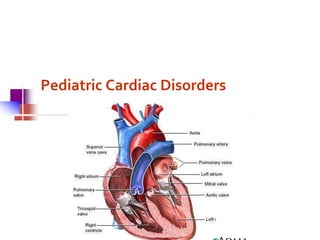
- 2. Fetal Circulation Main Blood Flow Placenta Umbilical Vein Liver Ductus Venosus Inferior Vena Cava Vena Cava Right Atrium Foramen Ovale Left Atrium Left Ventricle Aorta Body
- 3. Fetal Circulation Secondary Route: Right Atrium Right Ventricle Pulmonary Artery Ductus Arteriosus (so does not go to lungs) Aorta Body
- 4. Fetal Circulation Third route of blood flow Right Atrium Right Ventricle Pulmonary Artery Lungs (needs to perfuse the lungs and upper body with oxygen) Left Atrium Left Ventricle Aorta Body
- 5. Transition from Fetal Circulation to Pulmonary circulation The umbilical arteries and vein and the ductus venosus become non-functional Decreased pulmonary vascular resistance and increased pulmonary blood flow Increase in pressure of the left atrium, decrease pressure in right atrium, causing closure of foramen ovale. Pulmonary resistance is less than systematic resistance so there is left-to-right shunting resulting in closure of the ductus arteriosus.
- 7. Congestive heart failure The inability of the myocardium to circulate enough oxygenated blood to meet the demands of the body. When the heart fails, cardiac output is diminished. Heart rate, preload, contractitility, and afterload are affected. Peripheral tissue is not adequately perfused. Congestion in lungs and periphery develops.
- 8. Etiology and Pathophysiology Congenital defects – allow blood to flow from the left side of the heart to the right so that extra blood is pumped to the pulmonary system rather than through the aorta when the ventricle contracts. Obstructive congenital defects – restricts the flow of blood so the heart hypertrophies to work harder to force blood through the narrowed structures. The hypertrophied muscle becomes ineffective. Other defects which weaken the heart muscle.
- 9. Compensatory Mechanisms Stimulation of the sympathetic nervous system which releases norepinephrine from the adrenals. This stimulates blood vessels to constrict and an increase in the heart rate. Tachycardia increases venous return to the heart which stretches the myocardial fibers and increases preload. Only successful for short period of time. Increased renin and ADH secretion caused by decrease renal perfusion. Resultant increase in Na and H2O retention to increase fluid to the heart and leading to edema
- 10. Signs and Symptoms 1. Diaphoresis / sweating 2. Breathlessness –tachypnoea, coughing, crepitations 3. Tires easily 4. Poor feeding; poor weight gain, FTT 5. Hepatomegally 6. Cardiomegaly 7. Tachycardia; gallop rhythm 8. Only older children & adults develop signs of systemic congestion : oedema, orthopnoea, nocturnal dyspnoea, elevated JVP
- 11. Investigations Directed at finding the cause and quantifying function CXR- cardiomegaly, lungs are oligaemic/oedema Echocardiography- Congenital heart defects Arterial blood gas- reduced oxygen/ metabolic acidosis ECG, SERUM ELECTROLYTES
- 12. GENERAL MEASURES Bed reest- nurse in a semi – upright position Oxygen Diet- sufficient calorie intake Diuretics Digoxin Vasodilators e.g ACE inhibitors
- 13. Treatment of Congestive Heart Failure Medication Therapy Digitalis – increases contractility and decreases heart rate. ACE-inhibitors - arterial vasodilator / afterload reducing agent Diuretics - enhance renal secretion of sodium and water by reducing circulating blood volume and decreasing preload. Beta Blocker - increases contractility
- 14. Treatment of Congestive Heart Failure Diet – low sodium, small frequent feedings (be sure you can pick the right foods for a low NA diet. Nursing care: Measure intake and output – weighing diapers Observe for changes in peripheral edema and circulation If ascites present – take serial abdominal measurements to monitor changes. Skin care Turning schedule