2. Two distinct AOX activities were also purified from rat liver
and identified as NMN oxidases I and II (13, 14). The two
activities exhibited different Km values for the oxidation of
NMN to either its 2- or 4-pyridone. These two activities also
differed by pH optima, heat stability, and inhibitor sensitivity.
NMN oxidases I and II were found to possess distinct kinetic
parameters, Km and Vmax, for oxidation of several different
substrates, including benzaldehyde and NMN. Furthermore,
wide variation in Km has been observed between different
species and between individual rat strains (15–17). As was
found for mice, rat hepatic AOX could be purified to homoge-
neity to yield a native enzyme of 300 kDa that resolved into two
150-kDa subunits by SDS-PAGE (13, 14).
AOX genes are widely expressed phylogenetically and in
some organisms appear to arise from multigene families. Even
the Archae express MoCo enzymes related to AOX (18). Two
different AOX cDNA sequences were reported for corn plants
that were themselves 83% identical (19). Three cDNA se-
quences were reported for Arabidopsis thaliana (20), and to-
mato plants may also possess several AOX genes (21). Two
AOX genes have been identified in Drosophila melanogaster,
one encoding AOX and the other encoding the highly related
pyridoxal oxidase (PO) which was identified as an AOX (22–
24). Sequences for AOX or PO in Drosophila have not been
reported. Multiplicity in AOX genes was also reported for the
mouse where independently segregating loci appeared to en-
code AOX enzymes with distinct electrophoretic mobilities and
these were identified as AOX1 and AOX2 (9). Importantly, the
different isozymes appeared to segregate as different AOX ge-
netic loci under differential developmental and androgenic reg-
ulation. Human and bovine AOX sequences have been pub-
lished that are approximately 82% identical, and a small
fragment of a mouse AOX sequence was published (5, 25–27).
However, second copies of the vertebrate AOX genes have not
been cloned or sequenced. Therefore, while at least two or three
AOX genes appear to exist in plants and flys, they have not
been confirmed by sequence analysis in higher organisms. Fur-
thermore, Southern blot analysis of chromosomal DNA could be
interpreted to suggest that only a single AOX gene was present
in humans (28).
Molecular characterization of AOX from the rat has not been
reported. Because both forms of AOX appeared to be expressed
in rat liver, we have examined expression of AOX genes from
both male and female rat livers. We have confirmed the exist-
ence of two kinetically different forms of AOX in male and
female rats. Sequence analysis of the corresponding cDNAs
indicates that a single AOX gene is most likely activated in the
liver in male and female rats. By Northern blot analysis, male
and female rat liver RNA contained a single mRNA species that
did not exhibit induction by TP. Purified rat liver AOX from
males and females revealed a single 150-kDa band on SDS-
PAGE. Present experiments suggest that a primary difference
between male and female forms of AOX may lie in their respec-
tive redox states.
MATERIALS AND METHODS
RNA Purification and cDNA Synthesis—RNA was prepared from
organs of freshly killed Sprague-Dawley rats by quick freezing the
tissue in liquid nitrogen followed by extraction in guanidine isothionate
and phenol:chloroform:isoamyl alcohol (24:24:1) (29). Frozen tissues
were stored at Ϫ70 °C until use. Poly(A)ϩ
RNA was prepared by frac-
tionation on oligo(dT)-cellulose (Stratagene, La Jolla, CA). cDNA was
prepared by reverse transcription in a final volume of 20 l as follows.
1.0 g of poly(A)ϩ
RNA was mixed with diethyl pyrocarbonate-treated
water, 1.0 l of primer oligonucleotide at 20 M, 1.0 l of 10 mM
deoxyribonucleoside triphosphates (ACGT), 0.5 l of RNase inhibitor,
1.0 l of recombinant Moloney murine leukemia virus reverse tran-
scriptase (CLONTECH Laboratories, Palo Alto, CA), and 4.0 l of 5 ϫ
buffer (final conditions: 50 mM Tris-HCl, pH 8.3, 75 mM KCl, 3 mM
MgCl2). Reactions were incubated at 42 °C for 60 min and then heated
to 94 °C for 5 min to inactivate reverse transcriptase. Prior to use,
reactions were diluted to 100 l and 5 l was used for PCR amplifica-
tion. Reverse transcriptase reactions were stored at Ϫ70 °C.
3Ј and 5Ј RACE—A region from the middle of AOX1 was obtained by
PCR amplification of reverse transcribed male rat liver poly(A)ϩ
RNA
using synthetic oligonucleotides (Life Technologies, Inc., Gaitherburg,
MD) derived from a fragment of the mouse liver AOX1 sequence (27).
Nucleotides 1,682 through 2,217 were amplified with the oligonucleo-
tides MAO4RAT1 and MAO4RAT2 (Table I) to produce a single 535-
base pair fragment that was gel purified, sequenced in its entirety from
two directions, and cloned as pMID. The resulting sequence was used to
derive the unique sequence oligonucleotides for rat AOX1, FORINRAO,
and REVINRAO. 3Ј RACE was performed as follows. Male rat liver
poly(A)ϩ
RNA was reverse transcribed using the oligonucleotide 3Ј
RATRACE as a primer for reverse transcriptase. The resulting single
strand DNA was amplified by PCR using the oligonucleotides
MAO4RAT1 and 3Ј RATRACE. A second round of PCR amplification
was performed using the 5Ј nested oligonucleotide, REVINRAO, and 3Ј
RATRACE. A single product of 2,180 nt was obtained and sequenced
entirely from both directions. This fragment was cloned as p3Ј RA-
TRACE and showed 100% identity with the overlap region of pMID. 5Ј
RACE was performed as follows. Male rat liver poly(A)ϩ
RNA was
reverse transcribed using random hexamer oligonucleotides. The re-
sulting single strand DNA was amplified by PCR using the oligonucleo-
tides MAO4RAT2 and IVS22. A second round of PCR amplification was
performed using IVS22 and the nested oligonucleotide FORINRAO. A
single band of 1,930 nt was obtained, sequenced in its entirety from
both directions, and cloned as p5ЈRATRACE. This sequence revealed
100% identity in the region of overlap with pMID. The extreme 5Ј end
and upstream region of the male rat AOX1 cDNA was obtained by a
modified 5Ј RACE as follows. Male rat liver poly(A)ϩ
RNA was reverse
transcribed using random hexamer oligonucleotides. RNA was hydro-
lyzed in sodium hydroxide and the resulting single strand DNA was
subjected to two cycles of nested PCR, the first using an adapter ligated
oligonucleotide at the 5Ј end. Oligonucleotide 3ЈIVS11 was phosphoryl-
ated with polynucleotide kinase in the presence of ATP. Following
extraction with phenol:chloroform:isoamyl alcohol and ethanol precip-
itation the phosphorylated oligonucleotide was treated with dideoxya-
denosine triphosphate and terminal transferase from bacteriophage T4
to block elongation from the 3Ј end. Blocked, phosphorylated oligonu-
cleotide was then ligated to single strand DNA using bacteriophage T4
RNA ligase in the presence of hexamine cobalt chloride to produce
TABLE I
Oligonucleotides used for amplification of male and female rat AOX1
3Ј-RATRACE 5ЈCCCGGGGAATTCCTGCAGGTCGAC(T30)VN-3Ј
3Ј-RATUTR 5Ј-CCCGGGGAATTCCTGCAGGTCGACGCGTTCTGTAGTTGTTGAGCCAATCC-3Ј
MAO4RAT1 5Ј-CTGGAGTACATTAAAGTACCAGAATG-3
MAO4RAT2 5Ј-GTATTTCACCTTCAAGAATTTGATC-3Ј
FORINRAO 5Ј-GTTAGGATCAGAGGCTCCAAGTCTCGG-3Ј
REVINRAO 5Ј-CCGAGACTTGGAGCCTCTGATCCTAAC-3Ј
3Ј-IVS10 5Ј-GACTGGGCACAGACTGCTTTTGATG-3Ј
3Ј-IVS11 5Ј-CATCAAAAGCAGTCTGTGCCCAGTC-3Ј
IVS22 5Ј-CCGAGCTGCTCTTCTACGTGAACG-3Ј
RAT5 5Ј-CATCTCCTTCCTGAAATTCTGCCGATCC-3Ј
RAT6 5Ј-CCCGTGAGTCGGAGGTTCTTCCTCAGG-3Ј
3Ј-RREND 5Ј-CCCGGGGAATTCCTGCAGGTCGACTTT-3Ј
Rat Liver Aldehyde Oxidase 3879
byguestonFebruary1,2015http://www.jbc.org/Downloadedfrom
3. adapter-ligated single strand DNA. Adapter-ligated single strand-DNA
was subjected to first round amplification using the oligonucleotides 3Ј
IVS11 and RAT5. The resulting PCR products were subjected to a
second round of amplification using 3Ј IVS11 and the nested primer,
RAT6. The resulting 160-base pair DNA was cloned (p5ЈEND-male),
sequenced, and showed 100% identity with the overlap region of
p5ЈRATRACE. This sequence was inferred to contain the translation
initiation site and 49 nucleotides of 5Ј-untranslated region because it
showed excellent deduced amino acid sequence homology with human
and bovine AOX sequences, a single ATG was found to be in-frame with
the downstream sequence, and translational termination sequences
were observed upstream of this ATG and in the same reading frame.
DNA Sequence Analysis—Direct fluorescence sequence analysis was
performed on plasmid DNA or PCR products using oligonucleotide
primers designed to yield approximately 400 nt between primers and
approximately 100 nt of overlap. A list of sequencing oligonucleotides
and their sequences is available upon request. Prior to sequence deter-
mination, plasmids were prepared by alkaline lysis (29). PCR products
were purified from low melting point agarose by phenol extraction and
precipitation in ethanol. Sequences were determined using dideoxy-
nucleotide chain terminating system from Perkin-Elmer Applied Bio-
systems (Foster City, CA). Reactions used the ABI PRISMTM
Dye Ter-
minator cycle sequencing ready reaction kits (Perkin-Elmer). Sequence
reactions were fractionated on an ABI PRISM 310 DNA sequencer
equipped with a 43-cm microcapillary (Perkin-Elmer). All sequences
were determined from both directions and sequence data were compiled
manually.
Northern Blot Analysis and Quantitation—Northern blots were run
using formaldehyde-agarose gels and 5 g of poly(A)ϩ
RNA (6, 29). Hy-
bridization probes were isolated from the clones pMID, p3ЈRATRACE,
and p5ЈRATRACE by PCR amplification and agarose gel electrophore-
sis. Following isolation in phenol, DNA fragments were labeled by
random primed synthesis in the presence of [32
P]dATP for use as
hybridization probes. High stringency hybridization and washing were
conducted as described (6, 29). Following hybridization and autoradiog-
raphy, each 4,500-nt band was cut from the hybridization filter and
counted by liquid scintillation counting for quantitation. The remaining
filter was dissociated from residual 32
P and rehybridized with a -actin
specific probe. Actin hybridization was also quantitated by excising the
bands from the filter and liquid scintillation counting. Each AOX1
hybridization signal was normalized to the corresponding signal for
-actin after correction for background hybridization.
Castration and Hormone Supplementation—Castrated or sham cas-
trated male Sprague-Dawley rats were obtained from Charles Rivers
Laboratory (Wilmington, MA) following 1 week of recovery from sur-
gery. Surgery was performed when rats were 4 weeks of age. Rats were
maintained at Webb-Waring facilities for 1 additional week of equili-
bration. Testosterone proprionate was administered at a dose of 50
mg/kg body weight in corn oil by daily subcutaneous injection. Growth
hormone was administered at a dose rate of 0.05 IU/100 g of body
weight in a buffer composed of 30 mM NaHCO3, 150 mM NaCl, pH 8.25,
by subcutaneous injection twice daily. Sham castrated and castrated
controls received corn oil injection. Following 10 days of treatment, rats
were killed by sodium pentabarbitol administration. Organs were har-
vested immediately and dropped into liquid nitrogen for subsequent
RNA preparation. RNA was analyzed from individual organs with 4
rats in the sham controls, 4 rats in the castrated control group, 5 rats in
the castrated and testosterone supplemented group, and 5 rats in the
castrated and growth hormone supplemented group.
AOX Activity Assays—AOX activity and initial rate data were deter-
mined spectrophotometrically in a 1-ml reaction containing 50 mM
potassium phosphate buffer, pH 8.0, NMN at 5 mM or as needed, 10%
dimethyl sulfoxide, 250 international units of CAT, 5–50 M menadione
as required, and appropriate levels of purified or partially purified
enzyme. Initial rate data were obtained over a 5-min period. Formation
of the pyridone of NMN was monitored at 300 nm.
AOX Enzyme Purification and Characterization—AOX enzyme activ-
ity was purified from male and female Spraque-Dawley rat livers. After
removal of the liver, all procedures were performed at 4 °C or on ice
using ice-chilled buffers. Liver sections (10–20 g) were diced, rinsed
several times, homogenized, and dounced in 3 volumes of ice-cold 100
mM potassium phosphate, pH 7.5, containing 25 mM benzamidine hy-
drochloride, .2 mM phenylmethylsulfonyl fluoride, .1 mM EDTA. Homo-
genates were centrifuged for 1 h at 100,000 ϫ g. The supernatant was
brought to 5 mM DTT and incubated for 1 h. MnCl2 was then added to
the supernatant to a final concentration of 10 mM. The solution was
then centrifuged for 5 min at 17,000 ϫ g and the pellet was discarded.
Dry ammonium sulfate was added with stirring to achieve a final
concentration of 30%. The slurry was centrifuged and the pellet dis-
carded. The resulting supernatant was brought to 50% saturation with
ammonium sulfate, centrifuged, and the supernatant discarded. The
resulting pellet (50% pellet) was resuspended in 1/20 the original vol-
ume of potassium phosphate buffer. Acetone fractionation was subse-
quently achieved using acetone chilled with dry ice. Suspensions were
brought to 40% in chilled acetone, centrifuged, and the pellet discarded.
Supernatants were brought to 50% in acetone, centrifuged, and the
pellets collected. The 50% pellet was resuspended in the original vol-
ume of buffer and dry ammonium sulfate was added to achieve a 60%
saturation. The pellet was recovered by centrifugation and resuspended
in 1/20 of the original volume in 100 mM glycine, pH 9, containing 100
mM NaCl. After resuspension, insoluble debris was removed by centrif-
ugation and the solution was desalted on a 5 ϫ 15-cm Sephadex G-25
column in the above glycine buffer. The desalted solution was loaded
onto a 2.5 ϫ 10-cm benzamidine-Sepharose 6B (Pharmacia) column
equilibrated in the same buffer. The column was washed with 3 column
volumes of buffer and elution was achieved by flushing the column with
500 mM benzamidine hydrochloride. Elution was monitored at 436 nM
and the single eluting peak was collected and precipitated with ammo-
nium sulfate at 60% saturation. The pellet was stored at 4 °C for up to
1 day or was resuspended in a minimum volume of 100 mM potassium
phosphate, pH 7.5, and dialyzed against the same buffer overnight.
The OD 280/450 ratio was between 5 and 7 for male or female
preparations. Specific activity for NMN hydroxylation to the pyridone
was 100–250 nmol/min/mg for the female or male enzymes. Both en-
zyme preparations were inhibited to greater than 95% by inclusion of 50
M menadione. We observed persistent aggregation of the soluble pro-
tein fraction when preparations did not include treatment with DTT
early in the fractionation. Aggregation reduced the overall yield of AOX
enzyme to less than 0.1% of the starting activity. Reduction of the crude
lysate prior to MnCl2 treatment improved the overall yield to 7% of the
starting activity. SDS-PAGE analysis of the aggregated proteins sug-
gested no obvious bias for specific aggregated proteins. Furthermore,
reduction in 5 mM DTT was significantly more effective in preventing
aggregation than was 10 mM cysteine. Reduction of male or female liver
extracts permitted purification of both enzymes to homogeneity.
Preparation of Antibody to AOX—The amino-terminal decapeptide
comprising the sequence NH2-DRASELLFYV-COOH and the carboxyl-
terminal decapeptide comprising the sequence NH2-GSYVPWNIPV-
COOH were synthesized by Dr. Hans-Richard Rackwitz (German Can-
cer Research Center, Heidelberg, Germany). Antibody to the synthetic
oligo peptides was produced in rabbits by intravenous injection of 20 g
of peptide. Rabbits were boosted with peptide every 2 weeks. The IgG
fraction was prepared from serum by ammonium sulfate precipitation
following coagulation of the blood and sedimentation. This produced
two antisera preparations: AOX-NT (amino-terminal antibody) and
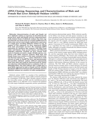
FIG. 1. PCR amplification, cloning, and sequence strategy for
male and female rat AOX1. The upper bar shows the deduced cDNA
structure for both male and female rat liver AOX1. PCR amplified and
cloned fragments are shown below. Note that the 3Ј RACE product for
the female has been truncated within the untranslated region and does
not comprise the entire untranslated region. Each cloned PCR fragment
was subjected to DNA sequence analysis using a battery of oligonucleo-
tides designed to encompass approximately 400 nucleotides between
oligonucleotide primers. Each fragment was sequenced entirely from
both directions. A list of sequence analysis primers and their sequences
is available upon request. The assembled cDNA sequences for both
male and female rat liver AOX have been deposited in the NCBI gene
bank data base.
Rat Liver Aldehyde Oxidase3880
byguestonFebruary1,2015http://www.jbc.org/Downloadedfrom
4. AOX-CT (carboxyl-terminal antibody).
Western Immunoblot Analysis—Protein was electrophoresed on SDS-
PAGE and transferred to polyvinylidine difluoride membranes (Bio-
Rad). Filters were sliced for staining with Commassie Brilliant Blue or
processed for immunoblot analysis. For reaction with antisera, filter
strips were blocked with gelatin overnight prior to reaction with pre-
immune sera, AOX-NT, or AOX-CT antisera. Antigen-antibody com-
plexes were detected by reaction with alkaline phosphatase streptavi-
din kit (Bio-Rad).
RESULTS
Different Forms of AOX Exist in Male and Female Rat Liv-
ers—AOX enzyme activity was measured in crude extracts of
FIG. 2. Alignment of deduced amino acid sequences. Amino acid
sequences for the four vertebrate AOXs have been aligned by multiple
FIG. 2—continued
Clustal analysis. Identical amino acids are boxed in black and biochem-
ically conserved differences are shown in gray. Regions thought to
mediate cofactor binding are indicated by the overline. Five sites within
the large MoCo-binding domain have been identified and are shown
individually. The 5 amino acid differences between male and female rat
liver AOXs have been indicated with an asterisk. The programs Clust-
alW, Boxshade, and Paint were used to create the figure.
Rat Liver Aldehyde Oxidase 3881
byguestonFebruary1,2015http://www.jbc.org/Downloadedfrom
5. male and female rat livers. Apparent Km (Km(app)) values were
determined from Lineweaver-Burk plots by measuring conver-
sion of NMN to its pyridone. Km(app) for male rat liver AOX was
538.8 M and Km(app) for the female was 1062.3 M, consistent
with previous reports showing two forms of AOX in livers from
rats and different forms of AOX in livers from male and female
mice. While no explanation for this difference has been pro-
duced, the two AOX genes identified in mice, AOX1 and AOX2,
suggested the possibility that two different AOX genes may be
expressed in rat liver.
cDNA Sequence Analysis of Male and Female Rat Liver
AOX1—Fig. 1 illustrates the PCR amplification strategy used
to obtain segments of male rat liver AOX1. DNA sequence of
the three PCR products was assembled to produce the male
rAOX1 cDNA. RAOX1 comprised 4,304 nucleotides, including
30 nt of polyadenylation, 210 nt of 3Ј-untranslated region, and
49 nt of 5Ј-untranslated region. A single open reading frame
was identified that encoded a protein of 1,333 amino acids and
a deduced mass of 147,009 Da. The deduced male rAOX1 pro-
tein exhibited 82% sequence identity with human AOX1 and
81% sequence identity with bovine AOX1. Multiple sequence
clustal analysis revealed conservation of co-factor domains cor-
responding to FeS I, FeS II, FAD, and five small domains
within the MoCo binding segment (Fig. 2).
Female rat liver AOX1 cDNA sequence was obtained using a
similar strategy with the exception that a unique sequence
oligonucleotide derived from the male sequence, 3Ј RATUTR,
was used to obtain the 3Ј RACE product. Thus, 47 nucleotides
of 3Ј-untranslated region was obtained for the female and this
does not include the polyadenylation site (Fig. 1). The assem-
bled cDNA sequence for female rat liver AOX1 encoded a de-
duced protein of 1,333 amino acids and 146,919 Da. The female
cDNA sequence was 99.8% identical to the male sequence and
the deduced protein sequence was 99.6% identical to the male
sequence. Of the 10 nucleotide differences detected between the
male and female rat liver AOX1 cDNA sequences described
here, five resulted in changes to the deduced amino acid se-
quences (Table II). However, while nucleotides 405 and 408
differ between male and female sequences reported here and to
the GenBank data base, these variations were also found be-
tween individual male clones and therefore do not represent
gender differences but differences between individual rats. The
full extent of individual variation was not determined and it
remains possible that all of the differences observed between
the two clones described may be attributed this cause alone.
Expression of Rat AOX1 mRNA—Fig. 3A shows Northern
blot analysis of poly(A)ϩ
RNA from male rat liver. Hybridiza-
tion probes were derived from each of the three male clones,
pMID, p5ЈRATRACE, and p3ЈRATRACE. The region between
nucleotides 1,682 and 2,217 of the male cDNA, corresponding
to the pMID hybridization probe, produced hybridization sig-
nals at approximately 4,500 and 2,500 nt. Hybridization probes
derived from both the p5ЈRATRACE and p3ЈRATRACE clones
produced predominantly a single band at 4,500 nt with weak
hybridization to the band at 2,500 nt. We conclude that the
predominant mRNA for rAOX1 detected by Northern blot anal-
ysis in males is approximately 4,500 nt, consistent with the
cDNA sequence assembled for rAOX1. The unexpected signal
at 2,500 nt may represent a cross-reactive species largely lo-
calized to the pMID region.
Hybridization probes derived from the p5ЈRATRACE pro-
duced predominantly a single band from both male and female
RNA (Fig. 3B). This RNA was also estimated to be 4,500 nu-
cleotides in size, and no difference in size or number of hybrid-
izing bands was detected between males and females.
Northern blot analysis of poly(A)ϩ
RNA from several differ-
ent tissues showed expression of a single 4,500-nt RNA for all
tissues examined (Fig. 3C). Different tissues did not show
variation in either the size or multiplicity of AOX RNAs. Var-
FIG. 3. Northern blot analysis of rat AOX1 expression. A, each of the three clones used to assemble male rat liver AOX1 was used as a
hybridization probe for independent Northern blots of male rat liver poly(A)ϩ
RNA. The major band at 4,500 nt corresponds to the AOX1 mRNA.
The band at 2,500 nt that has greater localization to the pMID region of AOX is assumed to represent a cross-reactive species. Although it has not
been excluded that this may represent a breakdown product of the larger RNA, it is too small to encode a full-length AOX. B, poly(A)ϩ
RNA from
male and female rat liver has been analyzed by Northern blot using the p5ЈRATRACE clone as a hybridization probe. No attempt is made here to
indicate a difference in abundance of the AOX mRNA between males and females. C, poly(A)ϩ
RNA from several different male rat tissues has been
analyzed by Northern blot using the AOX1 insert from p5ЈRATRACE as a hybridization probe.
TABLE II
Sequence differences in male and female rat AOX1
clones described here
The differences in cDNA sequence between the male and female rat
AOX1 clones are shown. nt-site refers to the specific base changed from
male to female using the A of the translational initiator as nucleotide
ϩ1. The nature of the base pair changed is shown along with the
change, if any, in the deduced amino acid sequence and the correspond-
ing amino acid number. Note, these changes reflect differences between
our clones and do not necessarily reflect consistent differences between
genders.
No. nt-Site Base pair change Amino acid change Amino acid site
1 133 G:C–A:T P–P
2 405 C:G–G:C A–G 119
3 408 G:C–T:A R–M 120
4 1,679 T:A–C:G L–L
5 1,994 A:T–G:C T–A 649
6 2,563 G:C–A:T L–L
7 2,872 T:A–C:G S–S
8 3,739 G:C–A:T Q–Q
9 3,875 C:G–T:A L–F 1,276
10 3,993 G:C–C:G R–T 1,315
Rat Liver Aldehyde Oxidase3882
byguestonFebruary1,2015http://www.jbc.org/Downloadedfrom
6. iation in the -actin control precludes drawing firm conclusions
at this point concerning relative levels of expression between
tissues.
RNA from male rats that had been sham castrated, cas-
trated, castrated and treated with TP, or castrated and treated
with growth hormone was analyzed by Northern blot. Fig. 4
shows that steady state RNA levels were only slightly affected
by any of these treatments. When hybridization signals were
quantitated and normalized to either OD 280 or to -actin
hybridization signal (Table III), we found no statistically sig-
nificant difference between groups in AOX1 mRNA abundance.
These data do not support significant regulation of rat AOX1
mRNA abundance by TP.
AOX Enzyme Purification and Characterization—AOX en-
zyme activity was purified to homogeneity from male rat livers
(Fig. 5A). These preparations produced a single band of 150
kDa by SDS-PAGE analysis. We were unable to sequence this
protein by direct Edman degradation suggesting that its amino
terminus was blocked, as was found for both the rabbit liver
and bovine liver AOXs (5, 26). Western immunoblot analysis of
partially purified AOX from male and female rat livers re-
vealed excellent reactivity of each enzyme preparation to this
synthetic peptide-derived antibody (Fig. 5B). Furthermore, re-
active proteins from male and female revealed a predominant
polypeptide of approximately 150 kDa with no evident differ-
ence in size between genders. (Fig. 5C).
Km(app) values for oxidation of NMN to its pyridone were
determined by analysis of Lineweaver-Burk plots. Fig. 6 and
Table IV show these results. As noted above, crude liver ex-
tracts from males produced Km(app) of 538.8 and 1062.3 M for
females. Reduction of the crude liver extract with 5 mM DTT
shifted Km(app) for males and females. Reduced male rat liver
produced Km(app) of 359.9 M and for the reduced female en-
zyme Km(app) was 354.5 M (Fig. 6B). Thus, male and female
forms of AOX can be converted to a form with indistinguishable
Km(app) values by chemical reduction in a crude lysate. Oxida-
tion of the reduced female preparation with 4,4Ј-DTDP con-
verted Km(app) back to a form similar to that obtained from the
untreated female preparations (Fig. 6B). Furthermore, AOX
purified from female rat liver through the post-benzamidine
stage could be reduced with DTT to yield an enzyme with
Km(app) of 261 M. Reoxidation of the reduced enzyme with
4,4Ј-DTDP converted the Km(app) to 1673 M (Fig. 6C). Thus,
the capacity to modulate Km(app) of rat liver AOX was main-
tained through purification of the enzyme and may therefore
reflect an intrinsic property of the enzyme.
DISCUSSION
In the present work have assembled full-length sequences for
AOX cDNAs from male and female rat liver. We examined
expression of AOX by Northern blot analysis from males and
females, from several tissues, and have examined the effect of
androgen regulation. We purified AOX from rat livers of males
and females and obtained a single 150-kDa band by SDS-PAGE
analysis consistent with the deduced amino acid sequences
from males or females of 1,333 amino acids and 147 kDa.
Antisera raised against synthetic decapeptides reacted with
AOX from males or females. Furthermore, Km(app) values for
crude extracts of male or female rat liver and post-benzamidine
purified AOX differed substantially but could be interconverted
by chemical reduction with DTT or oxidation with 4,4Ј-DTDP.
The deduced amino acid sequences for male and female rat
liver AOX are 81 and 82% identical to human and bovine AOXs
and are themselves 99.6% identical. They show excellent con-
servation in the domains attributed to iron, FAD, and MoCo
binding. By this criterion, the rat AOXs clearly belong to the
molybdenum iron-sulfur flavoproteins that include AOX, XDH,
and XO. Furthermore, the rat AOXs conform to the domain
models proposed for XDHs (7, 30, 31). Amino acids critical for
catalysis of this class of enzymes are conserved in the rat AOX
sequences as they are in most other AOXs and XDHs. In
particular, Glu-869 of the Mop enzyme is critical for catalysis
and is conserved in nearly all XDHs and AOXs (see Ref. 21 for
alignment of several sequences), including the two rat se-
quences reported here where it is found at amino acid 1265 in
MoCo domain 5 (Fig. 2).
We observed that the male and female cDNA sequences were
not identical. While several arguments could be advanced to
explain the differences, we suggest that the least tenable ar-
FIG. 4. Testosterone does not regulate steady state levels of rat
hepatic AOX. Poly(A)ϩ
RNA from individual male rat livers has been
analyzed by Northern blot. Individuals from the following four groups
have been used: castrated, sham castrated, castrated and testosterone
supplemented, castrated and growth hormone supplemented. -Actin
was used as a control to reveal uniform loading of RNA. Blots were first
probed with the AOX probe, the corresponding region cut from the filter
for counting, and the remaining filter was dissociated of all 32
P and
rehybridized with the -actin probe.
TABLE III
Quantitation of RNA hybridization
Hybridizing bands from the Northern blot shown in Fig. 4 were cut
from the filters and counted by liquid scintillation counting. Counts
from randomly selected regions were averaged and subtracted from
each signal to account for background radioactivity. Counts for each
AOX signal were then normalized to the -actin signal derived from the
same lane, and the normalized, background subtracted counts were
multiplied by 100. The number of animals in each group is shown by the
n. The mean and standard error of the mean (SE) were calculated for
each group. No statistical significance could be established between
group means.
Group n Mean S.E.
Sham castrated 4 69.0 10.3
Castrated control 4 86.6 11.4
Castrated testosterone 5 82.9 18.3
Castrated growth hormone 5 64.9 13.5
Rat Liver Aldehyde Oxidase 3883
byguestonFebruary1,2015http://www.jbc.org/Downloadedfrom
7. gument is for the expression of two different AOX genes in the
livers of male and female rats. Nucleotide differences between
males and females could arise during PCR amplification, clon-
ing, or sequence analysis since this is also PCR based. The
small number of differences observed between male and female
sequences could also arise from allelic differences between
males and females or simply from individual differences be-
tween rats. Sequence analysis of only one additional male
cDNA uncovered two of the changes (nt 405 and 408) found
between the male and female clones. The minimal number of
differences found between males and females coupled with the
individual variation already observed suggests that the differ-
ences are unlikely to represent expression of alternative genes.
Furthermore, sequence polymorphism is a well described phe-
nomenon in Drosophila XDH (32–34).
Multiplicity of AOX genes has been established for some
organisms (19–24), and in plants these data are supported by
multiple cDNA sequences of approximately 80% identity (19,
20). Thus, the observation that mice appear to express two
AOX genes in the liver was not surprising (9). However, genetic
data derived in the mouse have not been supported yet by
corresponding molecular data and few efforts have been made
to establish AOX gene copy number in vertebrates. Southern
blot analysis of chromosomal DNA failed to reveal second AOX
genes in several vertebrates under conditions in which at least
80% identity would have produced hybridization (28).
Northern blot analysis of AOX mRNA from male or female
rats, and from several different tissues, demonstrated expres-
sion of a single 4,500 nt RNA. We did not observe variation in
size or multiplicity of AOX mRNAs in the liver from males or
females where kinetically distinct forms of AOX were identi-
fied. Furthermore, castration and/or testosterone supplemen-
tation resulted in no significant alteration in AOX mRNA
abundance and we infer that testosterone does not appear to
exert significant regulation of AOX mRNA abundance or form
in the rat liver. We did observe surprisingly strong hybridiza-
tion to an RNA of 2,500 nt that could be localized to the region
from ϩ1,682 to ϩ2,217. While the identity of this RNA is
unknown, it is too small to encode an AOX of 150 kDa. Thus,
RNA analysis supports expression of a single AOX gene in the
liver where post-translational events may be important for
determining the differences between male and female kinetic
variants.
Interestingly, AOX-3 from A. thaliana encoded a protein of
only 568 amino acids truncated at the amino terminus (20). It
must differ from a true AOX because it cannot encode a protein
capable of binding the full set of co-factors. AOX-3 from A.
thaliana showed striking homology to AOX-1 and AOX-2 in the
MoCo-binding region, confirming that it is indeed a member of
the MH family. Confirmation that vertebrates encode such a
protein would be of great interest since it may represent the
RNA detected at 2,500 nt.
Since our observations suggested that rats express a single
AOX gene in the liver, we examined the possibility that redox
status might underlie the differences in male and female vari-
ants. We found that partially purified crude extracts of male
and female rat liver did indeed reveal different Km(app) variants
of AOX. Reduction of crude extracts from males or females with
DTT resulted in conversion to a single form. Subsequent reoxi-
dation of the reduced AOX with 4,4Ј-DTDP resulted in conver-
sion to a more female like Km(app). Reduction or oxidation of
post-benzamidine purified AOX also resulted in interconver-
sion between the two extremes of Km(app) suggesting that the
variants differed by their intrinsic oxidation state. Since DTT
directly reduces protein disulfides to thiols while 4,4Ј-DTDP
forms disulfides from thiols (35–38), we infer that manipula-
tion of thiol oxidation state can interconvert kinetic variants of
AOX.
Redox effects on XDH are well known. Conversion between
the NADϩ
dependent, D-form, and the oxygen dependent, O-
form, is a redox-dependent process reversible by chemical re-
duction (7, 39, 40). Furthermore, redox effects on XDH have
dramatic effects on the kinetic parameters of the enzyme (41).
AOX does not have an NADϩ
-dependent form of the enzyme,
and therefore conversion between D-form and O-form is irrel-
evant. While cysteine residues critical for D-form to O-form
conversion in XDH were not conserved in AOX (42), most of the
41 cysteine residues found in rat liver AOX are conserved with
vertebrate XDHs. These cysteine residues would be expected to
take part in the same biochemical reactivities as those found in
XDH. Since we observed that redox effects on Km(app) were
preserved from crude extracts through post-benzamidine-puri-
FIG. 5. Analysis of rat liver AOX1 protein. A, male rat liver AOX was purified to homogeneity from initially reduced extracts as described
under “Materials and Methods.” Purified AOX was analyzed by SDS-PAGE and stained with Ponceau S prior to subjecting the enzyme to
amino-terminal sequence analysis. B, antibody was raised against synthetic decapeptides from the amino (AOX-NT) and carboxy (AOX-CT)
termini. AOX was partially purified to retain numerous unrelated proteins, analyzed by SDS-PAGE (Crude AOX), and reacted to each antibody
as described under “Materials and Methods.” Preimmune, AOX-NT, and AOX-CT antisera were used at a 1:500 dilution. C, male and female rat
liver AOX was purified to homogeneity from initially reduced preparations. Enzymes were analyzed by SDS-PAGE and subjected to Western
immunoblot analysis using the AOX-CT antisera. Both male and female preparations migrated with apparent size of 150 kDa and both
preparations reacted to the AOX-CT antisera.
Rat Liver Aldehyde Oxidase3884
byguestonFebruary1,2015http://www.jbc.org/Downloadedfrom
8. fied enzyme and that these effects alone were sufficient to
explain the differences between male and female variants, we
infer that the different Km(app) variants result not from expres-
sion of alternative genes but from redox effects that may act
either on AOX itself or on a closely associated protein capable
of modulating Km(app) for AOX.
Our observations can be explained by the activity of the
hepatic microsomal monooxygenase. The cystamylating flavin
monooxygenase catalyzes oxidation of cysteamine, a thiol, to
cystamine, a disulfide, and this reaction provides a significant
source of disulfide responsible for maintaining the intracellular
thiol:disulfide potential (43, 44). The thiol:disulfide potential is
thought to reflect two ratios: the GSH:GSSG ratio and the
cysteamine:cystamine ratio. Protein oxidation state depends on
overall thiol:disulfide potential. Higher levels of monooxygen-
ase lead to a more oxidizing environment, while reduced mo-
nooxygenase levels result in a more reducing environment.
Significantly, monooxygenase from mice or rats is regulated by
testosterone in precisely the fashion that kinetic variants of
AOX are regulated (43). Male rat hepatic monooxygenase levels
are lower than female levels. Castration of male rats elevates
monooxygenase and leads to elevated cystamine and a more
oxidizing cytosol. Testosterone reverses this effect and restores
the reducing environment. Furthermore, testosterone treat-
ment of females lowers monooxygenase and elevates cysteam-
ine levels. These observations may explain both the effect of
testosterone and the ability to purify kinetically distinct forms
of AOX from male and female rats since neither thioltrans-
ferase nor glutathione reductase, the two other enzymes estab-
lishing thiol:disulfide potential, are regulated by testosterone
(43). We posit that AOX is sensitive to the thiol:disulfide po-
tential and that kinetic variants of AOX reflect the intracellu-
lar thiol:disulfide potential through thiol modification of AOX.
Activity of the monooxygenase could thereby regulate AOX
Km(app) by “setting” the thiol:disulfide potential leading to a
more oxidized form of AOX in the female or a more reduced
form of AOX in the male. This may have direct consequences
for ROS generation from AOX since the kinetically less efficient
enzyme may bias ROS generation for superoxide anion. This
argument does not require expression of a second AOX gene
and can account for the failure of testosterone to regulate AOX
gene expression in rats despite finding kinetically distinct
forms in males and females. AOX thiol modification could also
explain both novel electrophoretic variants (9) and the widely
variant kinetic characteristics of AOX from different organs
and different species (16, 45–47).
Acknowledgments—We thank Drs. Enrico Garrattini and Mineko
Terao (Milan, Italy) for discussion of these and other data.
REFERENCES
1. Hille, R., and Massey, V. (1985) in Molybdenum Enzymes (Spiro, T. G., ed) pp.
443–518, Wiley-Interscience Publishing Co., New York
2. Wooton, J. C., Nicolson, R. E., Cock, J. M., Walters, D. E., Burke, J. F., Doyle,
W. A., and Bray, R. C. (1991) Biochim. Biophys. Acta 1057, 157–185
3. Kisker, C., Schindelin, H., and Rees, D. C. (1997) Annu. Rev. Biochem. 66,
233–267
4. Berger, R., Mezey, E., Clancy K. P., Harta, G., Wright, R. M., Repine, J. E.,
Brown, M., Brownstein, M., and Patterson, D. (1995) Somatic Cell Mol. Gen.
21, 121–131
5. Wright, R. M., Vaitaitis, G. M., Weigel, L. K., Repine, T. B., McManaman, J. L.,
FIG. 6. Chemical reduction or oxidation abolishes differences
in Km(app) between male and female rat liver AOX. AOX activity
was purified from unreduced male and female rat livers through
(NH4)2SO4 fractionation and either treated with 5 mM DTT or not to
produce the crude native or crude reduced preparations. A separate
preparation was reduced initially and purified through benzamidine
fractionation to produce the post-benzamidine preparation. A, Lin-
eweaver-Burk plots were determined for male (squares) and female
(diamonds) preparations using NMN as the oxidizing substrate. B,
Lineweaver-Burk plots were determined for initially reduced male and
female crude extracts. In addition, the reduced female extract was then
treated with 1 mM 4,4Ј-DTDP to reoxidize it (open squares) and sub-
jected to Lineweaver-Burk analysis. C, female rat liver AOX was puri-
fied through benzamidine fractionation from an initially reduced ex-
tract. Enzyme was treated with 5 mM DTT or 1 mM 4,4Ј-DTDP to reduce
or oxidize the enzyme. Lineweaver-Burk analysis was then conducted
on the reduced (diamonds) or oxidized (squares) preparations.
TABLE IV
Effect of reduction and oxidation on Km(app) for crude and purified rat
liver AOX
Lineweaver-Burk plots shown in Fig. 6 were fitted by least squares
analysis. Km(app) were determined for each curve and the goodness of fit
r2
values were determined for each.
Km(app) r2
Crude native Male 371 0.65
Female 1385 0.94
Crude reduced Male-red 360 0.98
Female-red 354 0.94
Female-reox 1895 0.96
Post-benzamidine Reduced 261 0.83
Oxidized 1673 0.97
Rat Liver Aldehyde Oxidase 3885
byguestonFebruary1,2015http://www.jbc.org/Downloadedfrom
9. and Repine, J. E. (1995) Redox Report 1, 313–321
6. Wright, R. M., Weigel, L. K., Varella-Garcia, M., Vaitaitis, G. M., and Repine,
J. E. (1997) Redox Report 3, 135–144
7. Nishino, T. (1994) J. Biochem. (Tokyo) 116, 1–6
8. Yoshihara, S., and Tatsumi, K. (1997) Arch. Biochem. Biophys. 338, 29–34
9. Holmes, R. S. (1979) Biochem. Genet. 17, 517–527
10. Huff, S. D., and Chaykin, S. (1967) J. Biol. Chem. 242, 1265–1270
11. Gluecksohn-Waelsch, S., Greengard, P., Quinn, G. P., and Teicher, L. S. (1967)
J. Biol. Chem. 242, 1271–1273
12. Yoshihara, S., and Tatsumi, K. (1997) Biochem. Pharmacol. 53, 1099–1105
13. Ohkubo, M., and Fujimura, S. (1982) Biochem. Int. 4, 353–358
14. Ohkubo, M., Sakiyama, S., and Fujimura, S. (1983) Arch. Biochem. Biophys.
221, 534–542
15. Felsted, R. L., and Chaykin, S. (1967) J. Biol. Chem. 242, 1274–1279
16. Beedham, C., Bruce, S. E., Critchley, D. J., Al-Tayib, Y., and Rance, D. J.
(1987) Eur. J. Drug Met. Pharmokin. 12, 307–310
17. Sugihara, K., Kitamura, S., and Tatsumi, K. (1995) Biochem. Mol. Biol.
Internat. 37, 861–869
18. Bult, C. J., White, O., Olsen, G. J., Zhou, L., Fleischmann, R. D., Sutton, G. G.,
Blake, J. A., FitzGerald, L. M., Clayton, R. A., Gocayne, J. D., Kerlavage,
A. R., Dougherty, B. A., Tomb, J. F., Adams, M. D., Reich, C. I., Overbeek,
R., Kirkness, E. F., Weinstock, K. G., Merrick, J. M., Glodek, A., Scott, J. L.,
Geoghagen, N. S. M., Weidman, J. F., Fuhrmann, J. L., Nguyen, D.,
Utterback, T. R., Kelley, J. M., Peterson, J. D., Sadow, P. W., Hanna, M. C.,
Cotton, M. D., Roberts, K. M., Hurst, M. A., Kaine, B. P., Borodovsky, M.,
Klenk, H. P., Fraser, C. M., Smith, H. O., Woese, C. R., and Venter, J. C.
(1996) Science 273, 1058–1073
19. Sekimoto, H., Seo, M., Dohmae, N., Takio, K., Kamiya, Y., and Koshiba, T.
(1997) J. Biol. Chem. 272, 15280–15285
20. Hoff, T., Frandsen, G. I., Rocher, A., and Mundy, J. (1998) Biochim. Biophys.
Acta 1398, 397–402
21. Ori, N., Eshed, Y., Pinto, P., Paran, I., Zamir, D., and Fluhr, R. (1997) J. Biol.
Chem. 272, 1019–1025
22. Dickinson, W. J. (1970) Genetics 66, 487–496
23. Warner, C. K., Watts, D. T., and Finnerty, V. (1980) Mol. Gen. Genet. 180,
449–453
24. Warner, C. K., and Finnerty, V. (1981) Mol. Gen. Genet. 184, 92–96
25. Wright, R. M., Vaitaitis, G. M., Wilson, C. M., Repine, T. B., Terada, L. S., and
Repine, J. E. (1993) Proc. Natl. Acad. Sci. U. S. A. 90, 10690–10694
26. Li-Calzi, M., Raviolo, C., Ghibaudi, E., De-Gioia, L., Salmona, M., Cazzaniga,
G., Kurosaki, M., Terao, M., and Garattini, E. (1995) J. Biol. Chem. 270,
31037–31045
27. Bendotti, C., Prosperini, E., Kurosaki, M., Garattini, E., and Terao, M. (1997)
Neuro. Rep. 8, 2343–2349
28. Terao, M., Kurosaki, M., Demontis, S., Zanotta, S., and Garattini, E. (1998)
Biochem. J. 332, 383–393
29. Ausubel, F. M., Brent, R., Kingston, R. E., Moore, D. D., Seidman, J. G., Smith,
J. A., and Struhl, K. (1994) Current Protocols Molecular Biology, Green
Publishing and J. Wiley & Sons, New York
30. Sato, A., Nishino, T., Noda, K., Amaya, Y., and Nishino, T. (1995) J. Biol.
Chem. 270, 2818–2826
31. Glatigny, A., and Scazzocchio, C. (1995) J. Biol. Chem. 270, 3534–3550
32. Keith, T. P., Brooks, L. D., Lewontin, R. C., Martinez-Cruzado, J. C., and
Rigby, D. L. (1985) Mol. Biol. Evol. 2, 206–216
33. Riley, M. A., Kaplan, S. R., and Veuille, M. (1992) Mol. Biol. Evol. 9, 56–69
34. Comeron, J. M., and Aguade, M. (1996) Genetics 144, 1053–1062
35. Grassetti, D. R., and Murray, J. F., Jr. (1967) Arch. Biochem. Biophys. 119,
41–49
36. Eager, K. R., and Dulhunty, A. F. (1998) J. Membr. Biol. 163, 9–18
37. Cai, S., and Sauve, R. (1997) J. Membr. Biol. 158, 147–158
38. Zheng, S. Y., Xu, D., Wang, H. R., Li, J., and Zhou, H. M. (1997) Int. J. Biol.
Macromol. 20, 307–313
39. Waud, W. R., and Rajagopalan, K. V. (1976) Arch. Biochem. Biophys. 172,
354–364
40. Waud, W. R., and Rajagopalan, K. V. (1976) Arch. Biochem. Biophys. 172,
365–379
41. Saito, T., and Nishino, T. (1989) J. Biol. Chem. 264, 10015–10022
42. Nishino, T., and Nishino, T. (1997) J. Biol. Chem. 272, 29859–29864
43. Ziegler, D. M., Duffel, M. W., and Poulsen, L. L. (1980) Ciba Found. Symp. 72,
191–204
44. Tynes, R. E., and Hodgson, E. (1985) Arch. Biochem. Biophys. 240, 77–93
45. Krenitsky, T. A., Neil, S. M., Elion, G. B., and Hitchings, G. H. (1972) Arch.
Biochem. Biophys. 150, 585–599
46. Taylor, S. M., Stubley-Beedham, C., and Stell, J. G. P. (1984) Biochem. J. 220,
67–74
47. Beedham, C., Critchley, D. J., and Rance, D. J. (1995) Arch. Biochem. Biophys.
319, 481–490
Rat Liver Aldehyde Oxidase3886
byguestonFebruary1,2015http://www.jbc.org/Downloadedfrom
10. Repine
G. Riley, James L. McManaman and John E.
Richard M. Wright, Daniel A. Clayton, Mary
FEMALE FORMS OF HEPATIC AOX
MAY DISTINGUISH MALE AND
DIFFERENCES IN REDOX STATUS
Liver Aldehyde Oxidase (rAOX1):
Characterization of Male and Female Rat
cDNA Cloning, Sequencing, and
GENETICS:
SYNTHESIS, AND MOLECULAR
NUCLEIC ACIDS, PROTEIN
doi: 10.1074/jbc.274.6.3878
1999, 274:3878-3886.J. Biol. Chem.
http://www.jbc.org/content/274/6/3878Access the most updated version of this article at
.JBC Affinity SitesFind articles, minireviews, Reflections and Classics on similar topics on the
Alerts:
When a correction for this article is posted•
When this article is cited•
to choose from all of JBC's e-mail alertsClick here
http://www.jbc.org/content/274/6/3878.full.html#ref-list-1
This article cites 45 references, 17 of which can be accessed free at
byguestonFebruary1,2015http://www.jbc.org/Downloadedfrom