1. Probing the local conformational change
of a1-antitrypsin
JE-HYUN BAEK,1,2
HANA IM,3
UN-BEOM KANG,2,4
KI MOON SEONG,2
CHEOLJU LEE,4
JOON KIM,2
AND MYEONG-HEE YU1
1
Functional Proteomics Center, Korea Institute of Science and Technology, Seongbuk-gu, Seoul 136-791, Korea
2
Laboratory of Biochemistry, School of Life Sciences and Biotechnology, Korea University, Seongbuk-gu,
Seoul 136-701, Korea
3
Department of Molecular Biology, Sejong University, Gwangjin-gu, Seoul 143-747, Korea
4
Life Sciences Division, Korea Institute of Science and Technology, Seongbuk-gu, Seoul 136-791, Korea
(RECEIVED March 29, 2007; FINAL REVISION May 17, 2007; ACCEPTED May 22, 2007)
Abstract
The native form of serpins (serine protease inhibitors) is a metastable conformation, which converts into
a more stable form upon complex formation with a target protease. It has been suggested that movement
of helix-F (hF) and the following loop connecting to strand 3 of b-sheet A (thFs3A) is critical for such
conformational change. Despite many speculations inferred from analysis of the serpin structure itself,
direct experimental evidence for the mobilization of hF/thFs3A during the inhibition process is lacking.
To probe the mechanistic role of hF and thFs3A during protease inhibition, a disulfide bond was engineered in
a1-antitrypsin, which would lock the displacement of thFs3A from b-sheet A. We measured the inhibitory
activity of each disulfide-locked mutant and its heat stability against loop–sheet polymerization. Presence of a
disulfide between thFs3A and s5A but not between thFs3A and s3A caused loss of the inhibitory activity,
suggesting that displacement of hF/thFs3A from strand 5A but not from strand 3A is required during the
inhibition process. While showing little influence on the inhibitory activity, the disulfide between thFs3A and
s3A retarded loop–sheet polymerization significantly. This successful protein engineering of a1-antitrypsin is
expected to be of value in clinical applications. Based on our current studies, we propose that the reactive-site
loop of a serpin glides through between s5A and thFs3A for the full insertion into b-sheet A while a
substantial portion of the interactions between hF and s3A is kept intact.
Keywords: protein engineering; disulfide locking; a1-antitrypsin; serpin; conformational change; loop–
sheet polymerization
Supplemental material: see www.proteinscience.org
The serpins (SERine Protease INhibitors) are a super-
family of proteins that adopt a metastable conformation
required for their inhibitory activity (Huber and Carrell
1989; Stein and Carrell 1995). As a member of serpins,
a1-antitrypsin (a1-AT) also has a metastable native con-
formation and plays a physiological role in modulating
the activity of human leukocyte elastase in lung. Once the
protease recognizes the reactive-site loop (RSL) exposed
at one end of the a1-AT molecule, the scissile bond of the
RSL is cleaved while the protease is covalently attached
to the N-terminal part of the RSL (Loebermann et al.
1984; Wei et al. 1994). This event allows the RSL to
insert into the central b-sheet A between strands 3 and 5
(s3A and s5A) (Fig. 1; Loebermann et al. 1984; Baumann
et al. 1991), resulting in translocation of the target
protease from one pole of a1-AT to the opposite side
Reprint requests to: Myeong-Hee Yu, Functional Proteomics Center,
Korea Institute of Science and Technology, 39-1 Hawolgok-dong,
Seongbuk-gu, Seoul 136-791, Korea; e-mail: mhyu@kist.re.kr; fax:
+82-2-958-6919; or Joon Kim, School of Life Sciences and Biotech-
nology, Korea University, Anam-dong, Seongbuk-gu, Seoul 136-701,
Korea; e-mail: joonkim@korea.ac.kr; fax: +82-2-927-9028.
Article published online ahead of print. Article and publication date
are at http://www.proteinscience.org/cgi/doi/10.1110/ps.072911607.
1842 Protein Science (2007), 16:1842–1850. Published by Cold Spring Harbor Laboratory Press. Copyright Ó 2007 The Protein Society
2. with concomitant distortion of the active site of the
protease (Bruch et al. 1988; Stratikos and Gettins 1999;
Huntington et al. 2000).
Over the last two decades, the structural bases for the
inhibitory mechanism of a1-AT have been extensively
studied by various approaches. Through crystal structure
analysis, Loebermann et al. (1984) suggested that helix-F
(hF) and the following loop connecting to strand 3 of
b-sheet A (thFs3A) facilitate the insertion of RSL by
loosening their interactions with sheet A. Other biochem-
ical and crystallographic studies have also suggested that
a temporal opening of hF/thFs3A from b-sheet A may
be required for complete insertion of RSL (Bijnens et al.
2000; Gooptu et al. 2000; Wind et al. 2001). More
recently, thermodynamic analysis on a1-AT has shown
that hF/thFs3A has sufficient flexibility for insertion of
RSL (Cabrita et al. 2004; Tsutsui et al. 2006). Such a
conformational change of a1-AT occurring during inhibi-
tion process can also be observed without proteolytic
cleavage in the process of loop–sheet polymerization in
which RSL of one molecule inserts into the b-sheet A
of another molecule (Lomas et al. 1992; Skinner et al.
1998). Plasminogen activator inhibitor type 2, whose
regulatory mechanism depends on redox status, shows
polymerogenic behavior at the physiological state, possi-
bly through the displacement of hF/thFs3A induced by an
intrinsic disulfide bond (Wilczynska et al. 2003). How-
ever, reducing the disulfide bond, which may allow the
return of hF/thFs3A to the position covering b-sheet A,
abrogates loop–sheet polymerization. The results indicate
that hF/thFs3A in the native state plays a gatekeeping role
for preventing loop–sheet polymerization.
Although there are many suggestions, there is little
direct experimental evidence for the temporal displace-
ment of hF/thFs3A during the inhibition of protease.
Rather, it has mostly been inferred from the analysis of
serpin structure itself. To understand the mechanistic
role of hF/thFs3A, we have engineered several double-
cysteine variants of a1-AT and introduced a disulfide
bond to each mutant, which was expected to restrict the
movement of hF/thFs3A from b-sheet A. We investigated
how the designed disulfide locking influenced the inhibi-
tory activity and the loop–sheet polymerization.
Materials and Methods
Chemicals
Porcine pancreatic elastase (PPE), N-succinyl-(Ala)3-p-
nitroanilide, sodium iodoacetate, dithiothreitol, bis-ANS,
and glutathione (oxidized form) were purchased from
Sigma. Thiol and sulfide quantitation kit (T-6060) was
purchased from Molecular Probes. All other chemicals
were reagent-grade.
Recombinant a1-AT proteins and site-directed
mutagenesis
Site-specific mutations were induced by oligonucleotide-
directed mutagenesis at the target region and confirmed
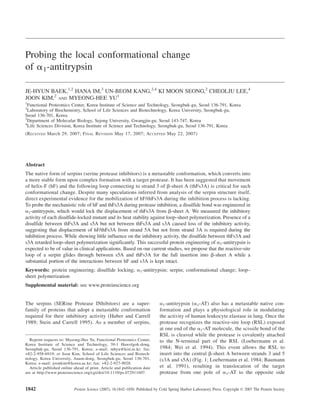
Figure 1. Structures of a1-AT. (A) The structure of native form (1QLP) is represented as a ribbon diagram with the position of six
engineered disulfide bonds. Left box, focused side view showing three engineered disulfide bonds between thFs3A and s5A (dotted
lines). Right box, focused side view showing two engineered disulfide bonds between thFs3A and s3A (dotted lines) and one
engineered disulfide between s3A and s5A (solid line). The balls in the structure indicate mutated residues to cysteine. (B) Structure of
a1-AT–trypsin complex (1EZX) showing that the RSL is inserted between s3A and s5A to form a new b-strand. Structures are color-
coded: hF/thFs3A, yellow; s3A, red; s4A, pink; s5A, orange; a1-AT, blue; trypsin, cyan.
Probing the local conformation change of serpin
www.proteinscience.org 1843
3. by DNA sequencing of the plasmid. All variants carried
the C232S mutation. Plasmids for a1-AT expression in
Escherichia coli and purification of recombinant a1-AT
protein were described previously (Kwon et al. 1995;
Kang et al. 2004). Protein concentration was determined
by measuring A280 in 6 M guanidine hydrochloride and
calculated from tyrosine and tryptophan content of the
a1-AT protein (Edelhoch 1967).
Disulfide bond formation and purification of a1-AT
Protein solution was loaded on a Q-Sepharose column
and washed with 50 mM tris-HCl buffer (pH 8.0). The
oxidation of double-cysteines to a disulfide was carried
out on column for 48 h at 4°C after buffer exchange with
the same buffer containing 1 mM oxidized glutathione
(GSSG). Protein was eluted with a linear-gradient of 0–
0.6 M NaCl. In case of low oxidation yield (K168C-
V337C), the eluted protein was treated with 10 mM
sodium iodoacetate (ICH2COONa) in 10 mM KH2PO4
buffer (pH 6.8) for 1 h at room temperature to add two
more negative charges onto the free thiols in reduced
form. The oxidized protein was purified by NaCl salt
gradients using a Mono-Q column on an A¨ KTA Explorer
100 instrument (Amersham Pharmacia). At the end of all
purification steps, samples were desalted using Sephadex
G-25 to exchange the buffer with 10 mM KH2PO4 buffer
(pH 6.5).
Gel electrophoresis
Nonreducing sodium dodecyl sulfate–polyacrylamide gel
electrophoresis (SDS-PAGE) was performed in a 10% gel
as described previously (Laemmli 1970). Prior to SDS-
PAGE, samples were boiled for 5 min in loading buffer
containing 2% SDS except reducing reagent. Non-dena-
turing PAGE was performed at 4°C without SDS in a 10%
gel using the method described previously (Goldenberg
and Creighton 1989). The gels were visualized by stain-
ing with Coomassie Brilliant Blue.
Determination of the yield of disulfide bond formation
Free thiols in engineered proteins were quantitated with a
colorimetric assay using the Thiol and Sulfide Quantita-
tion Kit (Molecular Probes) according to the manufac-
turer’s instructions. Briefly, 0.6 nmol of engineered
proteins was incubated with 2 mM cystamine followed
by 0.6 mg/mL papain-SSCH3 for 1 h incubation at room
temperature. The reaction mixture was then incubated
with 2.5 mM L-BAPNA for 1 h at room temperature, and
absorbance was measured at 410 nm. Thiol contents were
calculated from a calibration curve using standardized
cysteine solutions. Alternatively, the percentage of oxi-
dized form was measured by densitometric analysis for
both oxidized and reduced forms in nonreducing SDS-PAGE
using ImageQuant software (Molecular Dynamics).
Identification of disulfide bond by mass spectrometry
The protein band was excised, diced to small pieces,
washed with H2O, and destained with 50% acetonitrile.
After dehydration with acetonitrile, the gel pieces were
dried in the air and the protein was digested with 12.5 ng/
mL trypsin (Promega) in 25 mM ammonium bicarbonate
buffer (pH 8.0) at 37°C for overnight. To check the
existence of target mass for disulfide bonded tryptic
peptide, mass spectrum of tryptic digest was acquired
using MALDI-TOF instrument (Voyager DE-STR,
Applied Biosystems). For determination of disulfide
position, tryptic digest was loaded on a nanospray tip-
coupled capillary column (75 mm 3 16 cm) packed in-
house with Magic C18aq (Michrom BioResources) by
helium pressure cells. Peptides were eluted with a linear
gradient of 5%–40% solvent B over 30 min at 300 nL/min
(solvent A, 0.1% formic acid in water; solvent B, 0.1%
formic acid in 100% acetonitrile) using an Agilent Nano-
flow 1200 series HPLC system. We used a mass acquis-
ition method in which MS survey scan from 300 to 2000
m/z is followed by targeted MS/MS scans for the
disulfide-bonded tryptic peptide (888.73 m/z for the +3
charge state and 1332.59 m/z for the +2 charge state) on
an LTQ linear ion trap mass spectrometer (Thermofinni-
gan). Mass spectra were inspected manually.
Activity assay
Inhibitory activity of a1-AT was assessed by determining
the amount of a1-AT to completely inhibit 1 mol of PPE
(Seo et al. 2000). Various amounts of purified recombi-
nant a1-AT proteins were incubated in 50 mL of assay
buffer (30 mM phosphate, 160 mM NaCl, 0.1% poly-
ethylene glycol 8000, and 0.1% Triton X-100, pH 7.4)
with 100 nM PPE. After incubation at 37°C for 10 min,
the reaction mixture was diluted 10-fold with the assay
buffer and the residual protease activity was determined
using 1 mM N-succinyl-(Ala)3-p-nitroanilide as a sub-
strate.
Analysis of heat stability
a1-AT proteins were incubated at 0.2 mg/mL in 10 mM
KH2PO4 buffer (pH 6.5) for 8 h at 60°C. Aliquots were
removed at each time point (0, 0.25, 0.5, 1, 2, 3, 4, 6, and
8 h) and were rapidly cooled on ice. Samples were
analyzed by non-denaturing PAGE. The remaining inhib-
itory activity of each aliquot was also determined by the
above-mentioned activity assay.
Baek et al.
1844 Protein Science, vol. 16
4. Determination of association rate constants
The association rate constant for the interaction of a1-AT
with porcine pancreatic elastase was measured under
second-order conditions in a reaction mixture containing
equimolar concentrations (14 nM) of enzyme and inhib-
itor as previously described (Beatty et al. 1980).
Bis-ANS binding assay
Binding of bis-ANS was carried out with 1 mM a1-ATs for
effective analysis of biphasic change in bis-ANS fluores-
cence as previously described (James and Bottomley 1998).
The kinetic fluorescence measurements were performed in a
PerkinElmer SynergyHT-I spectrofluorimeter at 50°C and in
an LS50B spectrofluorimeter at 65°C. The rates of each
phase in fluorescence change were determined by fitting to a
single-exponential function.
Results
Selecting sites for disulfide bonds
To investigate the role of hF/thFs3A for inhibitory mecha-
nism, several disulfide bonds were designed and separately
introduced, through which the hF-thFs3A structure would be
constrained to the main body of a1-AT, b-sheet A. The X-ray
crystallographic structure for the native form of a1-AT (PDB
entry 1QLP); (Elliott et al. 2000) was examined to select
suitable sites. The sites were selected according to the
following criteria: (1) pairs of residues with one located at
hF or thFs3A and the other at s3A or s5A; (2) pairs of
residues whose Ca–Ca distances are within 5.0–9.5 A˚ ; and
(3) if possible, the residues for which single amino acid
substitution has been shown by previous investigations to
have minor or beneficial effects on the stability of a1-AT.
In our previous studies, the inhibitory activities for single
amino acid substitutions in the sites of K168, I169, L172,
V173, and F189 have been determined. Typically, K168I,
I169V, L172V, L172A, V173I, F189I, and F189V mutations
had a marginal effect on the stability, and some of the
mutations even stabilized the protein. As determined by
denaturant-induced equilibrium unfolding, the stability
change was ;0.0–1.9 kcal/mol, and the activity loss by the
mutations was <30% (Im et al. 1999; Seo et al. 2000; Lee
et al. 2001). On the basis of the criteria given, seven disulfide
bond sites were chosen. We added to the list one more
mutant, V185C–V333C, linking s5A and s3A, as a control
for detrimental effect on the activity. However, we excluded
the two double-mutant constructs, V161C–Y187C and
T165C–F189C, because of low-level expression in E. coli.
Finally, six disulfide bond sites were chosen as listed in Table
1. There is no native disulfide bond in human a1-AT, and the
single free cysteine at residue site 232 was replaced with
serine prior to engineering the disulfide bond at the desig-
nated sites.
Protein engineering for disulfide bond
The plasmids for the double-cysteine mutants were
constructed by site-directed mutagenesis, and the
recombinant mutant proteins were expressed in E. coli.
The six proteins were expressed in a sufficient quantity
for further purification. Oxidation of the two cysteines
in each mutant protein was carried out in the presence
of 1 mM GSSG. When a double-mutant was oxidized
and analyzed by nonreducing SDS-PAGE, another protein
band was detected that migrated faster than the reduced
form. For all double-mutants, the oxidized form migrated
faster than the corresponding free thiol form in non-
reducing SDS-PAGE (Fig. 2). The yield of disulfide bond
formation was also determined by densitometric analysis
and titration of free thiols using the thiol and sulfide
quantitation kit (Molecular Probes). The yield of the
oxidized form was >80% for all the constructs except
K168C–V337C, which yielded ;50% of the disulfide-
bonded form (Table 1). The oxidized form of K168C–
V337C was separated from the reduced form through ion-
exchange chromatography with a yield of 98% purity.
Mass-spectrometric analysis was carried out to con-
firm that the fast-migrating species truly contained the
engineered disulfide bond. The sequence and the position
of disulfide bond were confirmed by tandem mass
spectrometry (results are in the Supplemental material).
Activity assay of disulfide-locked a1-ATs
To analyze the effect of disulfide locking on the function
of a1-AT, we measured the inhibitory activities for all six
double-mutants in both reduced and oxidized states.
Table 1. Properties of double-cysteine mutants of a1-AT
Group Mutantsa
Ca–Ca
b
Locationc
Yield
(%)d
Relative activitye
2SH S–S
1 K168C–F189C 8.02 thFs3A–s3A 97 0.93 0.94
1 I169C–Y187C 8.36 thFs3A–s3A 99 0.36 0.38
2 K168C–V337C 8.47 thFs3A–s5A 98 0.56 (0.55) 0.01
2 L172C–V333C 8.25 thFs3A–s5A 80 0.79 0.16
2 V173C–K331C 9.13 thFs3A–s5A 95 0.73 0.04
3 V185C–V333C 5.08 s3A–s5A 90 0.34 0.03
a
One-letter codes for amino acids are used for mutations, which are
designated by the code for the wild-type residue followed by the residue
number and the code for the replacement residue.
b
The distance (A˚ ) between two a-carbons in the crystal structure of native
a1-AT (1QLP).
c
The nomenclature of Huber and Carrell was followed (Huber and Carrell 1989).
d
Purity of disulfide-bonded form.
e
Relative activity compared to the wild type. Activity in the parentheses
was measured after the oxidized protein was re-reduced with DTT. 2SH,
reduced form; S–S, oxidized form.
Probing the local conformation change of serpin
www.proteinscience.org 1845
5. According to the position of disulfide bond, the disulfide
pairs can be grouped into three categories: (1) K168C–
F189C and I169C–Y187C, in which disulfide connects
thFs3A to s3A; (2) K168C–V337C, L172C–V333C, and
V173C–K331C, in which disulfide connects thFs3A to
s5A; and (3) V185C–V333C, in which disulfide links s3A
and s5A (Table 1).
The first group of proteins showed no significant
changes in inhibitory activities upon formation of the
disulfide bond (Fig. 3; Table 1). The association rate
(kass) of the inhibitors with porcine pancreatic elastase
was not affected significantly either by the amino acid
substitutions or by disulfide formation (WT, 2.4 3 106
MÀ1
Á sÀ1
; reduced K168C–F189C, 2.9 3 106
MÀ1
Á sÀ1
;
oxidized K168C–F189C, 2.7 3 106
MÀ1
Á sÀ1
; reduced
I169C–Y187C, 2.3 3 106
MÀ1
Á sÀ1
; oxidized I169C–
Y187C, 2.5 3 106
MÀ1
Á sÀ1
). However, the second group
of proteins showed significant loss of inhibitory activity
by disulfide locking, as revealed by the completely
substrate-like behavior of the oxidized form (Fig. 3;
Table 1). For example, the K168C–V337C protein in
the reduced state had 56% activity compared to the wild
type, but its activity decreased significantly to <1% after
disulfide locking. When the disulfide bond was reduced
with dithiothreitol, the inhibitory activity was recovered to
the original level (55% of the wild-type activity), sug-
gesting that loss of activity is mainly attributed to disulfide
locking and not to some other effect such as protein
denaturation (Fig. 3; Table 1). Disulfide locking also
impaired the inhibitory activity of the third group,
V185C–V333C; the reduced form already showed activ-
ity loss (34% of the wild-type activity), but the activity
decreased even further by disulfide locking (Table 1).
Heat stability of the disulfide-locked a1-ATs
In order to test the role of hF/thFs3A region in gate-
keeping against loop–sheet polymerization, stability
against heat-induced aggregation was measured. Based
on the peptide insertion experiment that mimics the
sequence of RSL, previous studies showed that the
molecular mechanism of heat-induced aggregation of
a1-AT was loop–sheet polymerization (Lomas et al.
1992; Kwon et al. 1994; Kim et al. 1995; Dafforn et al.
1999). With the two proteins whose functional activity
was not affected by disulfide locking, heat-induced
aggregation was measured after incubation at 60°C for a
defined time by monitoring disappearance of monomeric
form in non-denaturing PAGE. As shown in Figure 4,
disulfide locking in the two mutant forms retarded the
disappearance of the monomeric form significantly. In
particular, the monomeric band of the oxidized K168C–
F189C remained up to 240 min, while that of the reduced
K168C–F189C was nearly gone after 30 min. The
measured remaining activity (Fig. 5) correlated well with
the amount of remaining monomeric form observed in
non-denaturing PAGE.
Serpin polymerization was shown to consist of two steps:
a monomeric conformational change followed by self-
association (James and Bottomley 1998; Dafforn et al.
1999). In order to assess the effect of disulfide locking on
the individual steps during polymerization, binding of bis-
ANS by a1-AT was measured under heat-induced denatur-
ing conditions. Since the two oxidized mutant proteins have
Figure 2. Nonreducing SDS-PAGE analysis of a1-AT variants. Four
representative double-cysteine mutants were analyzed on nonreducing
SDS-polyacrylamide gel. Lanes 1,2, K168C–F189C; lanes 3,4, I169C–
Y187C; lanes 5,6, K168C–V337C; lanes 7,8, V185C–V333C; lanes
1,3,5,7, reduced form; lanes 2,4,6,8, oxidized form.
Figure 3. Activity assay of a1-AT variants. A fixed amount of PPE was
mixed with varying concentrations of each a1-AT mutant, and the residual
protease activity was measured with N-succinyl-(Ala)3-p-nitroanilide as a
substrate. Inhibited fraction of the protease activity vs. molar ratio of a1-
AT to PPE is plotted. (Inset) Plot of inhibitory activity of K168C–V337C
with disulfide bond shown with extended x-axis. Slope values were derived
from the plot by linear regression analysis, and the inhibitory activity of
each mutant was calculated as slopemutant /slopewild-type. The resultant
activity values are summarized in Table 1. ), wild type; d, reduced form
of K168C–F189C; s, oxidized form of K168C–F189C; ., reduced I169C–
Y187C; P, oxidized I169C–Y187C; j, reduced K168C–V337C; u,
oxidized K168C–V337C; n, oxidized K168C–V337C that was re-reduced
with DTT.
Baek et al.
1846 Protein Science, vol. 16
6. quite different heat stability at 60°C (Fig. 4), two distinct
denaturation temperatures were applied for each mutant
(50°C for I169C–Y187C and 65°C for K168C–F189C).
Figure 6A shows that bis-ANS fluorescence of the wild type
and mutant a1-AT showed biphasic changes during heat
denaturation. In comparison to the wild-type kinetics,
I169C and Y187C substitutions by themselves did not alter
the kinetics of both phases significantly (Fig. 6A; 0.023
minÀ1
! 0.028 minÀ1
for the first phase and 3.31 3 10À3
minÀ1
! 2.59 3 10À3
minÀ1
for the second phase; data
summarized in Table 2). The disulfide locking in I169C–
Y187C retarded the first phase only slightly (0.028 minÀ1
! 0.024 minÀ1
) and retarded the second phase by twofold
(2.59 3 10À3
minÀ1
! 1.37 3 10À3
minÀ1
). The K168C
and F189C substitutions by themselves did not alter the
kinetics of both phases significantly at 65°C (Fig. 6B; Table
2; 0.738 minÀ1
! 0.708 minÀ1
for the first phase and 1.27
3 10À2
minÀ1
! 9.87 3 10À3
minÀ1
for the second phase).
The disulfide locking in K168C–F189C, however, retarded
both phases significantly (0.708 minÀ1
! 0.051 minÀ1
for
the first phase and 9.87 3 10À3
minÀ1
! 3.87 3 10À4
minÀ1
for the second phase).
Discussion
Conformational change of the proteins associated with
functional regulation has been of great interest in the
studies of protein structure and design. Serpins have
provided insights into the process because the metastable
native form undergoes a dramatic conformational tran-
sition to a stable state when they form a tight complex
with a target protease (Huber and Carrell 1989; Stein and
Carrell 1995; Huntington et al. 2000). Our previous
studies on stabilizing mutations of a1-AT revealed that,
while the metastability, caused by side-chain overpack-
ing, cavities, and surface hydrophobic pockets, is
distributed throughout the molecule, the interactions
critical to the activity were confined in the regions that
are mobilized during the complex formation with a target
Figure 4. Heat stability of wild-type and engineered proteins monitored
by non-denaturing PAGE. K168C–F189C and I169C–Y187C proteins in
their reduced and oxidized forms as well as the wild-type protein were
incubated at 60°C. At each designated time point, an aliquot was removed
and analyzed by non-denaturing PAGE. Wild-type and reduced proteins
show two bands on non-denaturing PAGE, while oxidized proteins that
have no free sulfhydryl group showed one band. This can be explained by
the fact that the pKa value of the sulfhydryl group on the cysteine residue
(;pH 8.3) is close to the pH value of the separating condition (pH 8.8).
Figure 5. Heat stability of wild-type and engineered proteins monitored
by inhibitory activity. The same aliquot as in Fig. 4 was used for activity
measurement. Inhibitory activity at each time point relative to the activity
at zero time of each protein is plotted as a function of incubation time: ),
wild type; d, reduced form of K168C–F189C; s, oxidized form of
K168C–F189C; ., reduced I169C–Y187C; P, oxidized I169C–Y187C.
Probing the local conformation change of serpin
www.proteinscience.org 1847
7. enzyme (Lee et al. 1996; Seo et al. 2000). One of the
regions various studies have focused on is the hF/thFs3A
region, which appears to be an obstacle during the RSL
insertion into the b-sheet A. In the present study, the
mechanistic role of hF/thFs3A during protease inhibition
was investigated by introducing several disulfide bonds
that could restrict the movement of thFs3A away from
s3A or s5A of b-sheet A.
The disulfide locking of thFs3A to s5A (group 2
mutants, K168C–V337C, L172C–V333C, and V173C–
K331C) impaired the inhibitory activity, but the release of
the lock restored the original activity (Fig. 3; Table 1).
These results suggest that restriction of the movement of
hF/thFs3A relative to s5A blocks the insertion of RSL
into b-sheet A. In contrast, the engineered disulfide bonds
between thFs3A and s3A (group 1 mutants, K168C–
F189C and I169C–Y187C) showed little effect on inhib-
itory activity of a1-AT (Table 1). Locking the b-sheet A
between s3A and s5A by disulfide (group 3 mutant,
V185C–V333C) affected inhibitory activity severely
(Table 1), which was expected because RSL has to be
inserted between the two strands to form a new strand,
s4A. The result shows that the disulfide locking method is
an effective way of preventing conformational change.
Taken together, our results suggest that temporal opening
of hF/thFs3A from s5A, but not from s3A, is necessary
for inhibition.
Previous studies have suggested that hF/thFs3A plays
an essential role in the protease inhibition mechanism
(Gooptu et al. 2000; Cabrita et al. 2002, 2004). It was
speculated that the movement of hF/thFs3A controls the
early conformational change (Cabrita et al. 2002, 2004;
Tsutsui et al. 2006). These proposals were based on the
interpretation of serpin structures, which were probed by
crystallography, denaturant-induced equilibrium unfold-
ing, or hydrogen-deuterium exchange under unfolding
conditions. In addition, Gettins hypothesized that full
displacement of the whole hF/thFs3A region and its
subsequent return during protease translocation were
essential for RSL insertion and the final protease crushing
(Gettins 2002). In our current study, however, we showed
through a direct experimental approach that such a drastic
detachment of hF is seemingly unnecessary for protease
inhibition, although the possibility of local unfolding
could not be excluded.
The current results are consistent with our previous data
on the structure–activity relationship of single-point muta-
tions. For the stabilizing mutations located at s5A and
facing toward thFs3A (substitutions at Lys331 and Lys335),
their stabilizing effect was correlated with activity loss
(average relative activity ¼ 0.65 6 0.11 of the wild-type
activity; Im and Yu 2000). Tighter interactions between s5A
and thFs3A caused by the mutations seem to interfere with
the insertion of RSL. However, stabilizing mutations
located on hF (K163T, G164V, and T165S) and s3A
Figure 6. Bis-ANS binding of wild-type and engineered proteins. (A) The
fluorescence change of bis-ANS followed by loop–sheet polymerization
process of a1-AT was monitored. (B) The excitation (bandwidth 5 nm) and
emission (bandwidth 5 nm) wavelengths used were 420 and 485 nm,
respectively. All proteins were incubated in 50 mM Tris-HCl, 50 mM NaCl
(pH 8.0), containing bis-ANS.
Table 2. Rate constants for the kinetic parameters of bis-ANS
binding during heat-induced denaturation
Protein
Rate (minÀ1
)a
First
phase
Folds
retardationb
Second
phase
Folds
retardationb
Temperature (50°C)
Wild type 0.023 — 3.31 3 10À3
—
Reduced I169C–Y187C 0.028 1.0 2.59 3 10À3
1.0
Oxidized I169C–Y187C 0.024 1.2 1.37 3 10À3
1.9
Temperature (65°C)
Wild type 0.738 — 1.27 3 10À2
—
Reduced K168C–F189C 0.708 1.0 9.87 3 10À3
1.0
Oxidized K168C–F189C 0.051 13.9 3.87 3 10À4
25.5
a
Rate constants were determined by kinetic analysis described as pre-
viously reported (James and Bottomley 1998).
b
Retardation by disulfide locking.
Baek et al.
1848 Protein Science, vol. 16
8. (A183V, A183I, F189I, and F189V) did not cause activity
loss (average relative activity ¼ 0.98 6 0.07 of the wild-
type activity); (Im et al. 1999; Seo et al. 2000). These results
suggest that mobilization between hF and s3A is not critical
in the inhibitory activity. The average B factor of thFs3A
(E166–D179) is 47.0, while that of hF (D149–T165) is
39.5 (PDB entry 1QLP). In addition, it was reported that
the hydrogen/deuterium exchange rate of thFs3A, which is
comparable to that of RSL, is faster than that of hF (Tsutsui
et al. 2006). Our results are consistent with all of these
previous structural studies. It is likely that RSL glides
through the gap between s5A and thFs3A during complex
formation with a target protease.
The hF/thFs3A region was considered critical in blocking
the loop–sheet polymerization (Bruce et al. 1994; Vleugels
et al. 2000; Wilczynska et al. 2003; Cabrita et al. 2004).
Loop–sheet polymerization, which has been reported for
several natural mutants of serpin, is a unique molecular
interaction between RSL of one molecule and the gap in the
b-sheet A of another (Aulak et al. 1993; Bruce et al. 1994;
Gooptu et al. 2000). Typically, the Z mutation of a1-AT
makes the protein very prone to the loop–sheet polymer-
ization, which impedes the folding and secretion of a1-AT
from liver cells into blood (Lomas et al. 1992; Yu et al.
1995). Various studies showed that molecular mechanism
of heat-induced aggregation of serpins is indeed loop–
sheet polymerization (Lomas et al. 1992; Kim et al. 1995;
Chang et al. 1997; Dafforn et al. 1999). Interestingly, the
two disulfide mutants (K168C–F189C and I169C–Y187C),
whose activities and association rate constants are little
affected by disulfide locking (Fig. 3; Table 1), showed a
substantial enhancement in heat stability upon disulfide
locking (Figs. 4, 5). The results indicate that disulfide
locking between s3A and thFs3A retards loop–sheet poly-
merization while it has little effect on loop insertion into its
own b-sheet during inhibition process.
Several studies further suggested that, based on the
biphasic changes of bis-ANS fluorescence during heat-
induced denaturation, the process of loop–sheet polymer-
ization consists of two steps: an initial opening of b-sheet A
of the molecule itself, and a following step of loop insertion
into b-sheet A of another molecule (James and Bottomley
1998; Dafforn et al. 1999). Disulfide locking of K168C–
F189C and I169C–Y187C showed stabilization effects in
both phases but more prominently toward the second phase
(Table 2). The stabilization effects of disulfide locking as
measured by bis-ANS binding kinetics (Fig. 6; Table 2)
are qualitatively consistent with those monitored by
non-denaturing PAGE (Fig. 4). Interestingly, the disulfide
locking of K168C–F189C, which showed a substantial
stabilization effect on the first phase of bis-ANS binding
(Fig. 6B), did not affect the activity (Fig. 3; Table 1). These
results indicate that the first phase of hydrophobic dye
binding in heat denaturation does not reflect the conforma-
tional change of the molecule during the inhibition process.
The results also imply that the basis of loop insertion during
the complex formation is different from that during loop–
sheet polymerization, as previously suggested (Cabrita
et al. 2004). These results are consistent with hF/thFs3A
exerting an obstructing effect on insertion of RSL from
another molecule but being ready for opening from s5A for
the RSL insertion during inhibitory complex formation.
The disulfide engineering of K168C–F189C mutant
a1-AT turned out to be a very useful outcome in practical
respects. Protein engineering through mutational ap-
proaches, such as single amino acid substitutions and
disulfide bridges, has been effectively applied to improve
protein properties for industrial or medical purposes
(Reiter et al. 1996; Hugo et al. 2002; Eijsink et al.
2004; McHugh et al. 2004). Patients with a1-AT defi-
ciency require replacement therapy with available protein
(Abusriwil and Stockley 2006). The oxidized form of
K168C–F189C turned out to be a very successful engi-
neering result, in the sense that detrimental loop–sheet
polymerization was abrogated while inhibitory activity
was affected little. This case of protein engineering is
expected to be of considerable value in replacement
therapy of a1-AT without risk of possible viral contam-
inations.
In conclusion, our results suggest that displacement of hF/
thFs3A from s5A but not from s3A is required for formation
of a stable complex of a serpin with a target protease in
inhibitory processes. It is likely that the conformational
change of hF/thFs3A involves a full detachment of thFs3A
away from s5A while keeping a substantial portion of the
interactions between hF and s3A intact. Restricting the
movement of thFs3A from strand s3A appears to reinforce
its gatekeeping role against loop–sheet polymerization. It
may be that the RSL of a1-AT glides from the side through
the gap between s5A and thFs3A for its full insertion into
b-sheet A during inhibitory complex formation.
Acknowledgment
This study was supported by a grant from the Functional
Proteomics Center, 21st Century Frontier R&D Initiatives of
the Korean Ministry of Science and Technology.
References
Abusriwil, H. and Stockley, R.A. 2006. a1-Antitrypsin replacement therapy:
Current status. Curr. Opin. Pulm. Med. 12: 125–131.
Aulak, K.S., Eldering, E., Hack, C.E., Lubbers, Y.P., Harrison, R.A., Mast, A.,
Cicardi, M., and Davis 3rd, A.E. 1993. A hinge region mutation in C1-
inhibitor (A436T) results in nonsubstrate-like behavior and in polymer-
ization of the molecule. J. Biol. Chem. 268: 18088–18094.
Baumann, U., Huber, R., Bode, W., Grosse, D., Lesjak, M., and Laurell, C.B.
1991. Crystal structure of cleaved human a1-antichymotrypsin at 2.7 A˚
resolution and its comparison with other serpins. J. Mol. Biol. 218:
595–606.
Beatty, K., Bieth, J., and Travis, J. 1980. Kinetics of association of serine
proteinases with native and oxidized a1-proteinase inhibitor and a1-
antichymotrypsin. J. Biol. Chem. 255: 3931–3934.
Probing the local conformation change of serpin
www.proteinscience.org 1849
9. Bijnens, A.P., Gils, A., Knockaert, I., Stassen, J.M., and Declerck, P.J. 2000.
Importance of the hinge region between a1-helix F and the main part
of serpins, based upon identification of the epitope of plasminogen
activator inhibitor type 1 neutralizing antibodies. J. Biol. Chem. 275:
6375–6380.
Bruce, D., Perry, D.J., Borg, J.Y., Carrell, R.W., and Wardell, M.R. 1994.
Thromboembolic disease due to thermolabile conformational changes of
antithrombin Rouen-VI (N187D). J. Clin. Invest. 94: 2265–2274.
Bruch, M., Weiss, V., and Engel, J. 1988. Plasma serine proteinase inhibitors
exhibit major conformational changes and a large increase in conforma-
tional stability upon cleavage at their reactive sites. J. Biol. Chem. 263:
16626–16630.
Cabrita, L.D., Dai, W., and Bottomley, S.P. 2004. Different conformational
changes within the F-helix occur during serpin folding, polymerization, and
proteinase inhibition. Biochemistry 43: 9834–9839.
Cabrita, L.D., Whisstock, J.C., and Bottomley, S.P. 2002. Probing the role
of the F-helix in serpin stability through a single tryptophan substitution.
Biochemistry 41: 4575–4581.
Chang, W.S., Whisstock, J., Hopkins, P.C., Lesk, A.M., Carrell, R.W., and
Wardell, M.R. 1997. Importance of the release of strand 1C to the poly-
merization mechanism of inhibitory serpins. Protein Sci. 6: 89–98.
Dafforn, T.R., Mahadeva, R., Elliott, P.R., Sivasothy, P., and Lomas, D.A. 1999.
A kinetic mechanism for the polymerization of a1-AT. J. Biol. Chem. 274:
9548–9555.
Edelhoch, H. 1967. Spectroscopic determination of tryptophan and tyrosine in
proteins. Biochemistry 6: 1948–1954.
Eijsink, V.G., Bjork, A., Gaseidnes, S., Sirevag, R., Synstad, B., van den
Burg, B., and Vriend, G. 2004. Rational engineering of enzyme stability.
J. Biotechnol. 113: 105–120.
Elliott, P.R., Pei, X.Y., Dafforn, T.R., and Lomas, D.A. 2000. Topography of a
2.0 A˚ structure of a1-AT reveals targets for rational drug design to prevent
conformational disease. Protein Sci. 9: 1274–1281.
Gettins, P.G. 2002. The F-helix of serpins plays an essential, active role in the
proteinase inhibition mechanism. FEBS Lett. 523: 2–6.
Goldenberg, D.P. and Creighton, T.E. 1989. Analysis of protein conformation
by gel electrophoresis. In Protein structure: A practical approach, pp. 225–
250. IRL Press, Oxford, UK.
Gooptu, B., Hazes, B., Chang, W.S., Dafforn, T.R., Carrell, R.W., Read, R.J.,
and Lomas, D.A. 2000. Inactive conformation of the serpin a1-antichymo-
trypsin indicates two-stage insertion of the reactive loop. Proc. Natl. Acad.
Sci. 97: 67–72.
Huber, R. and Carrell, R.W. 1989. Implications of the three-dimensional
structure of a1-AT for structure and function of serpins. Biochemistry 28:
8951–8966.
Hugo, N., Lafont, V., Beukes, M., and Altschuh, D. 2002. Functional aspects of
co-variant surface charges in an antibody fragment. Protein Sci. 11: 2697–
2705.
Huntington, J.A., Read, R.J., and Carrell, R.W. 2000. Structure of a serpin
protease complex shows inhibition by deformation. Nature 407:
923–926.
Im, H. and Yu, M.H. 2000. Role of K335 in the metastability and function of
inhibitory serpins. Protein Sci. 9: 934–941.
Im, H., Seo, E.J., and Yu, M.H. 1999. Metastability in the inhibitory mechanism
of human a1-AT. J. Biol. Chem. 274: 11072–11077.
James, E.L. and Bottomley, S.P. 1998. The mechanism of a1-AT polymer-
ization probed by fluorescence spectroscopy. Arch. Biochem. Biophys. 356:
296–300.
Kang, U.B., Baek, J.H., Ryu, S.H., Kim, J., Yu, M.H., and Lee, C. 2004. Kinetic
mechanism of protease inhibition by a1-AT. Biochem. Biophys. Res.
Commun. 323: 409–415.
Kim, J., Lee, K.N., Yi, G.S., and Yu, M.H. 1995. A thermostable mutation
located at the hydrophobic core of a1-AT suppresses the folding defect of
the Z-type variant. J. Biol. Chem. 270: 8597–8601.
Kwon, K.S., Lee, S., and Yu, M.H. 1995. Refolding of a1-AT expressed as
inclusion bodies in Escherichia coli. Biochim. Biophys. Acta 1247:
179–184.
Kwon, K.S., Kim, J., Shin, H.S., and Yu, M.H. 1994. Single amino acid
substitutions of a1-AT that confer enhancement in thermal stability. J. Biol.
Chem. 269: 9627–9631.
Laemmli, U.K. 1970. Cleavage of structural proteins during the assembly of the
head of bacteriophage T4. Nature 227: 680–685.
Lee, C., Maeng, J.S., Kocher, J.P., Lee, B., and Yu, M.H. 2001. Cavities of
a1-AT that play structural and functional roles. Protein Sci. 10: 1446–1453.
Lee, K.N., Park, S.D., and Yu, M.H. 1996. Probing the native strain in a1-AT.
Nat. Struct. Biol. 3: 497–500.
Loebermann, H., Tokuoka, R., Deisenhofer, J., and Huber, R. 1984. Human a1-
proteinase inhibitor. J. Mol. Biol. 177: 531–557.
Lomas, D.A., Evans, D.L., Finch, J.T., and Carrell, R.W. 1992. The mechanism
of Z a1-AT accumulation in the liver. Nature 357: 605–607.
McHugh, C.A., Tammariello, R.F., Millard, C.B., and Carra, J.H. 2004.
Improved stability of a protein vaccine through elimination of a partially
unfolded state. Protein Sci. 13: 2736–2743.
Reiter, Y., Brinkmann, U., Lee, B., and Pastan, I. 1996. Engineering antibody
Fv fragments for cancer detection and therapy: Disulfide-stabilized Fv
fragments. Nat. Biotechnol. 14: 1239–1245.
Seo, E.J., Im, H., Maeng, J.S., Kim, K.E., and Yu, M.H. 2000. Distribution of
the native strain in human a1-antitrypsin and its association with protease
inhibitor function. J. Biol. Chem. 275: 16904–16909.
Skinner, R., Chang, W.S., Jin, L., Pei, X., Huntington, J.A., Abrahams, J.P.,
Carrell, R.W., and Lomas, D.A. 1998. Implications for function and therapy of
a 2.9 A˚ structure of binary-complexed antithrombin. J. Mol. Biol. 283: 9–14.
Stein, P.E. and Carrell, R.W. 1995. What do dysfunctional serpins tell us about
molecular mobility and disease? Nat. Struct. Biol. 2: 96–113.
Stratikos, E. and Gettins, P.G. 1999. Formation of the covalent serpin
proteinase complex involves translocation of the proteinase by more than
70 A˚ and full insertion of the reactive center loop into b-sheet A. Proc.
Natl. Acad. Sci. 96: 4808–4813.
Tsutsui, Y., Liu, L., Gershenson, A., and Wintrode, P.L. 2006. The conforma-
tional dynamics of a metastable serpin studied by hydrogen exchange and
mass spectrometry. Biochemistry 45: 6561–6569.
Vleugels, N., Gils, A., Bijnens, A.P., Knockaert, I., and Declerck, P.J. 2000.
The importance of helix F in plasminogen activator inhibitor-1. Biochim.
Biophys. Acta 1476: 20–26.
Wei, A., Rubin, H., Cooperman, B.S., and Christianson, D.W. 1994. Crystal
structure of an uncleaved serpin reveals the conformation of an inhibitory
reactive loop. Nat. Struct. Biol. 1: 251–258.
Wilczynska, M., Lobov, S., Ohlsson, P.I., and Ny, T. 2003. A redox-sensitive
loop regulates plasminogen activator inhibitor type 2 polymerization.
EMBO J. 22: 1753–1761.
Wind, T., Jensen, M.A., and Andreasen, P.A. 2001. Epitope mapping for four
monoclonal antibodies against human plasminogen activator inhibitor type-
1. Eur. J. Biochem. 268: 1095–1106.
Yu, M.H., Lee, K.N., and Kim, J. 1995. The Z type variation of human a1-AT
causes a protein folding defect. Nat. Struct. Biol. 2: 363–367.
Baek et al.
1850 Protein Science, vol. 16