lam12373
- 1. ORIGINAL ARTICLE
Aeromonas piscicola AH-3 expresses an extracellular
collagenase with cytotoxic properties
A.S. Duarte1
, E. Cavaleiro1
, C. Pereira1,2
, S. Merino2
, A.C. Esteves1
, E.P. Duarte3
, J.M. Tomas2
and
A.C. Correia1
1 Department of Biology CESAM, University of Aveiro, Aveiro, Portugal
2 Departamento de Microbiologıa, Facultad de Biologıa, Universidad de Barcelona, Barcelona, Spain
3 Centre for Neurosciences and Cell Biology Department of Zoology, University of Coimbra, Coimbra, Portugal
Significance and Impact of the Study: Collagenases play a central role in processes where collagen
digestion is needed, for example host invasion by pathogenic micro-organisms. We identified a new col-
lagenase from Aeromonas using an integrated in silico/in vitro strategy. This enzyme is able to bind and
cleave collagen, contributes for AH-3 cytotoxicity and shares low similarity with known bacterial colla-
genases. This is the first report of an enzyme belonging to the gluzincin subfamily of the M9 family of
peptidases in Aeromonas. This study increases the current knowledge on collagenolytic enzymes bring-
ing new perspectives for biotechnology/medical purposes.
Keywords
AH-3, collagen interaction, cytotoxicity,
metalloprotease, microbial collagenase.
Correspondence
Ana Sofia Duarte, Department of Biology
CESAM, University of Aveiro, 3810-193
Aveiro, Portugal.
E-mail: asduarte@ua.pt
2014/1890: received 12 September 2014,
revised 10 November 2014 and accepted 26
November 2014
doi:10.1111/lam.12373
Abstract
The aim of this study was to investigate the presence and the phenotypic
expression of a gene coding for a putative collagenase. This gene (AHA_0517)
was identified in Aeromonas hydrophila ATCC 7966 genome and named colAh.
We constructed and characterized an Aeromonas piscicola AH-3::colAh
knockout mutant. Collagenolytic activity of the wild-type and mutant strains
was determined, demonstrating that colAh encodes for a collagenase. ColAh–
collagen interaction was assayed by Far-Western blot, and cytopathic effects
were investigated in Vero cells. We demonstrated that ColAh is a gluzincin
metallopeptidase (approx. 100 kDa), able to cleave and physically interact with
collagen, that contributes for Aeromonas collagenolytic activity and
cytotoxicity. ColAh possess the consensus HEXXH sequence and a glutamic
acid as the third zinc binding positioned downstream the HEXXH motif, but
has low sequence similarity and distinct domain architecture to the well-known
clostridial collagenases. In addition, these results highlight the importance of
exploring new microbial collagenases that may have significant relevance for
the health and biotechnological industries.
Introduction
During the past decades, substantiating evidence has been
gathered supporting the hypothesis that growth and pro-
liferation of pathogenic bacteria depend on the action of
proteolytic enzymes (Harrington 1996; Watanabe 2004).
Collagenases are involved in the degradation of extracellu-
lar matrixes of animal cells, due to their ability to digest
native and denatured collagen (Duarte et al. 2014). Colla-
genases and other collagen-degrading enzymes have been
implicated in the virulence of many pathogenic bacteria
(Lawson and Meyer 1992; Takeuchi et al. 1992; Matsush-
ita et al. 1999; Mukherjee et al. 2009).
The genus Aeromonas includes Gram-negative, faculta-
tive anaerobes, present in aquatic environments, food and
soil (Kingombe et al. 1999; Chacon et al. 2003; Carvalho
et al. 2012). Species of this genus have been described as
pathogens of humans, fish, invertebrates and insects
(Seshadri et al. 2006; Reith et al. 2008; Castro et al. 2010;
Janda and Abbott 2010; Li et al. 2011; Parker and Shaw
2011) and occasionally as symbionts of leeches and fish
(Janda and Abbott 2010; Silver et al. 2011).
Letters in Applied Microbiology 60, 288--297 © 2014 The Society for Applied Microbiology288
Letters in Applied Microbiology ISSN 0266-8254
- 2. In particular, Aeromonas hydrophila is associated with
gastroenteritis, wound diseases, soft tissue and burn infec-
tions, and sepsis, with lethal course in humans (Janda
and Abbott 2010). Virulence of Aer. hydrophila seems to
involve several extracellular molecules including entero-
toxins, hemolysins, elastases and other proteases (King-
ombe et al. 1999; Chacon et al. 2003; Seshadri et al. 2006;
Reith et al. 2008; Li et al. 2011; Parker and Shaw 2011).
Nevertheless, data on Aeromonas collagenases are scarce:
until now, only one report describes a collagenase in
Aeromonas veronii. This enzyme is involved in the pro-
gression of bacterial colonization and infection (Han
et al. 2008).
Several studies have demonstrated the ability of AH-3
—previously Aer. hydrophila and now known as Aeromo-
nas piscicola—to adhere and to invade host cells mediated
by the expression of a high number of virulence determi-
nants (Merino et al. 1992; Beaz-Hidalgo et al. 2009; Vil-
ches et al. 2009; Molero et al. 2011).
To investigate the function of the putative collagenase
in AH-3, we detected the gene in Aer. piscicola and con-
structed an AH-3 knockout mutant (AH-3::colAh). Phe-
notypic characterization of the mutant and the wild-type
strains included cytotoxicity evaluation and assessment of
collagenolytic activity and enzyme–substrate physical
interaction.
Results and discussion
An open reading frame (AHA_0517) of 2748 bp encoding
a 915 amino acids protein is annotated as a putative col-
lagenase in the genome of Aer. hydrophila ATCC 7966T
(accession number NC_008570). To characterize the role
of this putative collagenase, we designed and constructed
a knockout mutant by disrupting the AHA_0517 locus in
AH-3 by homologous recombination. Using specific
primers, we amplified by PCR a fragment of 904 bp from
the genomic DNA of Aer. piscicola AH-3 (accession num-
ber JQ639076). The nucleotide sequence of the amplicon
shared 88% similarity with a region with the same
length from AHA_0517. We hypothesized that the gen-
ome of Aer. piscicola AH-3, similarly to Aer. hydrophila
ATCC 7966T
, also contains the putative collagenase gene
(colAh).
To investigate this hypothesis, we performed mutagene-
sis of the genome targeted to the colAh region (Figure
S1). The 904-bp amplicon was inserted in the suicide
plasmid pFS100 to provide homologous recombination
with the genome, giving rise to the plasmid pFS-colAh.
By triparental mating, using Escherichia coli MC1061 as
donor, the construct was introduced into Aer. piscicola
AH-3. Sequence analysis of the mutant confirmed the
integration into the chromosomal DNA.
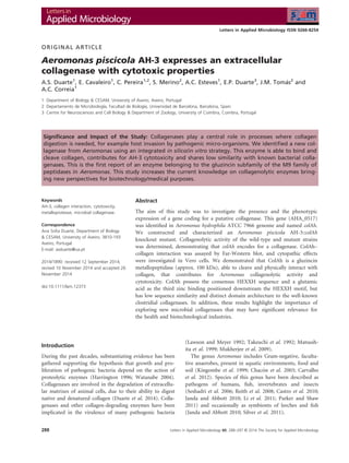
As seen by zymography, Aer. piscicola AH-3 and the
AH-3::colAh knockout mutant express several extracellular
gelatinases (Fig. 1a). Nevertheless, the mutant strain lacks
a gelatinase with an apparent molecular mass of approx.
100 kDa (Fig. 1a), corresponding to the molecular mass
of the product of the AHA_0517 gene.
The extracellular collagenolytic activities of the mutant
and of the wild-type strains were quantified by hydrolysis
of FALGPA substrate, a collagenase-specific synthetic pep-
tide (Van Wart and Steinbrink 1981). Results show that
AH-3::colAh cell-free supernatant (CFS) has a significant
lower collagenolytic activity (P 0Á001) when compared
to the wild-type strain (Fig. 1b), confirming that the
AHA_0517 gene is responsible for collagenolytic activity.
Both PMSF and 1,10-phenanthroline caused an inhibi-
tion of collagenolytic activity of AH-3 wild type and AH-
3::colAh. This inhibition pattern suggests the presence of
metallopeptidases and of serine peptidases.
Taken together, these results favour the hypothesis of
the presence of a gene in the genome of Aer. piscicola
AH-3 encoding an enzyme (ColAh) with relevant contri-
bution for the extracellular collagenolytic activity dis-
played by strain. The mutant strain still exhibited partial
collagenolytic activity on FALGPA hydrolysis assays; this
is an indication that other collagenolytic enzymes may
also be present in the extracellular medium.
The interaction between ColAh and type I collagen was
assessed by Far-Western blot (Fig. 1c). No protein–colla-
gen interactions were detected in colAh-deficient mutant
CFS, but in the wild strain, one band, with an apparent
molecular weight of %100 kDa, was detected confirming
that ColAh is able to physically interact with collagen.
This interaction suggests the presence of a collagen-bind-
ing domain on ColAh. This domain shares functional—
but not structural—similarity with the well-known colla-
gen recruitment domains, such as the CBD or the PKD-
like domains in clostridial collagenases (Eckhard et al.
2011, 2013; Duarte et al. 2014).
AH-3 CFS is highly cytotoxic, inducing the loss of
94Á1 % Vero cells’ viability (Fig. 2). This verotoxic effect
was significantly reduced (P 0Á001 at 1 : 4 CFS dilution)
for the AH-3::colAh mutant CFS, which promoted the loss
of 85Á7% cell viability. Regarding ColAh collagenolytic
activity and its cytotoxic effects, the results suggest that
ColAh may play a role in AH-3 infection mechanism,
degrading host collagen-rich matrices and favouring bacte-
rial penetration and migration in the host tissues.
SMART analysis of ColAh sequence shows the existence
of a signal peptide at the N-terminal of ColAh (with 23-
amino acid residues) and a presumable active site local-
ized at the C-terminal half of the core protein containing
an HEXXH motif (Fig. 3a). The HEXXH motif is a
metal-binding site (Bode et al. 1993; Rawlings and Barrett
Letters in Applied Microbiology 60, 288--297 © 2014 The Society for Applied Microbiology 289
A.S. Duarte et al. ColAh: Aeromonas collagenase
- 3. 1995; Wu and Chen 2011; Duarte et al. 2014), usually
found at the N-terminal half of the core protein of bacte-
rial collagenases. This HEXXH sequence and the gluta-
mate positioned 33–35 residues downstream the HEXXH
motif (Fig. 3b) were reported as the third zinc-binding
ligand of ColH collagenase (Clostridium histolyticum) and
are characteristic of the subfamily of gluzincin (Hooper
1994) of the MEROPS M9 family of peptidases. This
locates ColAh in the gluzincin subfamily of metallopep-
tidases.
The catalytic domain of ColAh (Met603-Phe871), pre-
dicted by SMART analysis, has a sequence identity of 51%
(67% similarity), 47% (66% similarity) and 50% (65% sim-
ilarity), respectively, with the sequences of the catalytic
domains of Vibrio, Shewanella and Myxococcus collagenases
(Fig. 3b), suggesting that ColAh may have a distinct speci-
ficity and/or a different mechanism of collagen digestion.
Modelling of amino acid sequences into 3D structures,
although surrounded by controversy, has gained increased
attention as it allows to predict protein structure and
function. 3D model of ColAh was made by I-TASSER ser-
ver utilities. This tool generates high-quality predictions
of 3D structure and biological function of protein mole-
cules from their amino acid sequences.
116·3
80·0
50·9
37·2
29·2
MW (kDa)
kDa 1 2 3
1 2
150
100
75
50
100
60
80
40
20
0
CFS CFS+PMSF
**
***
***
***
#
### ###
CFS+Phe CFS+100°C
Collagenolyticactivity(%)
(a)
(b)
(c)
Figure 1 Collagenase detection: (a) Gelatin
zymography of extracellular enzymatic activity
of Aeromonashydrophila strain AH-3 (lane 1)
and AH-3 mutant (AH-3::colAh; lane 2). The
collagenase expected migration position is
indicated by a black arrow. (b) FALGPA
extracellular hydrolytic activity AH-3 (grey bar)
and AH-3 mutant (white bar). Effect of
2 mmol lÀ1
PMSF, 10 mmol lÀ1
1,10-phenanthroline and thermal
denaturation (100°C) on the collagenolytic
activity of CFS. Data are expressed in
percentage of clostridial collagenase activity
(mean Æ standard error; n = 3). Statistical
significance of mutant AH-3 CFS
collagenolytic activity was determined using
Student’s t test. One-way ANOVA, followed by
a Dunnett’s multiple comparison test, was
used to determine the statistical significance
of inhibitors of AH-3 (*P 0Á05, **P 0Á01
and ***P 0Á001) or mutant AH-3 CFS
(#
P 0Á05, ##
P 0Á01 and ###
P 0Á001). (c)
Far-Western blot of AH-3 collagenase. CFS
from AH-3 (lane 1), AH-3 mutant (lane 2) and
collagen type I (positive control) (3) were
subjected to SDS. After electrophoresis,
proteins were transferred to a nitrocellulose
membrane and probed with collagen type I.
Bound proteins were detected by
chemiluminescence using an anti-collagen
type I antibody. ( ) AH-3 and ( ) AH-3
mutant.
Letters in Applied Microbiology 60, 288--297 © 2014 The Society for Applied Microbiology290
ColAh: Aeromonas collagenase A.S. Duarte et al.
- 4. Catalytic and noncatalytic domains of ColAh appear to
be independently organized, suggesting some flexibility
during macromolecular substrate recognition and catalysis
(Fig. 3c). To degrade fibrillar collagen—collagen in tissues
—collagenases must interact with insoluble collagen fibril
and then unwind the triple helix on tropocollagen to
expose the scissile peptide bond (Philominathan et al.
2009; Duarte et al. 2014). Analysis of ColAh showed the
presence of two internal repeats of approx. 120 residues
(RPT1 and RPT2; Fig. 3c). These repeated regions con-
tribute to the typical high molecular mass of collagenolyt-
ic enzymes and are suggested to participate in the
recognition of the macromolecular substrate (Ghuysen
et al. 1994; Philominathan et al. 2009). RPT1 sequence of
ColAh share no homology to the well-known bacterial
collagen-binding domains, but its relative position in the
overall sequence (Fig. 3c) and predicted secondary struc-
ture suggest an eventual participation as a collagen-bind-
ing domain.
As shown in Fig. 3c-2, the predicted three-dimensional
structure of the RPT1 sequence estimates an 18Á9-A-wide
cleft. The predicted width of ColAh RPT1 cleft is compat-
ible with the diameter of collagen triple helix (15 A), sim-
ilarly to the collagen-binding domain of clostridial
collagenases (Eckhard et al. 2009, 2011, 2013; Philomina-
than et al. 2009). Although the computational approach
supports the ColAh–collagen physical interaction and col-
lagenolysis, obtained by in vitro studies, it is vital that I-
TASSER data find experimental validation. Experimental
characterization of 3D structure of ColAh will be con-
ducted in the future.
The expression of extracellular collagenases by bacteria
may be related either to virulence or to nutrition, but in
both cases, the activity of these enzymes is dependent on
the capacity of these proteins to adhere and hydrolyse
collagens. Han and co-workers (Han et al. 2008) have
identified a gene involved in Aer. veronii pathogenesis
(corresponding to AHA_1043 in the genome of Aer.
hydrophila ATCC 7966T
). This gene codes for an enzyme
belonging to the U32 peptidase family. Unlike bacterial
collagenases (M9 family), U32-peptidases do not possess
the zinc-binding motif but, taking into account their
function in degrading collagen matrices, it is expected
that these peptidases may have similar physiological and
pathological roles, already demonstrated for the true
collagenases belonging to M9 family (Duarte et al. 2014).
Further studies are necessary to understand the relative
role of these distinct collagenolytic enzymes in bacterial
pathogenesis, namely in Aeromonas.
We have confirmed that Aer. piscicola AH-3 secretes a
100-kDa active collagenase belonging to the MEROPS
peptidase family M9 (PF01752), here named as ColAh. It
was possible to confirm that the enzyme hydrolyses and
physically interacts with collagen and that it shares the
typical motifs of gluzincins.
Although we have shown that the ColAh knockout
mutant is less cytotoxic than the wild-type strain, fur-
ther studies are needed to demonstrate the involvement
of this enzyme in infection processes by Aer. piscicola
AH-3.
As recently reviewed (Duarte et al. 2014), although
most bacterial collagenases are still uncharacterized, their
industrial applications are extensive. These enzymes have
been used in the food technology (Zhao et al., 2012), tan-
nery and meat industries (Dettmer et al., 2011; Kanth
et al., 2008) in the preparation of cells (Suphatharapra-
teep et al., 2011; Takagi et al., 2010), and in the produc-
tion of pharmaceutical compounds (Sakai et al. 1998)
and cosmetics (Demina 2009) or even in the (bio)restora-
tion of frescoes (Ranalli et al., 2005). The most important
area of application of bacterial collagenases is the health
industry: debridement of wounds and burns (Ramundo
and Gray 2008), cancer genetic therapy or electro-genetic
therapy (Cemazar et al. 2012; Kato et al. 2012), the treat-
ment of lumbar disc herniation (Chu 1987; Wu et al.
2009) and also the treatment of chronic total occlusions
(Strauss et al. 2003). Currently, bacterial collagenases are
accepted as therapy in several human diseases (Jordan
2008; Bayat 2010; Thomas and Bayat 2010), showing sig-
nificant results in the treatment of Dupuytren’s disease
(DD) and Peyronie’s disease (PD), until recently, were
generally treated by invasive surgical methods.
120
110
100
80
90
40
20
30
0
**
***
***
***
***
***
***
*** ***
1 1/4 1/6 1 1/4 1/6 1 1/4 1/6 1 1/4 1/6
10
Cellviability(%)
Figure 2 Evaluation of verotoxicity: Cytotoxicity of AH-3; AH-3::
colAh, Escherichia coli BL25 (negative control) and Aeromonas hydro-
phila ATCC 7966 (positive control) CFSs. Cytotoxicity was analysed for
differences with two-way ANOVA, followed by a Bonferroni post-test,
using a significance level of 0Á01 (**) or 0Á001 (***). Data are pre-
sented as mean Æ standard error of two independent experiments
performed in quadruplicate. (h) AH-3; ( ) AH-3::colAh; ( ) Aer. hy-
drophila ATCC 7966 and ( ) E. coli BL21
Letters in Applied Microbiology 60, 288--297 © 2014 The Society for Applied Microbiology 291
A.S. Duarte et al. ColAh: Aeromonas collagenase
- 5. The identification of novel enzymes involved in bacte-
rial infection mechanisms may lead to the development of
specific inhibition therapies. Also, the identification of
new bacterial collagenases has an enormous biotechnolog-
ical potential: the characterization of Aeromonas collagen-
ases surely deserves more attention.
Materials and methods
Bacterial strains, plasmids and growth conditions
The bacterial strains and plasmids used in this study are
listed in Table 1. Aeromonas strains were grown in tryptic
(a)
(b)
(c1) (c2)
1
RPT RPT
RPT PPC
PKD PPC
Peptidase_M9 Aeromonas spp
Shewanella violacea
Vibrio parahemolyticus
Peptidase_M9 Peptidase_M9
Peptidase_M9Peptidase_M9
Catalytic domain Collagen binding
domain
Double-Gly motif
Signal peptide
Catalytic domain
Repeated sequence 1
Repeated sequence 218.9Å
GG motif
Zinc ligands
HEXXH motif Glutamic acid
(third zinc ligand)
No significant
similarity
found
100 200
Figure 3 Protein alignment studies: (a) Comparison of ColAh (Aeromonas spp.) with other bacterial collagenases. SP—Signal peptide; RPT and
PKD domains—repeated sequences (generally associated with protein–protein interactions); PPC domain (bacterial prepeptidase C-terminal
domain). (b) Multiple sequence alignment of the catalytic centre of nine microbial collagenases from AH-3, Aeromonas salmonicida (A449),
Aeromonas hydrophila (ATCC 7966), Myxococcus xanthus (DK 1622), Shewanella piezotolerans WP3, Burkholderia pseudomallei (Pakistan 9),
Vibrio parahaemolyticus (K5030), Clostridium histolyticum and Clostridium perfringens. Collagenases’ amino acid sequence identity and similarity
values are indicated. (c) Ribbon diagram of ColAh. The signal peptide is shown in red, catalytic domain in pink, GG-motif in light green and zinc
ligands in teal (c1). The repeated sequences are shown in green and in blue (c2). It is predicted that this region is where triple helical collagens
binds. Protein structure and function was predicted using I-TASSER* and the image was prepared using the Pymol (http://www.pymol.org).
Letters in Applied Microbiology 60, 288--297 © 2014 The Society for Applied Microbiology292
ColAh: Aeromonas collagenase A.S. Duarte et al.
- 6. soy broth (TSB) or on tryptic soy agar (TSA). Escherichia
coli strains were grown on Luria-Bertani Miller broth
(LB-Miller) and LB-Miller agar (LA).
DNA amplification, plasmid and mutant construction
Primers A-COL-F1/A-COL-R1 (Table 2) were designed
according to the sequence of the putative collagenase gene
from Aer. hydrophila ATCC 7966 genome (accession
number NC_008570). They were used to amplify a 904-
bp DNA fragment of colAh (accession number JQ639076)
from Aer. piscicola, formerly Aer. hydrophila AH-3 (Beaz-
Hidalgo et al. 2009). The following amplification program
was used: one cycle at 94°C for 5 min, followed by 40
cycles of 94°C for 1 min, 60 °C for 1 min and 72°C for
4 min and a final extension at 72°C for 30 min. Ampli-
cons were purified, ligated into the plasmid pGEMâ
-T
Easy and transformed into E. coli DH5a. Transformants
were selected on LA containing 100 lg mlÀ1
ampicillin.
The plasmid construction was purified, and the insertion
was confirmed by sequencing with vector-specific primers
M13/SP6 (Promega, Madison, Wisconsin, USA).
Primers for mutant construction are indicated in
Table 2 (A3-COL-F1/A3-COL-R1) and were used to
amplify a 636-bp internal fragment of colAh from Aer. hy-
drophila AH-3 genomic DNA. The PCR product was
purified and cloned into pGEMâ
-T Easy as described
above, digested with EcoRI, ligated into the kpir replica-
tion-dependent suicide plasmid pFS100 (Hanahan 1983;
Rubires et al. 1997) and electroporated into E. coli
MC1061 (kpir). Transformants were grown in LA con-
taining 50 lg mlÀ1
kanamycin, at 30°C. To obtain the
knockout mutant, AH-3::colAh, triparental mating with
the mobilizing strain HB101/pRK2073 was used to trans-
fer the plasmid pFS-colAh from E. coli MC1061 to
Aer. hydrophila AH-405 (a spontaneous AH-3 rifampicin-
resistant mutant) (Ditta et al. 1985; Merino et al. 1992).
Transconjugants, selected on plates containing
50 lg mlÀ1
kanamycin and 100 lg mlÀ1
rifampicin at
30°C, contained the mobilized plasmid integrated onto
the chromosome by homologous recombination, leading
to two incomplete copies of the colAh gene (Figure S1).
Plasmid integration was verified by Southern blot using
middle stringency conditions (homology 75–100%) in
20% formamide hybridization buffer at 62°C, following
the manufacturer’s recommendations (Roche-Diagnostic).
A probe for colAh gene was generated by PCR using wild-
type AH-3 as template DNA and primers A1-COL-F1/A1-
COL-R1 (Table 2) and PCR conditions described above
were used, except that PCR DIG Labelling Mix (Roche)
was included in the reaction mixture instead of dNTPs.
Zymography analysis
After 18-h incubation in LB-Miller at 37°C, 5 ml culture
of each strain (wild type and knockout mutant) was
centrifuged at 8000 g for 20 min at 4°C to obtain the
cell-free supernatants (CFSs). CFSs were filtered (0Á20-
Table 1 Bacterial strains and plasmids
Strain or plasmid Relevant characteristics Source or reference
Escherichia coli DH5a FÀ
endA1 hsdR17 (rk
À
mk
+
) supE44 thi-1 recA1 relA1 gyr-A96 Φ80lacZDM15 Hanahan (1983)
MC1061 thi thr1 leu6 proA2 his4 argE2 lacY1 galK2 ara14 xyl5 supE44, kpir Rubires et al. (1997)
Aeromonas piscicola AH-3 O34, wild type Merino et al. (1992)
AH-405 AH-3, spontaneous RifR
Merino et al. (1992)
AH-3::colAh AH-3 colAh insertion mutant with pFS100, KmR
This study
Plasmids pRK2073 Helper plasmid, SpcR
Ditta et al. (1985)
pGEMâ
-T Easy PCR cloning vector, ApR
Promega
pFS100 pGP704 suicide plasmid, kpir-dependent, KmR
Rubires et al. (1997)
pFS-colAh pFS100 with an internal fragment of colAh, KmR
This study
Table 2 Primers used
Primer pair Sequence (50
–30
) Annealing temperature, °C Amplicon size, bp
A-COL-F1 50
-GGAAGGGGACAAGACCATCA-30
60 904
A-COL-R1 50
-CGTTGTTGAGCAGGAACAG-30
A3-COL-F1 50
-AGAGAGCCGAGTGCTCAAT-30
58 636
A3-COL-R1 50
-GCATCGCTGTAGTCACTGG-30
Letters in Applied Microbiology 60, 288--297 © 2014 The Society for Applied Microbiology 293
A.S. Duarte et al. ColAh: Aeromonas collagenase
- 7. lm-pore-size filter, Orange Scientific) prior to use and
stored at 4°C until use for no longer than 24 h.
For zymography analysis, CFSs were incubated at 25°C
for 10 min with sample buffer (100 mmol lÀ1
Tris-HCl,
pH 8Á8; 4% SDS; 20% glycerol) in a 1 : 1 ratio (v:v).
Proteins were separated by electrophoresis at 4°C in
gelatine–polyacrylamide gels (Sarmento et al. 2009). After
electrophoresis, proteins were renatured in Triton X-100
[2Á5 % (v/v)] for 30 min at room temperature. The gels
were then incubated at 30°C for 16 h in reaction buffer
(50 mmol lÀ1
Tris, 5 mmol lÀ1
NaCl, 10 mmol lÀ1
CaCl2, 0Á001 mmol lÀ1
ZnCl2, pH 7Á6). Afterwards, the
gels were stained [1% Coomassie Blue R-250 (Sigma-
Aldrich, Madrid, Spain), 50% ethanol, 10% acetic acid]
and distained in 25% ethanol and 10% acetic acid. Gela-
tinolytic activity was detected by the presence of clear
bands on a blue background.
Collagenolytic activity
Collagenase activity was measured by hydrolysis of the
synthetic peptide FALGPA (2-furanacryloyl-Leu-Gly-Pro-
Ala) (Van Wart and Steinbrink 1981). The reaction mix-
ture consisted of 1% FALGPA (F5135, Sigma) (v/v) in
50 mmol lÀ1
Tricine, 400 mmol lÀ1
NaCl, 10 mmol lÀ1
CaCl2, 0Á02% NaN3, pH 7Á5, according to Van Wart and
Steinbrink (1981). CFSs were prepared as described for
zymography and incubated FALGPA at 25°C for 24 h.
The absorbance of at least three independent experiments
was determined at 345 nm in a NanoDrop (Thermo Sci-
entific). The Cl. histolyticum collagenase (Sigma) was pre-
pared in cold deionized water (10 mg mlÀ1
) and used as
positive control in a final concentration of 500 nmol lÀ1
,
according to the manufacturer’s instructions (Sigma).
Values are expressed in percentage: a decrease in absor-
bance of approx. 0Á500 OD345 unit corresponds to com-
plete hydrolysis (100%) of FALGPA substrate by
clostridial collagenase. The influence of protease inhibi-
tors on collagenolytic activity was assessed [1,10-phenan-
throline (10 mmol lÀ1
) and PMSF (2 mmol lÀ1
)].
Thermo-inactivation of collagenolytic activity of CFSs was
carried out at 100°C for 5 min.
Far-Western blot analysis
CFSs were prepared as described for zymography and
fractionated on a 10 % SDS-PAGE. Proteins were trans-
ferred onto nitrocellulose membranes that were subse-
quently incubated with human collagen type I
(250 lg mlÀ1
in 100 mmol lÀ1
sodium phosphate buffer,
pH 7Á4). Membranes were blocked with skim milk in
TBS-T [10 mmol lÀ1
Tris-HCl at pH 8Á0, 150 mmol lÀ1
NaCl, 0Á5% (v/v) Tween] and afterwards were incubated
overnight with anti-collagen type I primary antibody
(Novus Biologicals, UK). Detection was achieved using a
horseradish peroxidase-linked secondary antibody (ECL
kit, GE Healthcare) according to the manufacturer’s
instructions.
Resazurin-based cytotoxicity assay
CFSs were prepared as described for zymography. Vero
cell growth in tissue culture flasks was performed as
described previously (Ammerman et al. 2008; Cruz et al.
2013). Afterwards, 100 ll of a suspension of Vero cells in
DMEM (Dulbecco’s modified Eagle medium, Gibco) sup-
plemented with 10% FBS (Foetal Bovine Serum, Gibco)
was distributed into a 96-well tissue culture plate
(2 9 104
cells per well) and incubated for 24 h (Æ80%
confluent monolayer) at 37°C in 5% CO2 atmosphere.
Serial dilutions [1 : 4; 1 : 16 (v:v)] of CFS in Phosphate
Buffered Saline (PBS) were made, and an aliquot of
100 ll of filtered supernatants was added to each well.
The microtiter plates were incubated at 37°C in 5% CO2
for 48 h. After cell treatment, the medium was removed
by aspiration and 50 ll of DMEM with 10% resazurin
(0Á1 mg mlÀ1
in PBS) was directly added to each well.
The microtiter plates were incubated at 37°C in 5% CO2
until reduction of resazurin (Al-Nasiry et al. 2007). The
absorbance was read at 570 and 600 nm wavelength in a
microtiter plate spectrophotometer (Infinite 200, Tecan
i-control). The CFS preparations that induced cytopathic
effect at least up to 1 : 16 dilution in 50% or more cells
were recorded as a cytotoxic positive result as described
before (Sha et al. 2002; Ghatak et al. 2006). Aeromonas
hydrophila ATCC 7966 and E. coli BL21 (DE3) were used
as positive and negative controls, respectively. Each sam-
ple was tested in two independent experiments performed
in quadruplicate.
Data analysis
Cytotoxicity and activity data were expressed as means of
at least 3 replicates Æ standard error. Statistical signifi-
cance of differences of collagenolytic activity was deter-
mined using Student’s t-tests or by one-way analysis of
variance (ANOVA), followed by a Dunnett’s multiple com-
parison test. Cytotoxicity was analysed for differences
with two-way ANOVA, followed by a Bonferroni post-test,
using a significance level of 0Á01 or 0Á001.
DNA sequencing and analysis
Sequencing reactions were carried out using the ABI
Prism dye terminator cycle sequencing kit (Perkin Elmer).
The DNA sequence was translated in all six frames, and
Letters in Applied Microbiology 60, 288--297 © 2014 The Society for Applied Microbiology294
ColAh: Aeromonas collagenase A.S. Duarte et al.
- 8. the deduced amino acid sequences of all open reading
frames (ORFs) were compared with sequences from the
nonredundant GenBank and EMBL databases using the
BLAST (Altschul et al. 1997) tool. ClustalW was used for
multiple sequence alignments (Figure S2 Thompson et al.
1994).
Protein structure and function prediction
The deduced amino acid sequence of colAh gene was
analysed using the Simple Modular Architecture Research
Tool (SMART; Letunic et al. 2008). The 3D model of the
ColAh (residues 1–915) was predicted using ab initio
modelling. In this study, the I-TASSER method (Ambrish
et al. 2010), a protein structure modelling approach based
on an algorithm consisting of consecutive steps of thread-
ing, fragment assembly and iteration, was used to obtain
structure with the lowest energy. Function insights were
derived by matching the predicted models with protein
function databases. Images were produced using PyMOL
(DeLano 2002).
Nucleotide sequence accession number
The nucleotide sequence of colAh from Aer. piscicola
AH-3 was deposited in the GenBank under the accession
number JQ639076.
Acknowledgements
This work was supported by European Funds through
COMPETE and by National Funds through the Portu-
guese Science Foundation (FCT) within project PEst-C/
MAR/LA0017/2013. The author also wish to acknowledge
FCT for grants to AC Esteves, AS Duarte and E Cavaleiro
(FCT; BPD/38008/2007, BPD/46290/2008 and BD/47502/
2008). Part of this work was also supported by Plan Nac-
ional de I+D+I and FIS grants (Ministerio de Educacion,
Ciencia y Deporte and Ministerio de Sanidad, Spain) and
by Generalitat de Catalunya (Centre de Referencia en Bio-
tecnologia).
Conflict of Interest
No conflict declared.
References
Al-Nasiry, S., Geusens, N., Hanssens, M., Luyten, C. and
Pijnenborg, R. (2007) The use of Alamar Blue assay for
quantitative analysis of viability, migration and invasion of
choriocarcinoma cells. Hum Reprod 22, 1304–1309.
Altschul, S.F., Madden, T.L., Sch€affer, A.A., Zhang, J., Zhang,
Z., Miller, W. and Lipman, D.J. (1997) Gapped BLAST
and PSI-BLAST: a new generation of protein database
search programs. Nucleic Acids Res 25, 3389–3402.
Ambrish, R., Kucukural, A. and Zhang, Y. (2010) I-TASSER: a
unified platform for automated protein structure and
function prediction. Nat Protoc 5, 725–738.
Ammerman, N.C., Beier-Sexton, M. and Azad, A.F. (2008)
Growth and maintenance of Vero cell lines. Curr Protoc
Microbiol 11, A.4E.1–A.4E.7.
Bayat, A. (2010) Connective tissue diseases: a nonsurgical
therapy for Dupuytren disease. Nat Rev Rheumatol 6, 7–8.
Beaz-Hidalgo, R., Alperi, A., Figueras, M. and Romalde, J.
(2009) Aeromonas piscicola sp. nov. isolated from diseased
fish. Syst Appl Microbiol 32, 471–478.
Bode, W., Gomis-R€uth, F.X. and St€ockler, W. (1993) Astacins,
serralysins, snake venom and matrix metalloproteinases
exhibit identical zinc-binding environments
(HEXXHXXGXXH and Met-turn) and topologies and
should be grouped into a common family, the
‘metzincins’. FEBS Lett 331, 134–140.
Carvalho, M.J., Martınez-Murcia, A., Esteves, A.C., Correia, A.
and Saavedra, M.J. (2012) Phylogenetic diversity, antibiotic
resistance and virulence traits of Aeromonas spp. from
untreated waters for human consumption. Int J Food
Microbiol 159, 230–239.
Castro, G.A., Lopes, C.O., Leal, C.A.G., Cardoso, P.G., Leite,
R.C. and Figueiredo, H.C.P. (2010) Detection of type-III
secretion system genes in Aeromonas hydrophila and their
relationship with virulence in Nile Tilapia. Vet Microbiol
144, 371–376.
Cemazar, M., Golzio, M., Sersa, G., Escoffre, J.M., Coer, A.,
Vidic, S. and Teissie, J. (2012) Hyaluronidase and
collagenase increase the transfection efficiency of gene
electrotransfer in various murine tumors. Hum Gene Ther
23, 128–137.
Chacon, M.R., Figueras, M.J., Castro-Escarpulli, G., Soler, L.
and Guarro, J. (2003) Distribution of virulence genes in
clinical and environmental strains of Aeromonas spp.
Antonie Van Leeuwenhoek 84, 269–278.
Chu, K.H. (1987) Collagenase chemonucleolysis via epidural
injection – a review of 252 cases. Clin Orthop Relat 215,
99–104.
Cruz, A., Areias, D., Duarte, A., Correia, A., Suzuki, S. and
Mendo, S. (2013) Aeromonas molluscorum Av27 is a
potential tributyltin (TBT) bioremediator: phenotypic and
genotypic characterization indicates its safe application.
Antonie Van Leeuwenhoek 104, 385–396.
DeLano, W. (2002) The PyMOL molecular graphics system.
http://www.pymol.org.
Demina, N.S. (2009). Cosmetic product for removing rough
skin and reducing wrinkles comprises the microbial
collagenase preparation ultralysin. WO2009002208-A1
WORU000522 27 Sep 2007 RU2355383-C2 RU123728.
Letters in Applied Microbiology 60, 288--297 © 2014 The Society for Applied Microbiology 295
A.S. Duarte et al. ColAh: Aeromonas collagenase
- 9. Dettmer, A., Ayub, M.A.Z. and Gutterres, M. (2011) Hide
unhairing and characterization of commercial enzymes
used in leather manufacture. Braz J Chem Eng 28, 373–80.
Ditta, G., Schmidhauser, T., Yakobson, E., Lu, P., Liang, X.W.,
Finlay, D.R., Guiney, D. and Helinski, D.R. (1985)
Plasmids related to the broad host range vector, pRK290,
useful for gene cloning and for monitoring gene
expression. Plasmid 13, 149–153.
Duarte, A.S., Correia, A. and Esteves, A.C. (2014) Bacterial
collagenases - A review. Crit Rev Microbiol, doi: 10.3109/
1040841X.2014.904270. In press.
Eckhard, U., Sch€onauer, E., Ducka, P., Briza, P., N€uss, D. and
Brandstetter, H. (2009) Biochemical characterization of the
catalytic domains of three different Clostridial collagenases.
Biol Chem 390, 11–18.
Eckhard, U., Sch€onauer, E., N€uss, D. and Brandstetter, H.
(2011) Structure of collagenase G reveals a chew-and-
digest mechanism of bacterial collagenolysis. Nat Struct
Mol Biol 18, 1109–1114.
Eckhard, U., Sch€onauer, E. and Brandstetter, H. (2013)
Structural basis for activity regulation and substrate
preference of clostridial collagenases G, H and T. J Biol
Chem 288, 20184–20194.
Ghatak, S., Agarwal, R.K. and Bhilegaonkar, K.N. (2006)
Comparative study of cytotoxicity of Aeromonas spp. on
four different cell lines. Comp Immunol Microbiol Infect
Dis 29, 232–240.
Ghuysen, J., Lamotte-Brasseur, J., Joris, B. and Shockman, G.
(1994) Binding site-shaped repeated sequences of bacterial
wall peptidoglycan hydrolases. FEBS Lett 342, 23–28.
Han, H.J., Taki, T., Kondo, H., Hirono, I. and Aoki, T. (2008)
Pathogenic potential of a collagenase gene from Aeromonas
veronii. Can J Microbiol 54, 1–10.
Hanahan, D. (1983) Studies on transformation of Escherichia
coli with plasmid. J Mol Biol 166, 557–580.
Harrington, D.J. (1996) Bacterial collagenases and collagen-
degrading enzymes and their potential role in human
disease. Infect Immun 64, 1885–1891.
Hooper, N.M. (1994) Families of zinc metalloproteases. FEBS
Lett 354, 1–6.
Janda, J.M. and Abbott, S.L. (2010) The genus Aeromonas:
taxonomy, pathogenicity, and infection. Clin Microbiol Rev
23, 35–73.
Jordan, G.H. (2008) The use of intralesional clostridial
collagenase injection therapy for Peyronie’s disease: a
prospective, single-center, non-placebo-controlled study.
J Sex Med 5, 180–187.
Kanth, S.V., Venba, R., Madhan, B., et al. (2008) Studies on
the influence of bacterial collagenase in leather dyeing.
Dyes Pigments 76, 338–47.
Kato, M., Hattori, Y., Kubo, M. and Maitani, Y. (2012)
Collagenase-1 injection improved tumor distribution and gene
expression of cationic lipoplex. Int J Pharm 423, 428–434.
Kingombe, C.I., Huys, G., Tonolla, M., Albert, M.J., Swings, J.,
Peduzzi, R. and Jemmi, T. (1999) PCR detection,
characterization, and distribution of virulence genes in
Aeromonas spp. Appl Environ Microbiol 65, 5293–5302.
Lawson, D.A. and Meyer, T.F. (1992) Biochemical
characterization of Prophyromonas gingivalis collagenase.
Infect Immun 60, 1524–1529.
Letunic, I., Doerks, T. and Bork, P. (2008) SMART 6: recent
updates and new developments. Nucleic Acids Res 37,
D229–D232. http://smart.embl-heidelberg.de/
Li, Y., Liu, Y., Zhou, Z., Huang, H., Ren, Y., Zhang, Y., Li, G.,
Zhou, Z. et al. (2011) Complete genome sequence of
Aeromonas veronii strain B565. J Bacteriol 193, 3389–3390.
Matsushita, O., Jung, C.M., Katayama, S., Minami, J.,
Takahashi, Y. and Okabe, A. (1999) Gene duplication and
multiplicity of collagenases in Clostridium histolyticum.
J Bacteriol 181, 923–933.
Merino, S., Camprubı, S. and Tomas, J.M. (1992) Effect of
growth temperature on outer membrane components and
virulence of Aeromonas hydrophila strains of serotype O:34.
Infect Immun 60, 4343–4349.
Molero, R., Wilhelms, M., Infanzon, B., Tomas, J.M. and
Merino, S. (2011) Aeromonas hydrophila motY is essential
for polar flagellum function, requires coordinate
expression of motX and Pom proteins. Microbiology 157,
2772–2784.
Mukherjee, J., Webster, N. and Llewellyn, L.E. (2009)
Purification and characterization of a collagenolytic
enzyme from a pathogen of the great barrier reef sponge,
Rhopaloeides odorabile. PLoS One 4, e7172–77.
Parker, J.L. and Shaw, J.G. (2011) Aeromonas spp. clinical
microbiology and disease. J Infect 62, 109–118.
Philominathan, S.T., Koide, T., Hamada, K., Yasui, H., Seifert,
S., Matsushita, O. and Sakon, J. (2009) Unidirectional
binding of clostridial collagenase to triple helical
substrates. J Biol Chem 284, 10868–10876.
Ramundo, J. and Gray, M. (2008) Enzymatic wound
debridement. J Wound Ostomy Continence Nurs 35, 273–
280.
Ranalli, G., Alfano, G., Belli, C., et al. (2005) Biotechnology
applied to cultural heritage: biorestoration of frescoes
using viable bacterial cells and enzymes. J Appl Microbiol
98, 73–83.
Rawlings, N.D. and Barrett, A.J. (1995) Evolutionary families
of metallopeptidases. Meth Enzymol 248, 183–228.
Reith, M.E., Singh, R.K., Curtis, B., Boyd, J.M., Bouevitch, A.,
Kimball, J., Munholland, J., Murphy, C. et al. (2008) The
genome of Aeromonas salmonicida subsp. salmonicida
A449: insights into the evolution of a fish pathogen. BMC
Genom 9, 427.
Rubires, X., Saigı, F., Pique, N., Climent, N., Merino, S.,
Albertı, S., Tomas, J.M. and Regue, M. (1997) A gene
(wbbL) from Serratia marcescens N28b (O4) complements
the rfb-50 mutation of Escherichia coli K-12 derivatives.
J Bacteriol 179, 7581–7586.
Sakai, Y., Yamato, R., Onuma, M., Kikuta, T., Watanabe, M.
and Nakayama, T. (1998) Non-antigenic and low allergic
Letters in Applied Microbiology 60, 288--297 © 2014 The Society for Applied Microbiology296
ColAh: Aeromonas collagenase A.S. Duarte et al.
- 10. gelatin produced by specific digestion with an enzyme
coupled matrix. Biol Pharm Bull 21, 330–334.
Sarmento, A.C., Lopes, H., Oliveira, C.S., Vitorino, R., Samyn,
B., Sergeant, K., Debyser, G., van Beeumen, J. et al. (2009)
Multiplicity of aspartic proteinases from Cynara
cardunculus L. Planta 230, 429–439.
Seshadri, R., Joseph, S.W., Chopra, A.K., Sha, J., Shaw, J., Graf,
J., Haft, D., Wu, M. et al. (2006) Genome sequence of
Aeromonas hydrophila ATCC 7966T
: jack of all trades. J
Bacteriol 188, 8272–8282.
Sha, J., Kozlova, E.V. and Chopra, A.K. (2002) Role of various
enterotoxins in Aeromonas hydrophila-induced
gastroenteritis: generation of enterotoxin gene-deficient
mutants and evaluation of their enterotoxic activity. Infect
Immun 70, 1924–1935.
Silver, A.C., Williams, D., Faucher, J., Horneman, A.J.,
Gogarten, J.P. and Graf, J. (2011) Complex evolutionary
history of the Aeromonas veronii group revealed by host
interaction and DNA sequence data. PLoS ONE 6, e16751.
Strauss, B.H., Goldman, L., Qiang, B., Nili, N., Segev, A.,
Butany, J., Sparkes, J.D., Jackson, Z.S. et al. (2003)
Collagenase plaque digestion for facilitating guide wire
crossing in chronic total occlusions. Circulation 108, 1259–
1262.
Suphatharaprateep, W., Cheirsilp, B. and Jongjareonrak, A.
(2011) Production and properties of two collagenases from
bacteria and their application for collagen extraction. N
Biotechnol 28, 649–55.
Takagi, M., Yoshioka, H. and Wakitani, S. (2010) A mass
separation of chondrocytes from cartilage tissue utilizing
an automatic crushing device. J Biosci Bioeng 109, 73–4.
Takeuchi, H., Shibano, Y., Morihara, K., Fukushima, J., Inami,
S., Keil, B., Gilles, A.M., Kawamoto, S. et al. (1992)
Structural gene and complete amino acid sequence of
Vibrio alginolyticus collagenase. Biochem J 281, 703–708.
Thomas, A. and Bayat, A. (2010) The emerging role of
Clostridium histolyticum collagenase in the treatment of
Dupuytren disease. Ther Clin Risk Manag 6, 557–572.
Thompson, J.D., Higgins, D.G. and Gibson, T.J. (1994)
CLUSTAL W: improving the sensitivity of progressive
multiple sequence alignment through sequence weighting,
position specific gap penalties and weight matrix choice.
Nucleic Acids Res 22, 4673–4680.
Van Wart, H.E. and Steinbrink, D.R. (1981) A continuous
spectrophotometric assay for Clostridium histolyticum
collagenase. Anal Biochem 113, 356–365.
Vilches, S., Jimenez, N., Tomas, J.M. and Merino, S. (2009)
Aeromonas hydrophila AH-3 type III secretion system
expression and regulatory network. Appl Environ Microbiol
75, 6382–6392.
Watanabe, K. (2004) Collagenolytic proteases from bacteria.
Appl Microbiol Biotechnol 63, 520–526.
Wu, J.W. and Chen, X.L. (2011) Extracellular metalloproteases
from bacteria. Appl Microbiol Biotechnol 92, 253–262.
Wu, Z., Wei, L.X., Li, J., Wang, Y., Ni, D., Yang, P. and
Zhang, Y. (2009) Percutaneous treatment of non-
contained lumbar disc herniation by injection of oxygen-
ozone combined with collagenase. Eur J Radiol 72, 499–
504.
Zhao, G.Y., Zhou, M.Y., Zhao, H.L., et al. (2012)
Tenderization effect of cold-adapted collagenolytic protease
MCP-01 on beef meat at low temperature and its
mechanism. Food Chem 134, 1738–44.
Supporting Information
Additional Supporting Information may be found in the
online version of this article:
Figure S1. Directed mutagenesis by homologous
recombination of AH-3.
Figure S2. Alignment of deduced amino acid sequences
of the ColAh from Aeromonas hydrophila (AH-3) with
collagenase family protein from Aer. salmonicida (GENE
ID: 4997592 ASA_3723) and putative collagenase from
Aer. hydrophila (GENE ID: 4487009 AHA_0517).
Letters in Applied Microbiology 60, 288--297 © 2014 The Society for Applied Microbiology 297
A.S. Duarte et al. ColAh: Aeromonas collagenase