LOGIQ e Ultrasound: Compact Portability
•
2 likes•1,222 views

The LOGIQ e ultrasound system combines the performance of a cart-based system with the portability of a laptop. It is designed for general and specialty imaging applications. Key features include a 15-inch high resolution LCD display, lithium ion battery, and docking cart for extended use. The system offers B-mode, M-mode, Doppler, and specialty imaging modes along with calculation packages and reporting tools.

Recommended
More Related Content
What's hot
What's hot (17)
Similar to LOGIQ e Ultrasound: Compact Portability
Similar to LOGIQ e Ultrasound: Compact Portability (20)
More from Madhuka Perera
More from Madhuka Perera (20)
Recently uploaded
Recently uploaded (20)
LOGIQ e Ultrasound: Compact Portability
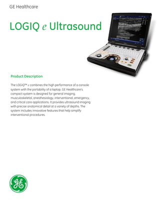
- 1. LOGIQ e Ultrasound Product Description The LOGIQ™ e combines the high performance of a console system with the portability of a laptop. GE Healthcare’s compact system is designed for general imaging, musculoskeletal, anesthesiology, interventional, emergency, and critical care applications. It provides ultrasound imaging with precise anatomical detail at a variety of depths. The system includes innovative features that help simplify interventional procedures. GE Healthcare
- 2. General Specification Console Dimensions Height 70 mm (2.75 in) console only 100 mm (3.94 in) with handle Width 295 mm (11.61 in) console only 343 mm (13.50 in) with handle Length 346 mm (13.62 in) console only 375mm (14.76 in) with handle Weight with Battery approximately 5.2 Kg (11.5 lbs) Console Electrical Power Voltage: 100-240 V AC Frequency: 50/60 Hz Power: Max. 130 VA Console Design Laptop Style Lithium Ion Battery Integrated Solid State Drive CPU – Intel Duo Core Docking Cart Dimensions Height: 810-950 mm (26.6-31.2 in) Width: 470 mm (15.4 in) Depth: 617 mm (20.2 in) Weight: 53 kg (116.8 lbs.) without accessories Isolation Cart Dimensions Height: 830-1130 mm (32.7-44.5 in) Width: 540 mm (21.3 in) Depth: 510 mm (20.1 in) Weight: 30.5 kg (67.1 lbs) without accessories User Interface Operator Keyboard Alphanumeric Keyboard Ergonomic Hard Keys Backlight Keys Display Screen 15 in High-Resolution Color LCD Resolution: 1024 x 768 Horizontal/Vertical viewing angle: +/-80 degree Brightness Adjustment Integrated Speakers Audio Volume Adjustment Interactive Dynamic Software Menu Console Interfaces DC Power Input USB 2.0 (3) LAN 10/100/1000 BaseT Docking Cart Connector HDMI Docking Cart Interfaces AC Power Input DVI USB 2.0 (4) Speakers 3 Probe Port (optional) Isolation Cart Interfaces AC Power Input 3 probe Port (optional) System Overview Applications Abdomen Cardiac Gynecology Musculoskeletal
- 3. System Overview (Continued) Applications (Continued) Obstetrical Nerve Block Pediatric Small Parts Urological Vascular Rheumatology Emergency Medicine Cardiac intra-operative Transducer Types Convex Array: C1-5-RS Microconvex Array: 8C-RS, E8C-RS Linear Array: 9L-RS, 12L-RS, L4-12t-RS, L8-18i-RS, L10-22-RS Phased Array: 3Sc-RS, 6S-RS TEE: 6Tc-RS Operating Modes B-Mode M-Mode Anatomical M-Mode/Color M-Mode (AMM) (optional) Color Flow Mode (CFM) Power Doppler Imaging (PDI) High-Res PDI (optional) Continuous Wave Doppler (CWD) (optional) Pulse Wave Doppler (PWD) Tissue Velocity Image/Tissue Velocity Doppler (TVI/TVD) (optional) Needle Recognition (optional) Standard Features Integrated Solid State Drive Automatic Optimization CrossXBeamTM Standard Features (Continued) Speckle Reduction Imaging (SRI-HD) Virtual Convex/Virtual Apex Fine Angle Steer HD Zoom (Write Zoom) Coded Harmonic Imaging (CHI) Raw Data Processing Quicksave On-board User Manual (Help) InSiteTM ExC capability Loop storage—from live scanning and from memory Patient Information Database Customizable User Interface Full M&A Calculation Package with Real Time Auto Doppler Calculations Vascular Calcs Cardiac Calcs OB Calcs and Tables Fetal Trending Multi Gestational Calcs Musculoskeletal and Hip Dysplasia Calcs Gynecological Calcs Urological Calcs Renal Calcs Small Parts Calcs Rheumatology Calcs Pediatric Calcs Report Writer Package Software Options easy3D LOGIQ View Needle Recognition Stress Echo eSmart Trainer Auto IMT DICOM® 3.0 Connectivity/Encrypted DICOM
- 4. System Overview (Continued) Software Options (Continued) Flow Quantitative Analysis Patient Follow-up Tool with fusion M-Color Flow Mode (CM) Anatomical M-Mode (AMM) TVI/TVD High-Res PDI Ophthalmic Hardware Options Footswitch Printer CWD USB ECG (AHA/IEC) Barcode Scanner External DVD R/W storage Cart Options Docking Cart 3 probe port Extended Life Battery Isolation Cart 3 probe port Storage Basket Storage drawer Display Modes Simultaneous Capability B/PW or TVI/TVD B/CFM or PDI B/M or AMM Dual B (B/B) Dual B + CFM or PDI Needle Recognition + CFM (PDI) Real-time Triplex Mode Selectable Alternating Modes Needle Recognition B/M B/PW B/CW B + CFM (PDI)/M B + CFM (PDI)/PW B + CFM (PDI)/CW 3D – Mode Display Modes (Continued) Multi Image Split Screen Live and/or frozen B + B/CFM or PDI Independent Cine playback Conventional or wide screen display Conventional or wide screen display Zoom: Read/Write Zoom Colorized Image Colorized B Colorized M Colorized PW Colorized CW Timeline Display Independent Dual B/PW/CW Display Display Formats: Top/Bottom or Side/Side selectable Format Size: Vert1/3 B; Vert1/2 B; Vert2/3 B; Horiz1/2 B; Horiz1/4 B; TL Only format, switchable after freeze Update mode: timed based on sweep Quad Screen Display access from Split Screen Virtual Convex Display Annotation Institution/Hospital Name Date: 3 types selectable MM/DD/YY, DD/MM/YY, YY /MM/ DD Time: 2 types selectable 24 hours, 12 hours Operator Identification Patient Name: First, Last, & Middle Patient Identification: 64 characters Gestational Age from LMP/EDD/GA/BBT Power Output Readout MI: Mechanical Index TIS: Thermal Index Soft Tissue TIC: Thermal Index Cranial (Bone) TIB: Thermal Index Bone System Status (real-time or frozen) Probe Orientation Marker: Coincides with orientation marking on the image monitor
- 5. System Overview (Continued) Display Annotation (Continued) Image Preview Gray/Color Bar Cine Gauge Measurement Summary Window Measurement Results Window: pre-settable display location Probe Type Application Name Imaging Parameters by Mode (current mode/see below) Focal Zone Markers Body Pattern B Scale Markers: 2 types; Depth/Width M Scale Markers: 2 types; Time/Depth, Time Image Management Menu: Menu, Delete, and Image Manager Image Palette Caps Lock: On/Off System Messages Display Trackball Functionality Status: Scroll, M&A (Measurement and Analysis), Position, Size, Scan Area Width, and Tilt Battery Status Biopsy Guide Line and Zone Heart Rate System Parameters System Setup User Programmable Preset Capability Factory Default Preset Data Factory Default Application Data Languages setup for UI: Brazilian Portuguese, Chinese, Danish, Dutch, English, Finnish, French, German, Greek, Italian, Japanese, Norwegian, Russian, Spanish and Swedish Languages for Manuals: Brazilian Portuguese, Chinese, Czech, English, French, German, Italian, Japanese, Spanish, Bulgarian, Croatian, Danish, Dutch, Estonian, Finnish, Greek, Hungarian, Indonesian, Korean, Latvian, Lithuanian, Norwegian, Polish, Portuguese, Romanian, Russian, Serbian, Slovakian, Swedish, Kazakh, Traditional Chinese, and Turkish Operation Error Message Display System Boot Up: < 25 sec Probe Loading: < 5 sec B-Mode Brightness mode. Real time displays of a two dimensional cross section of a three-dimensional soft tissue structure. Ultrasound echoes of different intensities are mapped to different gray scale or color values in the display. Scan Parameters B/M Acoustic Output: 0-100%; 2%, 5%, 10% increments Image Reverse: On/Off B Colorize: 9 types Thermal Index: TIC, TIS, TIB Focus Number: probe dependent, 8 in maximum Line Density: 5 increments: probe dependent Frame Average: 6 increments Edge Enhance: 8 increments Angle (deg): probe dependent, 10-131 degrees Gray Scale Map: 12 types Gain: 0–90 dB, 1 dB increments Dynamic Range: 36–96 dB, 3-6 dB increments Harmonics: on/off Virtual Convex: on/off Depth: 0.2-33 cm: probe dependent Focus Depth: 7-9 increments: probe dependent Rejection: 6 increments Frequency: 3-5 increments: probe dependent Color Flow Mode (CFM) or Color Doppler A real-time two-dimensional cross-section image of blood flow. Color gradient used to represent directional blood flow (velocity, variance, power and/or direction) prioritized over amplitude. Scan Parameters Base Line Invert: On/Off CF/PDI Focus Depth: 6 steps default pre-settable CF/PDI Acoustic Output: 0-100%; 2%, 5%, 10% increments Packet Size: 8-24: probe dependent
- 6. System Parameters (Continued) Color Flow Mode (CFM) or Color Doppler (Continued) Scan Parameters Line Density: 5 increments Frame Average: 7 increments PRF: 0.3K–22K Hz: probe dependent Spatial Filter: 6 steps Gain: 0–40 dB, 0.5 dB increments Wall Filter: 4 steps: application and probe dependent Angle/Width (deg, mm): probe dependent CF/PDI Vertical Size (mm): default pre- settable CF/PDI Center Depth (mm): default pre- settable CF/PDI Frequency: 2–4 steps: probe dependent CF/PDI Focal Number: 1 Color Map: 14 types at most: application and probe dependent Color Threshold: 10–100%, 10% increments Power Doppler Imaging Mode (PDI) Color gradient used to represent blood flow using amplitude shift vs. velocity shift (Color Doppler). Prioritizes amplitude over direction. Scan Parameters PDI Map: 14 types CF/PDI Acoustic Output: 0-100%; 2%, 5%, 10% increments Packet Size: 8-24: probe dependent Spatial Filter: 6 steps Frame Average: 7 steps: probe dependent PRF: 0.3K-11.4K Hz: depth dependent Power Threshold: 10–100%, 10% increments CF/PDI Vertical Size: default pre-settable CF/PDI Center Depth: default pre-settable CF/PDI Focal Number: 1 Gain: 0–40 dB, 0.5 dB increments Wall Filter: 4 increments: probe dependent CF/PDI Frequency: 2–4 increments: probe dependent High-Res PDI (Optional) Provides better hemodynamics visualization by combining effects of B-mode and color flow Doppler using a proprietary equation. Scan Parameters High-Res PDI Map: 11 types High-Res PDI Acoustic Output: 0-100%; 2%, 5%, 10% increments Packet Size: 8-20: probe dependent Spatial Filter: 6 steps Frame Average: 7 steps: probe dependent PRF: 0.2K-25K Hz: depth dependent Power Threshold: 10–100%, 10% increments High-Res PDI Focal Number: 1 Gain: 0–40 dB, 0.5 dB increments Wall Filter: 4 increments: probe dependent High-Res PDI Frequency: 2–3 increments: probe dependent Available on 9L-RS, 12L-RS, L4-12t-RS, L8-18i-RS and L10-22-RS probes M-Mode/Anatomical M-Mode (Optional) Motion mode. Soft tissue structure is presented as scrolling display, with depth on the Y-axis and time on the X-axis. Anatomical M-Mode (AMM) Allows M-Mode on stored 2D cine clip. Facilitates arrhythmia assessment and cardiac measurements. Scan Parameters Sweep Speed: 8 increments M Color: 9 types M/PW Display Format: V-1/3B, V-1/2B, V-2/3B, H-1/2B, H- 1/4B, TL Only B/M Acoustic Output: 0-100%; 2%, 5%, 10% increments Rejection: 6 increments Gray Scale Map: 12 types M Gain: +/- 20dB delta from B, 1dB increments
- 7. System Parameters (Continued) PW/CW Mode Pulse Wave Doppler (PW), Continuous Wave Doppler (CW) are used for displaying the speed, direction, and spectral content of blood flow at selected anatomical sites. Scan Parameters Maximum and Minimum Velocity Scales PW Max: 870 cm/s, 19,800 Hz Min: 15 cm/s, 700 Hz CW Max: 1,460 cm/s, 40,000 Hz Min: 40 cm/s, 2,100 Hz Gray Scale Map: 8 types Base Line: 5–95% SV Gate: 1, 2, 3, 4, 5, 6, 7, 8, 10, 12, 14, 16 mm: application dependent Angle Correct: +/- 90°, 1° step Spectral Color: 6 types PW Sweep Speed: 8 increments Invert: On/Off M/PW Display Format: V-1/3B, V-1/2B, V-2/3B, H-1/2B, H- 1/4B, TL Only PW Acoustic Output: 0–100%, 10% increments Spectral Averaging: 5 increments pre-settable Rejection: 15 increments Gain: 0-85 dB, 1 dB increments Wall Filter: 5.5-5,000, 500 Hz, 22 increments: probe and application dependent PW Angle Steer: 0 +/- 10, 15, 20° PRF: 700–19,800 Hz with PW, 2,100–40,000 Hz with CW Sample Volume Depth: 29.9 increments default pre-settable Audio Volume PW Frequency 2–4 steps: probe dependent M-Color Flow Mode Overlays color on the M-mode trace Coded Harmonic Imaging (Tissue Harmonics) (CHI) Enhances near field resolution for improved small parts imaging as well as far field penetration. Diminishes low frequency amplitude noise and provides clarity to needle, anatomy and motion TVI/TVD (Optional) Tissue Velocity Imaging calculates and color-codes the velocities in tissues Tissue Velocity Doppler provides spectral information for selected Doppler sample. eSmart Trainer (Optional) Provides modules showing basic scanning techniques with graphics of probe position, anatomy and example clinical images. Patient Follow-up Tool with fusion (Optional) For monitoring a patient condition over time. Automatically recalls the imaging parameters, comments and body patterns to be identical to your previous exam. Provides an alert if you use a different transducer than last time. Works in B-Mode, Color Mode and PDI Quick Save Single button push sends single image or entire patient exam to memory stick or network Report Writer (Optional) On-board reporting package automates report writing Formats various exam results into a report suitable for printing or reviewing on a standard PC Exam results include patient info, exam info, measurements, calculations, images, and comments Standard templates provided and allows for customization. Needle Recognition Mode (Optional) Provides accurate display of the needle, anatomy and motion even in Color and Power Doppler. Includes ability to adjust needle gain and angle. Available on all linear probes
- 8. System Parameters (Continued) 3D (Optional) Acquisition of Color data provides Automatic rendering of B mode and Color Flow Mode images in 3D. 3D Landscape 3D Movie Automatic Optimization Auto Tissue Optimization: ATO Auto Color Flow Optimization: ACO Auto Spectrum Optimization: ASO CrossXBeam Provides 3, 5, 7 or 9 angles of spatial compounding Live Side by Side DualView Display Compatible with: Color Mode, PW, SRI-HD, Coded Harmonic Imaging, Virtual Convex SRI-HD Speckle Reduction Imaging provides multiple levels of speckle reduction Compatible with/ B-Mode, Color, and 3D imaging LOGIQ View (Optional) Extended field of view imaging that allows viewing and measurement of anatomy that is larger than would fit in a single image possible. Requires manual sweep over anatomy of interest. Renders a panoramic image up to 60 cm, in long axis. It also allows you to see a wider field of view for comparing normal to abnormal anatomy. Virtual Convex Provides wider FOV in the far field Available on linear probes Virtual Apex Provides wider FOV in the near field Available on Sector probes Measurements and Calculations B-Mode Calcs Distance Circumference (Ellipse/Trace) Area (Ellipse/Trace) % Stenosis Angle between 2 lines Ratios Depth from Probe Surface M-Mode Calcs Distance Time Slope Heart Rate Doppler Calcs Velocity Frequency Time Acceleration Heart Rate Auto Doppler Trace function with automatic calcs Time averaged max/mean velocity Ratios PI (Pulsatility Index) RI (Resistivity Index) Vascular Measurements/Calculations Upper/Lower Artery/Vein Summary Worksheet Obstetrics Measurements/Calculations Gestational Age Calculation Multi-Gestational Calculation EFW Calculation Summary Worksheet Fetal Trend Graph
- 9. Measurements and Calculations (Continued) Gynecology Measurements/Calculations Ovarian Follicle Measurements Summary Worksheet Urology Measurements/Calculations Volume Measurements Summary Worksheet Musculoskeletal Measurements/Calculations Labeled measurements Cardiac Measurements/Calculations Ventricle, Atrium, Value Measurements Auto IMT. Automated measurement of the intimae media thickness of common carotid artery Summary Worksheet Quantitative Flow Analysis (Optional) Helps quantify and evaluate the blood flow within a region of interest, to assist with diagnosis and monitoring. Probes (all optional) C1-5-RS Wide Band Convex Applications: Abdomen, Obstetrics, Gynecology, Urology, Pediatric/Neonatal, Nerve Block, MSK, ED (FAST, Pleural) Imaging Frequency: 2.0–5.0 MHz Biopsy Guide: Multi-angle, disposable with a reusable bracket 8C-RS Wide Band Microconvex Applications: Abdomen, Basic Cardiac, Vascular, Pediatric/Neonatal, Small Parts, Nerve Block, MSK, Rheuma, ED (FAST, Pleural, Ophthalmic) Imaging Frequency: 4.2–11 MHz Biopsy Guide Not Available E8C-RS Wide Band Microconvex Applications: Obstetrics, Gynecology, Urology Imaging Frequency: 4.2–10MHz Biopsy Guide: Fixed Angle; Reusable Bracket, Disposable Sleeve 3Sc-RS Wide Band Phased Array Applications: Abdomen, Obstetrics, Gynecology, Basic Cardiac, Vascular, Urology, Pediatric/Neonatal, ED (FAST, Pleural, Orbits) Frequency: 1.7–4.0 MHz Biopsy Guide Available: Multi Angle, Reusable Bracket, Disposable Sleeve. 6S-RS Wide Band Phased Array Applications: Abdomen, Basic Cardiac, Vascular, Urology, Pediatric/Neonatal, ED (FAST, Pleural) Imaging Frequency: 3.0–7.0 MHz Biopsy Guide Not Available 9L-RS Wide Band Linear Applications: Abdomen, Vascular, Pediatric/Neonatal, Small Parts, Nerve Block, MSK, Rheuma, ED (Fast, Pleural) Imaging Frequency: 3.0–9.0 MHz Biopsy Guide: Multi Angle; Reusable Bracket, Disposable Sleeve 12L-RS Wide Band Linear Applications: Vascular, Pediatric/Neonatal, Small Parts, Nerve Block, MSK, Rheuma, ED (Fast, Pleural, Ophthalmic) Imaging Frequency: 4.2–13.0 MHz Biopsy Guide Multi Angle and Out-of-Plane; Reusable Bracket, Disposable Sleeve L4-12t-RS Wide Band Linear With Buttons Applications: Vascular, Pediatric/Neonatal, Small Parts, Nerve Block, MSK, Rheuma, ED (Fast, Pleural, Ophthalmic) Imaging Frequency: 4.2–13.0 MHz Biopsy Guide Multi Angle and Out-of-Plane; Reusable Bracket, Disposable Sleeve
- 10. Probes (all optional) (Continued) L8-18i-RS Wide Band Linear Applications: Vascular, Small Parts, Nerve Block, MSK, Rheuma, ED (Fast, Pleural) Imaging Frequency: 6.7–18.0 MHz Biopsy Guide Not Available L10-22-RS Wide Band Linear Applications: Vascular, Small Parts, Nerve Block, MSK, Rheuma Imaging Frequency: 10.0–22.0 MHz Biopsy Guide Not Available 6Tc-RS Wide Band Convex Applications: Cardiac intra-operative Imaging Frequency: 3–8 MHz Biopsy Guide: Not Available Safety Conformance LOGIQ e is: Certified to AAMI/ANSI ES60601-1:2005/(R)2012 Certified to CAN/CSA-C 22.2 No.60601-1:08 by an SCC accredited Test Lab CE Marked to Council Directive 93/42/EEC on Medical Devices Compliant with DIRECTIVE 2012/19/EU on Waste Electrical and Electronic Equipment (WEEE) requirement. Conforms to the following standards: IEC 60601-1 Electrical medical equipment IEC 60601-1-2 Electromagnetic compatibility IEC 60601-1-6 Medical Electrical Equipment—Part 6: General Requirements for safety—Usability IEC60601-2-37: Particular requirements for the safety of ultrasonic medical diagnostic and monitoring equipment ISO 10993 Biological evaluation of medical devices NEMA UD3 Acoustic output display (MI, TIS, TIB, TIC)
- 12. About GE Healthcare GE Healthcare provides transformational medical technologies and services to meet the demand for increased access, enhanced quality and more affordable healthcare around the world. GE (NYSE: GE) works on things that matter - great people and technologies taking on tough challenges. From medical imaging, software & IT, patient monitoring and diagnostics to drug discovery, biopharmaceutical manufacturing technologies and performance improvement solutions, GE Healthcare helps medical professionals deliver great healthcare to their patients. GE Healthcare 9900 innovation Drive Wauwatosa, WI 53226 U.S.A. 888 526 5144 www.gehealthcare.com DOC1720483 July 2015 ©2015 General Electric Company — All rights reserved. General Electric Company reserves the right to make changes in specifications and features shown herein, or discontinue the product described at any time without notice or obligation. Contact your GE Representative for the most current information. Not all features or specifications described in this document may be available in all probes and/or modes. GE Medical Systems Ultrasound & Primary Care Diagnostics, LLC, a General Electric Company, doing business as GE Healthcare. GE, GE Monogram, CrossXBeam, InSite, and LOGIQ are trademarks of General Electric Company or one of its subsidiaries. DICOM is the registered trademark of the National Electrical Manufacturers Association for its standards publications relating to digital communications of medical information. All other third party trademarks are the property of their respective owner. GE Healthcare, a division of General Electric Company. Europe GE Healthcare Beethovenstr. 239 D - 42655 Solingen T 49 212 2802 0 F 49 212 2802 28 Asia GE Healthcare Clinical Systems ASIA 1105-1108 Maxdo Center 8 XingYi Road, Shanghai 200336 T 86 21 5257 4640 F 86 21 5208 0582