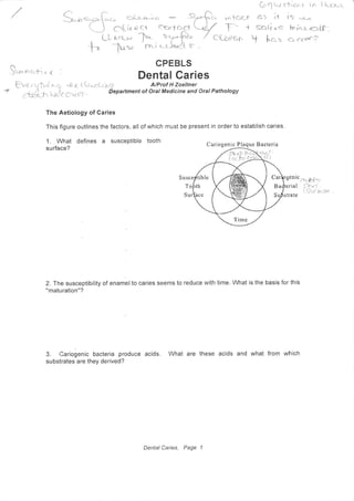
1. The Aetiology of Caries
This figure outlines the factors, all of which
1. What defines a suscePtible tooth
surface?
Dental Garies
Susce
must be present in order to establish caries.
Cariogenic Plaque Bacteria
G.iU" s'f.,o^r. rn I L-CL}
?- 11 lS "-r_r,
Sb(i cc h i,u-q=iS:
Y hct" ft 6'6"enl
1<t-tf
r c(r
stnttr*b*'t'
rial i):+f/-frc'r*se
CPEBLS
$**".-+, o,< :
,q. "t 6 t) tt*rr*r hO A/Prof H Zoellner
I_OICCV pf - Qepartment of Oral Medicine and Oral Pathology
T
S rate
2.fhe susceptibility of enamelto caries seems to reduce with time. What is the basis for this
"maturation"?
3. Cariogenic bacteria produce acids. What are these acids and what from which
substrates are they derived?
tible
Dental Caries, Page 1
2. how are they made?
iCCne
} nA t,'fur-rt e -s. &-t S-t-r cr6g '.{}r,-}rs_,*_f s &ciet s ? .
(-)- e*u--.ntz
gi tc, cr,evt r f7',tf
*'S,f*t*
t
G;ti."J. i'"
h
s'c,cchc.r cus=(
eh;t*" * frL,rcc,n f*r
^4ffiJi'Tiffi.F['-l-Bocteo.q rdLqs Ccia ?.rcc1r*c1'cn : ri-lrni,r//'-'- r-
bi&e',a
5 what is the signiricanl
",q,*J"tjil#"t1;".,hn '8"'#'fl 'tf 5ffi1f? $ocaries? &{ r'!c'(tq-.s? fit (?cr(t({
?.g*sccc$*"'<s, ai*.-Jx Scr'r:Syc-rt j^g;Qctr P*
*'* b&janq
'c Gl- iogz-,*; o fr..u *'r , o , (Drt ch T*SI*),fi2]-r o }:octe, ,o ,
(ituch
]::Agtlcc
ctJci.ts f*f C!*6ri1"1 . d{+*t i..-- c{
strcoes { q-*-r co,a
- -t=ef{-,t{ .
4. Some species of plaque bacteria, including Streptococcus mutans not only generate
acids, but also produce extracellular polysaccharides. What are these polysaccharides and
5SLUL"
6. What species other than Streptococcus mutans are thought to be important in caries?
-{
'?(
I e.f,o
C
horc(ti
a{ b{- cc( n s.
7. What is the acquired pellicle and how does it contribute to the formation of plaque?
Dental Caries, Page 2
3. L What are the factors in saliva inhibiting caries?
n Co'o , T&;- i F -
--<
r,rt',t*ng(eb"cLrun" .
-{
^1l
A an;a"$,[.*ty,,'i^ ,ll*"',1* I
+'
3.ff"t
" L+' **'- Jar.ys
+ ( 't Ec.fqnt-s
a * !*t iuc,'21
"-,X(csp
C^*o?* acflr;t-X) '
r rcLr€
S sardfury jcc-
Aruti.' [-relcterib-(
o-t{ tos-U f
hact&-,r.i
9. Caries does not progress at the same rate in all individuals or sites within the same
mouth. Some patients for example display rampant caries while others show only slowly
progressive caries or even have arrested lesions. Why is this so?
"t .$c,aat + t clu Q c'triorr c"*e 'Cor*-.*, lncryrci:
Og st<-,a- .
i-
c{-l
*r( --l( .-t-")*r'1 <-(r '
n.)
c-i--- ^.jh.*r, nr{c -t" 'ltc"cre-n'{l-t
x'€"f^ c11"L-1^'Tuslo * *541*r-$ist r3-:
rre ji* t'lli - it K o']. 'FcT'r,'ro"l
() .-{-tr.o.(}-* I
Dental Caries, Page 3
4. 10' what is the pattern of caries in patients with xerostomia and why?
cL11'comparetheclinicalimagesofdentalcariesprovided.
lnthespaceprovided,oulinethe essential differences betweei tfre two images.
suggest in which of the two cases, caries is likely to continue to progress more quickly.
Explain your answer.
1""g""- a, Ch"-o)
a.l)o e-0 e d.
itB
llA ^ ,,-s*LtDtr-
[/
la^f yr-plc;tr, moJ t
,-/
*-ec ,nc---ciriacrt *-r icq
al
cr c*lv q
*--Lc s.S C. C'cec.f
tr
F
fotu"
o (;. *.pr?T-c^;.^.{)*f n cr s,.o_.i#,* *
de;,k ; c o( rrc^,,. /ar.p.o- Cc-vJfc-1 .--
'dl
Dental Caries, page 4
5. f"puohc zc';
, tra{r , io r'c;s rd,,
c{*rllr. ?t-rrr
";c,irc-}"t t }clu
rec-CFtf-y c-.Li'i,
ci--r-**+.n-^- 1
Enamel Caries
12. This is a diagram illustrating
enamel caries. Label the: Surface of
the enamel, Translucent zone, Dark
zone, Body of lesion, Surface zone
f,-., r -j ---r-^'Ci,
4.)
i JJ:l-;
"-.-. .r3
!
J* 1); -'.r'^,-: Jr rr*! r
14. ln the dark zone:f'trq.'r,l^' " :. .1. r
#l{lu
'".T: '.-t-,'-l-i a.
-i-: . r
I "r"
l) What percentage of volume of lhe tissue is apparently porous?
p:t1 ,/',
--rr- '-fr*
#.HfrS*tu.ci*
ii) There is great variability in pore size in this zone with some pores being
"tLffJktdinthe translucent zone and others being much larger. How do you explain this observati?+
Kn *.'{r"Lr cil; ? h*i -it'i
' I 'J hr t l'-i--r' ci-L'6' ^!-
" ''''
,-,rr'#-?L,''1r rI E{ 1;"i.'tr
t 'Fn-, o -jr" , . -rr,n*,- -fi: -:'r.,r ;i"'. ' '/+-;,'*
wn -l:?:*::;:_,{...,
rf + ri..,r.,
, i*' J.. ,..'/./,, --^ -.,{ -7r.,-:.
hr^t i'oi ' ctr -'i+t*,i*a_..iL(. !i y'
Dental Caries, Page 5 {.{ (, lyG;' S'Lr**" <J ,rl.
[. (tc"_r- tr-tj
/)r,,at (.r{,-.ir;
;--; ,^i it l' ,.
"
r J..., ,
' ; r-r,,-"- )
--]
<-, ft"-*t""
6. 15. ln the bodY of the lesion:
l) What percentage volume of the tissue is apparentlY Porous?
*1=es s lYr-irr"A-n r"ut
s *t' c,c,rit
"Slr.
.#t?.#ffi?S :,
Many crystals in this zone are larger than in normal enam
CGtn". i-a Cn tclt' c ct Ctr
"*T
iii) The body of the lesion often becomes stained' what is the source of this stain?
6z( ff
16. The surface zone is relatively unaffected by caries. How do you explain this?
f61 c.,rtfoct tl'/ -tv:'(iva '-/ l-
///-
*--t h'rt A'
Cn-t s o go-", c*;r cq lti-^"r'*-l*- t=.t' t6--fi. -t
.t*1-' .$
, lcr-u
ti ,.tf -{r"c" I H:-rY*')
z
TUC.
l.
fe
17. Draw the shaPe of
i) a smooth surface enamel lesion and
ii).an enamel lesion confined to the
fissure.
What is the reason for the difference
in the shape of these two lesions?
Dental Caries, Page 6
7. 18.
ln enamel caries:
Why is the dark zohe "dark" and how is its width affected by the rate of caries?
CL
19. The given diagram illustrates a carious lesion as it progresses
the dentino-enamel junction. Label each of the four zones of
progresses.
from the earliest stage to
the enamel lesion as it
D
>.F-
:zex/
s r^*t ci*
"TPu-*&h;h "
l)At i,vhat stage of the enamel lesion does the lesion have a "white" appearance?
dYoqr C rLtu +0 htra $ Pf fu rt'bn
<*----r{-
"rir,t*te- qfrc.c'er.( <. -+' *}ot--F" *:: 5'ii) At which stage does the lesion sometirhSs become stained? h ltu-hclt s' 2E7- rr+",
7. J nOqr( {-Lnctr; nr"tn-nd. -|2-{ace. <2c,.-c. q-Dri-r-
J) ' S-$ C c.+cc,.r s S{-c..t'rv
-
qtb-au,^ rp$e .
'F"i6'?":Htn"o"ffi,r:ffi--hifl
fi nction,theresion"[,"uorraterary
along the junction. What colour does the enamel acquire at this stage?
Mcre
ee
orsC.u. oI. DfT N(%{i*-r.'ch
s"Qupt-'ar, JL V)-''"-.*^hJ
(ar *,i)
Dental Caries, Page 7
8. (' t-r*'&1, ffif;m),""., n".tli" n""#**i;ffi;;;;;7-- i i
24. Onefundamental difference between dentine caries and enamel caries is that because
dentine is cellular, the odontoblasts can respond to the caries irritant' Define in turn:
i) Sclerotic dentine
cc"'Lb rYii) Dead tracts
iii) Reactionary (secondary) dentine
25. This is a diagram of a
dentine carious lesion.
Label the: Enamel, Sound
dentine, Clinical cavitY, Zone
of sclerosis, Zone of
demineralisation, Zone of
invasion, Zone of Destruction,
Reactionary Dentine.
' ;*i
(.lrfci*,r)
'cj;51<.cft;
Dentat Caries, Page 9
9. iv) How do you explain these changes in colour?
cL 20. Examine the virtual micrograph of the ground section provided.
http: i /slidebox. ucc. usvd, ed u . auldsb/s napshotViewer. oh o?snaoshotlD= 48a30eeb6b3da4f50 b4Bfc4f3 1 c7f899
Draw the lesion in the space provided and label zones identified. ls this what you expect to
see? How do you explain any differences?
Dentine Caries
21. Dentine caries is mediated by bacteria invading the tissue. What structures do the
bacteria exploit to invade and penetrate the dentine?
22. How does this affect the pathway of a developing dentinal carious lesion?
Dental Caries, Page I
10. 26. How does the zone of sclerosis form?
27 . Nhal is the zone of demineralisation and what sort of bacteria are probably responsible
for forming this zone?
2g. What is the zone of invasion and what properties would bacteria in this area need to
have?
S-!e-
frlui'ocr.s T
*'ff
Qts
.j-"
29. Draw bacteria in'the zone of invasion using the
diagram provided as a guide. Label.dentinal tubules,
pioneer organisms and liquefaction foci.
30. Draw bacteria in the zone of destruction, labelling:
dentinal tubules, organisms, liquefaction foci and
transverse clefts.
ry;FT.':- {t*i}Sa -t'jsfi"''-.,
or ff"ti.,")
bcvcte-n., cc*n ,
Dentatcaries' Pase 10
-l-^ iv'c-Ct.a I a1'e rA-d hcOOCks S,
.Z
P"f cu^rLJ Jr'huJ-t: =
:h*^t('YSe
j
u4 , tu_
tf,
Cl3ruq mo, Fcr- Eara 66p 1. k>tr
c&ne etr */
"61-(.j-U(.l((+(+nc! . )
![how are they made?
iCCne
} nA t,'fur-rt e -s. &-t S-t-r cr6g '.{}r,-}rs_,*_f s &ciet s ? .
(-)- e*u--.ntz
gi tc, cr,evt r f7',tf
*'S,f*t*
t
G;ti."J. i'"
h
s'c,cchc.r cus=(
eh;t*" * frL,rcc,n f*r
^4ffiJi'Tiffi.F['-l-Bocteo.q rdLqs Ccia ?.rcc1r*c1'cn : ri-lrni,r//'-'- r-
bi&e',a
5 what is the signiricanl
",q,*J"tjil#"t1;".,hn '8"'#'fl 'tf 5ffi1f? $ocaries? &{ r'!c'(tq-.s? fit (?cr(t({
?.g*sccc$*"'<s, ai*.-Jx Scr'r:Syc-rt j^g;Qctr P*
*'* b&janq
'c Gl- iogz-,*; o fr..u *'r , o , (Drt ch T*SI*),fi2]-r o }:octe, ,o ,
(ituch
]::Agtlcc
ctJci.ts f*f C!*6ri1"1 . d{+*t i..-- c{
strcoes { q-*-r co,a
- -t=ef{-,t{ .
4. Some species of plaque bacteria, including Streptococcus mutans not only generate
acids, but also produce extracellular polysaccharides. What are these polysaccharides and
5SLUL"
6. What species other than Streptococcus mutans are thought to be important in caries?
-{
'?(
I e.f,o
C
horc(ti
a{ b{- cc( n s.
7. What is the acquired pellicle and how does it contribute to the formation of plaque?
Dental Caries, Page 2](data:image/gif;base64,R0lGODlhAQABAIAAAAAAAP///yH5BAEAAAAALAAAAAABAAEAAAIBRAA7)