

Recommended
More Related Content
What's hot
What's hot (20)
Similar to Physiology of Cornea
Similar to Physiology of Cornea (20)
More from Tukezban Huseynova, MD
More from Tukezban Huseynova, MD (15)
Recently uploaded
Recently uploaded (20)
Physiology of Cornea
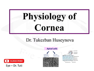
- 1. Physiology of Cornea Dr. Tukezban Huseynova Eye – Dr. Tuti Apical cells Small light cells (young cells) Dark cells (mature cells)
- 2. Part 1
- 4. Epithelium, 5-7 cells layers Apical Cells, 3-4 cells layer Wing cells, 1-3 cells layer Basal cells, 1 cells layer
- 5. Apical cells These cells finally degenerate and are sloughed from the corneal surface vThis process results In turnover of the entire Epithelium every 7 days Basal cells Wing cells
- 6. Apical cells Small light cells (young cells) Dark cells (mature cells)
- 7. Wing cells
- 8. Basal cells Major keratin pair K3 & K12 Minor keratin pair K14 & K5 ! The basal cell are distinguished from limbal stem cells by the expression of major Keratin pair
- 9. Basement Membrane vLamina Lucida (anteriorly, 23nm) vLamina densa (apposed to Bawmen’s layer) vReticular lamina (within to Bawmen’s layer)
- 10. Cell Adhesion in the Epithelium
- 11. Cell Adhesion in the Epithelium
- 12. Cell Adhesion in the Epithelium Junctional complexes of the cornea Gap junction Tight junction
- 13. Limbus Limbal epithelium 0.5 – 1 mm Basal layer Nonkeratinized, stratified squamous corneal cells Lies in superficial corneal epithelial layers Terminal differentiated cells Lies in the suprabasal layers Postmitotic cells Transient amplifying cell Migrate centrally within cornea to occupy its basal layer 64-kD keratin expression Stem cells Devision or Defferentiation Maintenance of the Corneal epithelium
- 14. Maintenance of the Corneal epithelium Y Y X X + Y = Z Z X, Y, Z Hypothesis of corneal epithelial maintenance. X - proliferation of basal cells Y - centripetal movement of cells Z - cell loss from the surface
- 15. Epithelium wound healing Injury Latent Phase Cell migration Cell proliferation Cell adhesion 3 steps of corneal healing
- 16. Epithelium wound healing Transient Amplifying cells (TA) Step 1: Cell migration
- 17. Epithelium wound healing Step 2: Cell proliferation Limbal Stem Cells Migration Proliferation and movement toward surface Migration
- 18. Epithelium wound healing Fonte: João Alfredo Kleiner DVM, MSc Clinical Case Patient: 11 y.o., female Complaints: severe ocular pain in the right eye Diagnosis: superficial traumatic ulcer Treatment: topical antibiotics, artificial tears clockwise vortex (whorl pattern)
- 19. Epithelium wound healing Hurrycane keratopathy
- 20. Epithelium wound healing Step 3: Cell adhesion Migratory epithelial cells Basal Lamina Adhesion plaqes
- 21. Clinical Manifestation of Abnormalities in Epithelium structure v Epithelial basement membrane dystrophy (dot changes) v Epithelial basement membrane dystrophy (fingerprints changes)
- 22. The main corneal biochemical components ü Fibronectin–integrin system Epithelial movement
- 23. The main corneal biochemical components ü Fibronectin–integrin system Epithelial movement ü Hyaluronan
- 24. The main corneal biochemical components ü Fibronectin–integrin system Epithelial movement ü Hyaluronan ü Proteolytic enzymes
- 25. The main corneal biochemical components ü Fibronectin–integrin system Epithelial movement ü Hyaluronan ü Proteolytic enzymes Cytokines and growth factors ü Epidermal growth factor
- 26. The main corneal biochemical components ü Fibronectin–integrin system Epithelial movement ü Hyaluronan ü Proteolytic enzymes Cytokines and growth factors ü Epidermal growth factor ü Transforming growth factor-β
- 27. The main corneal biochemical components ü Fibronectin–integrin system Epithelial movement ü Hyaluronan ü Proteolytic enzymes Cytokines and growth factors ü Epidermal growth factor ü Transforming growth factor-β ü Basic fibroblast growth factor (bFGF)
- 28. The main corneal biochemical components ü Fibronectin–integrin system Epithelial movement ü Hyaluronan ü Proteolytic enzymes Cytokines and growth factors ü Epidermal growth factor ü Transforming growth factor-β ü Basic fibroblast growth factor (bFGF) ü Interleukins
- 29. Part 2
- 31. Modified region of anterior stroma Characteristics Thickness is 8 – 14 μm Acellular homogeneous zone It lacks fibroblast therefore after injury it is unable to regenerate- replaced by course scar tissue Anterior surface of this layer is smooth The collagen fibres in Bowman's layer are synthesized and secreted by stromal keratocytes
- 32. Prevents keratocytes metaplasia to fibroblast and scar formation Anchoring site for epithelial cells to ensure stability Function Prevents stromal keratocytes from exposure to epithelial growth factors Tough acellular layer provide mechanical supports E – Epithelium S – Stromal Stromal collagen fibers B – Bowman's layer Bar = 2 μm
- 33. III. STROMA
- 34. Stromal Structure Made up of 200 – 250 lamellas of collagen fibers The lamellas formed of bundles of collagen fibers They stretch from limbus to limbus At the limbus they turn and run circumferentially forming an annulus 1.5 to 2.0 mm wide around the cornea (which maintains the curvature of the cornea) This has profound implication for possible alterations of stromal structure during refractive or cataract surgery that may lead to possible refractive errors
- 36. Proteoglycans The extrafibrillar material sometimes called the Ground Substance of the stroma, is largely made up of CD/DS Decorin (type IV) is the only CD/DS proteoglycan in the cornea. More abundant in the anterior cornea than in the posterior. KS More abundant in the posterior stroma. ü Lumican ü Keratocan ü Mimecan v CD/DS - chondroitin sulfat/dermatan sulfat v KS - keratan sulfat
- 37. Proteoglycans Function Confer hydrophilic properties of stroma Maintains corneal transparency Helps in regular spacing of collagen fibers to ensure transparency
- 38. Keratocytes Cellular component of the corneal stroma Turn over every 2 to 3 days Synthesize collagen The cells are activated when the stroma is damaged Differentiate into fibroblast during wound healing There is a gap junction between cells
- 39. Corneal Transparency and Stromal Function Interwoven fibrous collagen The regular arrangement of collagen mechanical strength transparency of this tissue Fibers of regular diameter Interfibrillar distance less than a Wavelength of light Avascularity Relative dehydration
- 40. Interactions between Proteogycans and Collagen in the Stroma Granular Dystrophy
- 41. Interactions between Proteogycans and Collagen in the Stroma Granular Dystrophy Lattice Dystrophy
- 42. Interactions between Proteogycans and Collagen in the Stroma Granular Dystrophy Lattice Dystrophy Macular type 1 Dystrophy
- 44. Part 3
- 46. A The anterior part 3mm P The posterior part which is secreted after birth Thickness Note! There is a distinct structural difference between fetal & postnatal components at birth: 3 – 4 µm at childhood: about 5 µm at adult: 10 – 12 µm
- 47. Hassal-Henle warts Descemet’s membrane abnormalities
- 48. Hassal-Henle warts Corneal Guttata Descemet’s membrane abnormalities
- 49. Hassal-Henle warts Corneal Guttata Descemet’s membrane abnormalities Posterior Embryotoxon
- 50. Spreading endothelial cells Descemet’s wound healing Injury Synthesis of fresh basal lamina
- 51. V. ENDOTHELIUM
- 53. Endothelial Cell Dencity, ECD At birth: about 6000 cells/mm² At adults: about 2500 cells/mm²
- 54. Endothelial Cell Dencity, ECD The "+" indicates a larger cell, the "-" indicates a smaller cell, and the numbers indicate the number of sides forming the cell
- 55. Ultrastructural features 3-D view of deep cornea showing part of endothelium, DM, Stroma
- 56. Nutrition of Endothelium Endothelium gets its nutrition & O₂ from aqueous Essential nutrients (such as glucose & amino acids) pass across its surface to supply the cellular needs of all the corneal layers
- 57. Fluid regulation of Endothelium Providing a barrier function to the ingress of salt and metabolites to the stroma Actively reducing the osmotic pressure of stroma by metabolically pumping the bicarbonate ions out of the stroma to aqueous The state of relative clearence of stroma is maintained by this delicate monolayer of cells by two ways
- 58. Endothelial Repair Physical & chemical damage to endothelium results in loss of cells Neighbor cells move over to fill the gap by sliding process and enlargement of cells occur Thus, after injury, the endothelial cell density falls, the cell area increases and the cell height decreases