SlideShare a Scribd company logo
1 of 59
Mucocele
• Salivary ducts, especially those of the minor salivary glands, are
occasionally traumatized, commonly by lip biting, and severed
beneath the surface mucosa. Subsequent saliva production may
then extravasate beneath the surface mucosa into the soft
tissues.
• Over time, secretions accumulate within the tissues and produce
a pseudocyst (without a true epithelial lining) that contains thick,
viscous saliva. These lesions are most common in the mucosa of
the lower lip and are known as mucoceles
1
2
• The second most common site of mucocele formation is the
buccal mucosa.
• Mucocele formation results in an elevated, thinned, stretched
overlying mucosa that appears as a vesicle filled with a clear or
blue-gray mucus.
• The patient frequently relates a history of the lesion filling with
fluid, rupture of the fluid collection, and refilling of these lesions.
• Some instances of mucocele formation regress spontaneously
without surgery. For persistent or recurrent lesions, the preferred
treatment consists of excision of the mucocele and the associated
minor salivary glands that contributed to its formation
3
A, Excision of mucocele of right
lower lip.
B, Gross specimen of intact
mucocele is demonstrated.
4
• Usually; local anesthesia is administered via a mental nerve
block, and an incision is made through the mucosa.
• Careful dissection around the mucocele may permit its
complete removal; however, in many cases the thin lining
ruptures and decompresses the mucocele before removal.
• The regional associated minor salivary glands are removed
as well and sent for histopathologic evaluation. The
recurrence rates of mucoceles may be as high as 15% to
30% after surgical removal, possibly caused by incomplete
removal or repeat trauma to the minor salivary glands.
5
6
• The most common lesion of the sublingual gland is the ranula,
which may be considered a mucocele of the sublingual salivary
gland.
• Ranulas result from mucous retention in the sublingual gland
ductal system or mucous extravasation as a result of ductal
disruption.
• The two types of ranulas are the simple ranula and the plunging
ranula.
• The simple ranula is confined to the area occupied by the
sublingual gland in the sublingual space, superior to the
mylohyoid muscle.
7
Left floor of mouth
ranula
Bilateral floor of mouth
ranula.
8
• The progression to a plunging ranula occurs when the
lesion extends beyond the level of the mylohyoid muscle
into the submandibular space .
• Ranulas may reach a larger size than mucoceles because
their overlying mucosa is thicker and because trauma that
would cause their rupture is less likely in the floor of the
mouth.
• As a result, a plunging ranula has the potential to extend
through the mylohyoid muscle into the neck and to
compromise the airway, resulting in a medical emergency.
9
Right plunging ranula through the mylohyoid muscle seen on
computed tomography scan (arrow).
10
• The differential diagnosis of a floor of mouth swelling includes ranula,
lymphoepithelial cyst, epidermoid or dermoid cyst, salivary gland tumors (e.g.
, mucoepidermoid carcinoma) , and mesenchymal tumors (e.g. , lipoma,
neurofibroma, or hemangioma) .
• The differential diagnosis of a midline neck mass includes thyroid
enlargement (i. e . , goiter or tumor) , thyroglossal duct cyst, dermoid cyst,
and plunging ranula.
• The differential diagnosis of a lateral neck mass includes lymphadenopathy,
epidermoid cyst, lipoma, infectious mononucleosis, metastatic carcinoma,
lymphoma, salivary gland tumors (e.g. , submandibular gland or tail of the
parotid gland), submandibular gland sialadenitis, lymphoepithelial cyst,
sarcoidosis, tuberculosis, cat-scratch disease, cystic hygroma, carotid body
tumor, or plunging ranula.
• The usual treatment of the ranula is marsupialization, in which a portion of
the oral mucosa of the floor of the mouth is excised, along with the superior
wall of the ranula. 11
A, Ranula in the right
floor of mouth caused by
accumulation of
sublingual gland
secretions in soft tissues
resulting from rupture of
salivary duct.
B, Diagram of
marsupialization
incision.
C, Marsupialization of
ranula, with excision of
oral mucosa and superior
wall of ranula.
D, Completion of
marsupialization of left
floor of mouth ranula with
placement of
circumferential
sutures.
E, Diagram of completed
marsupialization.
12
• Subsequently, the ranula wall is sutured to the oral
mucosa of the floor of the mouth and allowed to
heal by secondary intention.
• The preferred treatment for recurrent or persistent
ranulas is excision of the ranula and sublingual
gland via an intraoral approach; several recent
studies have indicated that this might be
appropriate for initial therapy.
13
14
SALIVARY GLAND INFECTIONS
• Infections of the major salivary glands can be acute or chronic
and are commonly, but not always, related to obstructive disease,
especially in the submandibular gland (obstruction leads to
infection) .
• The cause of acute suppurative sialadenitis of the parotid gland
usually involves a change in fluid balance that is likely to occur in
patients who are elderly, debilitated, malnourished, dehydrated, or
plagued with chronic illness.
• In these cases, gland infections are usually bilateral. The mean
age of occurrence of infections is 60 years, with a slight male
predilection. 15
• Salivary gland infections may be caused by a variety of
organisms, including aerobic and anaerobic bacteria,
viruses, fungal organisms, and mycobacteria. In most
cases, mixed bacterial flora is responsible for sialadenitis.
• The Single most common organism implicated in salivary
gland infection is Staphylococcus aureus because this
organism normally colonizes around ductal orifices.
• In addition, during instances of decreased or slowed
salivary flow (i.e., obstruction or dehydration) , retrograde
influx of S. aureus into the ductal system and gland
occurs and results in infection.
16
• The clinical characteristics of acute bacterial
salivary gland infections include rapid onset of
swelling in the preauricular (parotid gland) or
submandibular regions, with associated erythema
and pain.
• Palpation of the involved gland reveals no flow or
elicits a thick, purulent discharge from the orifice of
the duct.
17
A, Left parotid gland infection.
B, Right acute bacterial parotitis with erythema. This infection is extremely
painful and may indicate another serious illness. Treatment requires
hospitalization, intravenously administered antibiotics, and possibly surgical
drainage. 18
Purulent discharge from left parotid duct is demonstrated in
patient with an infection involving parotid gland.
19
• Treatment of bacterial salivary gland infections includes
symptomatic and supportive care, including intravenous
fluid hydration, antibiotics, and analgesics.
• Initial empirical antibiotics should be aimed at the most
likely causative organism, S. aureus, and should include a
cephalosporin (first generation) or antistaphylococcal
semisynthetic penicillin (oxacillin or dicloxacillin) .
• Culture and sensitivity studies of purulent material should
be obtained to aid in selecting the most appropriate
antibiotic for each patient.
20
• Antibiotics should be administered intravenously in high doses for
the majority of these patients, who ordinarily require
hospitalization.
• On most occasions, surgery consisting of incision and drainage
becomes necessary in the management of salivary gland
infections. Untreated infections may progress rapidly and can
cause respiratory obstruction, septicemia, and eventually, death.
• In some instances of recurrent salivary gland infection, the
repeated insults result in irreversible functional impairment of
gland function, and excision of the gland may be indicated.
21
• Viral parotitis, or mumps, is an acute, nonsuppurative
communicable disease. Before routine vaccination (e.g.,
measles, mumps, and rubella vaccine) against the
disease began, viral parotitis occurred in epidemics during
the winter and spring.
• Differentiation of viral from bacterial salivary gland
infection is important because viral infections are not the
result of obstructive disease and require different
treatment, not including antibiotics.
22
• Mumps is characterized by a painful, nonerythematous
swelling of one or both parotid glands that begins 2 to 3
weeks after exposure to the virus (incubation period) .
• This disease occurs most commonly in children between
ages 6 and 8. The signs and symptoms of mumps include
preauricular pain and swelling, fever, chills, and
headache.
23
• Viral parotitis usually resolves in 5 to 12 days after its
onset. Supportive and symptomatic care for fever,
headache, and malaise with antipyretics, analgesics, and
adequate hydration treats viral parotitis.
• Complications of the disease include meningitis,
pancreatitis, nephritis, orchitis, testicular atrophy, and
sterility in approximately 20% of young males affected.
24
NECROTIZING SIAlOMETAPlASIA
• Necrotizing sialometaplasia is a reactive, nonneoplastic
inflammatory process that usually affects the minor salivary
glands of the palate. However, it may involve minor salivary
glands in any location.
• Necrotizing sialometaplasia is of unclear origin but is thought to
result from vascular infarction of the salivary gland lobules.
• Potential causes of diminished blood flow to the affected area
include trauma, local anesthetic injection, smoking, diabetes
mellitus, vascular disease, and pressure from a denture
prosthesis. The usual age range of affected patients is between
23 and 66 years.
25
• Lesions usually appear as large ( 1 to 4 cm) , painless or
painful, deeply ulcerated areas lateral to the palatal
midline and near the junction of the hard and soft palate.
• Although lesions are usually unilateral, bilateral
involvement may occur. Some patients may report a
prodromal flulike illness before the onset of the ulceration.
26
A, Necrotizing sialometaplasia of posterior palate with ulceration.
27
• This condition is of considerable concern because,
clinically and histologically, it resembles a malignant
carcinoma (squamous cell or mucoepidermoid
carcinoma).
• The appropriate diagnosis and management of this
disease relies on evaluation by an oral maxillofacial
surgeon and pathologist who are familiar with this entity
because the result of a misdiagnosis may be extensive,
unwarranted surgical resection.
28
• Helpful histologic criteria for distinguishing necrotizing
sialometaplasia from a malignant process include the
maintenance of the overall salivary lobular morphology, the
generally nondysplastic appearance of the squamous islands or
nests, and evidence of residual ductal lumina within the epithelial
nests.
• The ulcerations of necrotizing sialometaplasia usually heal
spontaneously within 6 to 10 weeks after their onset and require
no surgical management.
29
SJOGREN'S SYNDROME
• Sjogren's syndrome (SS) is a multisystem disease process with a
variable presentation. The two types of SS are
• (1) primary SS, or sicca syndrome, characterized by xerostomia
(dry mouth) and keratoconjunctivitis sicca (dry eyes) ; and
• (2) secondary SS, which is composed of primary SS and an
associated connective tissue disorder, most commonly
rheumatoid arthritis.
• Although the cause of SS is unknown, there appears to be a
strong autoimmune influence. SS shows a female predilection of 9
: 1 , with more than 80% of affected individuals being females with
a mean age of 50 years.
30
Sjogren's syndrome showing
(A) dry eyes
(keratoconjunctivitis sicca) and
(B) dry mouth (xerostomia).
31
• Generally, the first symptoms to appear are arthritic complaints,
followed by ocular symptoms, and late in the disease process,
salivary gland symptoms.
• The involvement of the salivary and lacrimal glands results from a
lymphocytic replacement of the normal glandular elements. The
xerostomia results from a decreased function of the major and
minor salivary glands, with the parotid gland being the most
sensitive.
• The diagnosis of SS is suggested by the patient's complaints and
by a variety of abnormal immunologic laboratory tests.
32
• The oral component of SS may be diagnosed using
salivary flow rate studies and sialograms that can show
acinar destruction.
• The use of a labial minor salivary gland biopsy, as
mentioned previously, currently is considered to be highly
accurate in aiding the diagnosis.
• The histopathologic changes seen in the minor glands are
similar to those in the major glands (parotid).
• Keratoconjunctivitis sicca is suggested by the patient's
complaints and a Schirmer's test for lacrimal flow.
33
Schirmer's test for dry eyes in a patient with Sjogren's
syndrome. Filter paper is placed in the ocular fornix and observed for
"wetting" to a certain distance within a specific time limit.
34
• The treatment for SS includes symptomatic care with artificial
tears for the dry eyes and salivary substitutes for the dry mouth.
• Additionally, the medication pilocarpine (Salagen) or the Biotene
products may be useful to stimulate salivary flow from the
remaining functional salivary gland tissue
35
TRAUMATIC SALIVARY
GLAND INJURIES
• Traumatic injuries, particularly lacerations, involving the salivary
glands and their ducts may accompany a variety of facial injuries,
including fractures.
• Injuries that occur in proximity to one of the major salivary glands
or ducts require careful evaluation.
36
• Facial lacerations may involve not only the gland and its
ductal system but also branches of the facial nerve and
branches of major facial vessels.
• These structures require meticulous attention for
appropriate diagnosis and prompt repair.
• Usually, facial nerve lacerations that are anterior to a
vertical line from the lateral canthus of the eye to the
mental foramen are not amenable to surgical repair.
37
A, Diagram showing the
position of Stensen's
duct along a line drawn
from the tragus to the
middle of the upper lip.
B, Injuries to the terminal
branches of the facial
nerve anterior to line do
not require repair, and
function usually returns.
38
• Stensen's ductal repair may include ductal anastomoses, in which the
proximal and distal portions of the duct are identified, a plastic or metal
catheter is placed as a stent, and the duct is sutured over the stent.
• The catheter usually remains in place for 10 to 14 days for epithelialization of
the duct to occur.
• Additionally, nerve anastomoses may be required and performed by placing
epineurial sutures, using magnification, to reapproximate the nerve stumps.
• The lacerations are closed in a usual layered fashion, after debridement of
the soft tissue wounds to cleanse the site of entrapped particles, such as
glass or dirt.
• Potential sequelae of trauma involving the major salivary glands include
infection, facial paralysis, cutaneous salivary gland fistula, sialocele
formation, and duct obstruction as a result of scar formation, with eventual
glandular atrophy and decreased function. The involved gland may
eventually require surgical removal.
39
40
NEOPLASTIC SALIVARY GLAND DISORDERS
• Although a comprehensive discussion of salivary gland neoplasms is
beyond the scope of this chapter and many other sources are available
for this information, a brief review of several important aspects of the
more common lesions is warranted.
• Salivary gland tumors occur much more commonly in the major glands
(80% to 85%) as opposed to the minor glands ( 15% to 20%) .
• Additionally, between 75% and 80% of major gland tumors are benign,
whereas 50% to 55% of minor gland tumors are benign. The
overwhelming majority of salivary tumors occur in the parotid gland,
and the majority of those are benign (mostly pleomorphic adenomas) .
41
42
Benign Salivary Gland Tumors
• The pleomorphic adenoma, or benign mixed tumor, is the most
common salivary gland tumor. The mean age of occurrence is 45
years, with a male-to-female ratio of 3:2 .
• In the major glands the parotid gland is involved in more than 80%
of cases; in the minor glands the most common intraoral site is
the palate.
• Pleomorphic adenomas are usually slow growing, painless
masses. The histopathologic examination shows two cell types:
(1) the ductal epithelial cell and (2) the myoepithelial cell, which
may differentiate along a variety of cell lines (pleomorphic means
many forms) . 43
• A connective tissue capsule exists, which may be incomplete.
The treatment involves complete surgical excision with a
margin of normal uninvolved tissue.
• Parotid lesions are treated with removal of the involved lobe
along with the tumor. Recurrence is possible in rare occasions,
as well as a small risk (5%) of malignant transformation to a
carcinoma ex pleomorphic adenoma.
44
Pleomorphic adenomas. A, Palate. B to D,
Parotid gland. E, Submandibular gland.
45
• Warthins tumor, or papillary cystadenoma lymphomatosum,
almost exclusively affects the parotid gland, specifically the tail of
the parotid gland.
• The peak incidence is in the sixth decade of life, with a male-to-
female ratio of 7 : 1 . This lesion presents as a slow-growing, soft,
painless mass .
• Warthin's tumor is believed to be caused by entrapped salivary
epithelial rests within developing lymph nodes.
• The histopathologic examination shows an epithelial component
in a papillary pattern and a lymphoid component with germinal
centers. The treatment of this lesion is simple surgical excision,
and recurrence is rare.
46
Warthin's tumor of the tail of the parotid gland.
47
• The monomorphic adenoma is an uncommon solitary lesion
composed of one cell type, affecting predominantly the upper lip
minor glands (canalicular adenoma) and the parotid gland (basal
cell adenoma) .
• The mean age of occurrence is 61 years, and the lesion usually
presents as an asymptomatic, freely movable mass. The
histopathologic examination reveals an encapsulated lesion
composed of one type (monomorphic) of salivary ductal epithelial
cell. The treatment is simple surgical excision.
48
Monomorphic (canalicular) adenoma of the left upper
lip/vestibule.
49
Malignant Salivary Gland Tumors
• The mucoepidermoid carcinoma is the most common malignant
salivary gland tumor.
• The tumor makes up 10% of major gland tumors (mostly parotid)
and 20% of minor gland tumors (mostly palatal) . This lesion may
occur at any age, but the mean age is 45 years. The male-to-
female ratio is 3 : 2 .
• The clinical presentation is a submucosal mass that may be
painful or ulcerated. The mass may appear to have a bluish tinge
because of the mucous content contained within the lesion.
50
A, Mucoepidermoid
carcinoma of the
palate (note bluish
tinge from mucin
content).
B, Mucoepidermoid
carcinoma of
the palate with
ulceration.
51
• An intraosseous form of mucoepidermoid carcinoma may present
as a multilocular radiolucency of the posterior mandible.
• The histopathologic examination shows three cell types: (1)
mucous cells, (2) epidermoid cells, and (3) intermediate (clear)
cells.
• The proportion of each cell type helps to grade the
mucoepidermoid carcinoma as high-, intermediate-, or low-grade
lesions.
• The higher the grade, the more predominance of epidermoid cells
and pleomorphism, lack of mucous cells and cystic areas, and
overall more aggressive behavior.
52
• The treatment of low-grade lesions is wide surgical excision with a
margin of uninvolved normal tissue; high grade lesions require
more aggressive surgical removal with margins, and possibly,
local radiation therapy.
• The low-grade lesions have a 95% 5-year survival rate, whereas
the high-grade lesions have less than a 40% 5-year survival rate.
53
A, Central mucoepidermoid
carcinoma of the right
retromolar pad minor salivary
glands.
B, Panoramic radiograph
showing an underlying
multilocular radiolucency.
54
• The polymorphous low-grade adenocarcinoma is the second most common
intraoral salivary gland malignancy. This lesion was first described in 1983;
before its identification, many cases were probably misdiagnosed as adenoid
cystic carcinoma.
• The most common site is the junction of the hard and soft palates. The male-
to-female ratio is 3 : 1 , with a mean age of 56 years. These tumors present
as slow-growing, asymptomatic masses that may be ulcerated. The
histopathologic examination shows many cell shapes and patterns
(polymorphous) .
• Patients experience an infiltrative proliferation of ductal epithelial cells in an
"Indian file" pattern. This lesion shows a predilection for invasion of
surrounding nerves. The treatment of this tumor is wide surgical excision,
with a relatively high recurrence rate of 14%.
55
Polymorphous low-grade adenocarcinoma of the
palate.
56
• The adenoid cystic carcinoma is the third most common intraoral
salivary gland malignancy, with a mean age of 53 years and a
male-to-female ratio of 3:2 .
• Approximately 50% of these tumors occur in the parotid gland,
whereas the other 50% occur in the minor glands of the palate.
• These tumors present as slow-growing, nonulcerated masses,
with an associated chronic dull pain.
57
Adenoid cystic carcinoma of the palate.
58
• Occasionally, parotid lesions may result in facial paralysis
as a result of facial nerve involvement
• The histopathologic examination demonstrates an
infiltrative proliferation of basaloid cells arranged in a
cribriform (Swiss cheese) pattern. As seen in the
polymorphous low-grade adenocarcinoma, there may be
perineural invasion.
• The treatment is wide surgical excision, followed in some
cases by radiation therapy The prognosis is poor despite
aggressive therapy
59

More Related Content

What's hot (20)

Oroantral fistula
Oroantral fistulaOroantral fistula
Oroantral fistula
 
Oroantral communication & fistula
Oroantral communication & fistulaOroantral communication & fistula
Oroantral communication & fistula
 
Lefort 1 fracture
Lefort 1 fracture Lefort 1 fracture
Lefort 1 fracture
 
Gingiva
GingivaGingiva
Gingiva
 
Pre prosthetic surgery
Pre prosthetic surgeryPre prosthetic surgery
Pre prosthetic surgery
 
Salivary glands disorders i
Salivary glands disorders iSalivary glands disorders i
Salivary glands disorders i
 
Odontogenic Cysts
Odontogenic CystsOdontogenic Cysts
Odontogenic Cysts
 
Gingival inflammation & clinical features of gingivitis
Gingival inflammation & clinical features of gingivitisGingival inflammation & clinical features of gingivitis
Gingival inflammation & clinical features of gingivitis
 
Principles of management and prevention of Odontogenic Infections
Principles of management and prevention of Odontogenic Infections Principles of management and prevention of Odontogenic Infections
Principles of management and prevention of Odontogenic Infections
 
Management of oral cyst
Management of oral cystManagement of oral cyst
Management of oral cyst
 
ANUG
ANUGANUG
ANUG
 
Management of oroantral fistula
Management of oroantral fistulaManagement of oroantral fistula
Management of oroantral fistula
 
Developmental disturbances of tongue
Developmental disturbances of tongueDevelopmental disturbances of tongue
Developmental disturbances of tongue
 
Midface fractures
Midface fracturesMidface fractures
Midface fractures
 
periapical radiolucencies
 periapical radiolucencies periapical radiolucencies
periapical radiolucencies
 
Management of impacted3rd molar
Management of impacted3rd molarManagement of impacted3rd molar
Management of impacted3rd molar
 
Bsso
BssoBsso
Bsso
 
Management of Mandibular Fractures
Management of Mandibular FracturesManagement of Mandibular Fractures
Management of Mandibular Fractures
 
Exodontia
Exodontia Exodontia
Exodontia
 
Mandibular Angle Fractures
Mandibular Angle FracturesMandibular Angle Fractures
Mandibular Angle Fractures
 

Similar to Lecture 5 Diagnosis and management of salivary gland disorders Part 2

Diseases of-salivary-glands
Diseases of-salivary-glands Diseases of-salivary-glands
Diseases of-salivary-glands Weam Faroun
 
Salivary Glands Disorders
Salivary Glands DisordersSalivary Glands Disorders
Salivary Glands DisordersHadi Munib
 
Salivary Gland Diseases - A Summary.pptx
Salivary Gland Diseases - A Summary.pptxSalivary Gland Diseases - A Summary.pptx
Salivary Gland Diseases - A Summary.pptxssusere4339d
 
Pediatric soft tissue lesions/certified fixed orthodontic courses by Indian d...
Pediatric soft tissue lesions/certified fixed orthodontic courses by Indian d...Pediatric soft tissue lesions/certified fixed orthodontic courses by Indian d...
Pediatric soft tissue lesions/certified fixed orthodontic courses by Indian d...Indian dental academy
 
Seminar 14-15-salivary gland-diseases
Seminar  14-15-salivary gland-diseasesSeminar  14-15-salivary gland-diseases
Seminar 14-15-salivary gland-diseasesIndian dental academy
 
Abdominal tuberculosis
Abdominal tuberculosisAbdominal tuberculosis
Abdominal tuberculosisVikas Kumar
 
Cholecystitis & carcinoma of gallbladder
Cholecystitis & carcinoma of gallbladder Cholecystitis & carcinoma of gallbladder
Cholecystitis & carcinoma of gallbladder Baiti Basheer
 
Salivary gland diseases
Salivary gland diseasesSalivary gland diseases
Salivary gland diseasesEsraa Bahjat
 
Tuberculosis of peripheral lymph nodes
Tuberculosis of peripheral lymph nodesTuberculosis of peripheral lymph nodes
Tuberculosis of peripheral lymph nodesLadi Anudeep
 
Diseases of salivary glands
Diseases of salivary glandsDiseases of salivary glands
Diseases of salivary glandsYaqoob Marri
 
Non plaque induced gingivitis
Non plaque induced gingivitisNon plaque induced gingivitis
Non plaque induced gingivitispardishaddadi1
 
Head and Neck Space Infections.POWERPOINT
Head and Neck Space Infections.POWERPOINTHead and Neck Space Infections.POWERPOINT
Head and Neck Space Infections.POWERPOINTdrskbarla
 
Diverticulosis and diverticular disease
Diverticulosis and diverticular diseaseDiverticulosis and diverticular disease
Diverticulosis and diverticular diseaseDoha Rasheedy
 
Salivary Gland Diseases.pptx
Salivary Gland Diseases.pptxSalivary Gland Diseases.pptx
Salivary Gland Diseases.pptxEidleMohamedsaed
 
Granulomatous disease of nose
Granulomatous disease of noseGranulomatous disease of nose
Granulomatous disease of noseRITURAJANMBBS
 
PHYSIOTHERAPY IN PELVIC INFLAMMATORY DISEASE.pptx
PHYSIOTHERAPY IN PELVIC INFLAMMATORY DISEASE.pptxPHYSIOTHERAPY IN PELVIC INFLAMMATORY DISEASE.pptx
PHYSIOTHERAPY IN PELVIC INFLAMMATORY DISEASE.pptxMuskan Rastogi
 

Similar to Lecture 5 Diagnosis and management of salivary gland disorders Part 2 (20)

Diseases of-salivary-glands
Diseases of-salivary-glands Diseases of-salivary-glands
Diseases of-salivary-glands
 
Salivary Glands Disorders
Salivary Glands DisordersSalivary Glands Disorders
Salivary Glands Disorders
 
Salivary Gland Diseases - A Summary.pptx
Salivary Gland Diseases - A Summary.pptxSalivary Gland Diseases - A Summary.pptx
Salivary Gland Diseases - A Summary.pptx
 
Pediatric soft tissue lesions/certified fixed orthodontic courses by Indian d...
Pediatric soft tissue lesions/certified fixed orthodontic courses by Indian d...Pediatric soft tissue lesions/certified fixed orthodontic courses by Indian d...
Pediatric soft tissue lesions/certified fixed orthodontic courses by Indian d...
 
Seminar 14-15-salivary gland-diseases
Seminar  14-15-salivary gland-diseasesSeminar  14-15-salivary gland-diseases
Seminar 14-15-salivary gland-diseases
 
Abdominal tuberculosis
Abdominal tuberculosisAbdominal tuberculosis
Abdominal tuberculosis
 
Cholecystitis & carcinoma of gallbladder
Cholecystitis & carcinoma of gallbladder Cholecystitis & carcinoma of gallbladder
Cholecystitis & carcinoma of gallbladder
 
E.N.T 5th year, 3rd lecture (Dr. Sherko)
E.N.T 5th year, 3rd lecture (Dr. Sherko)E.N.T 5th year, 3rd lecture (Dr. Sherko)
E.N.T 5th year, 3rd lecture (Dr. Sherko)
 
salivary gland disorder.pptx
 salivary gland disorder.pptx salivary gland disorder.pptx
salivary gland disorder.pptx
 
Salivary gland diseases
Salivary gland diseasesSalivary gland diseases
Salivary gland diseases
 
Tuberculosis of peripheral lymph nodes
Tuberculosis of peripheral lymph nodesTuberculosis of peripheral lymph nodes
Tuberculosis of peripheral lymph nodes
 
Diseases of salivary glands
Diseases of salivary glandsDiseases of salivary glands
Diseases of salivary glands
 
Non plaque induced gingivitis
Non plaque induced gingivitisNon plaque induced gingivitis
Non plaque induced gingivitis
 
Infectious disease p3
Infectious disease p3Infectious disease p3
Infectious disease p3
 
Head and Neck Space Infections.POWERPOINT
Head and Neck Space Infections.POWERPOINTHead and Neck Space Infections.POWERPOINT
Head and Neck Space Infections.POWERPOINT
 
Diverticulosis and diverticular disease
Diverticulosis and diverticular diseaseDiverticulosis and diverticular disease
Diverticulosis and diverticular disease
 
Salivary Gland Diseases.pptx
Salivary Gland Diseases.pptxSalivary Gland Diseases.pptx
Salivary Gland Diseases.pptx
 
Granulomatous disease of nose
Granulomatous disease of noseGranulomatous disease of nose
Granulomatous disease of nose
 
PHYSIOTHERAPY IN PELVIC INFLAMMATORY DISEASE.pptx
PHYSIOTHERAPY IN PELVIC INFLAMMATORY DISEASE.pptxPHYSIOTHERAPY IN PELVIC INFLAMMATORY DISEASE.pptx
PHYSIOTHERAPY IN PELVIC INFLAMMATORY DISEASE.pptx
 
L15 amebiasis
L15 amebiasisL15 amebiasis
L15 amebiasis
 

More from Lama K Banna

The TikTok Masterclass Deck.pdf
The TikTok Masterclass Deck.pdfThe TikTok Masterclass Deck.pdf
The TikTok Masterclass Deck.pdfLama K Banna
 
دليل كتابة المشاريع.pdf
دليل كتابة المشاريع.pdfدليل كتابة المشاريع.pdf
دليل كتابة المشاريع.pdfLama K Banna
 
Investment proposal
Investment proposalInvestment proposal
Investment proposalLama K Banna
 
Lecture 3 facial cosmetic surgery
Lecture 3 facial cosmetic surgery Lecture 3 facial cosmetic surgery
Lecture 3 facial cosmetic surgery Lama K Banna
 
lecture 1 facial cosmatic surgery
lecture 1 facial cosmatic surgery lecture 1 facial cosmatic surgery
lecture 1 facial cosmatic surgery Lama K Banna
 
Facial neuropathology Maxillofacial Surgery
Facial neuropathology Maxillofacial SurgeryFacial neuropathology Maxillofacial Surgery
Facial neuropathology Maxillofacial SurgeryLama K Banna
 
Lecture 2 Facial cosmatic surgery
Lecture 2 Facial cosmatic surgery Lecture 2 Facial cosmatic surgery
Lecture 2 Facial cosmatic surgery Lama K Banna
 
Lecture 12 general considerations in treatment of tmd
Lecture 12 general considerations in treatment of tmdLecture 12 general considerations in treatment of tmd
Lecture 12 general considerations in treatment of tmdLama K Banna
 
Lecture 10 temporomandibular joint
Lecture 10 temporomandibular jointLecture 10 temporomandibular joint
Lecture 10 temporomandibular jointLama K Banna
 
Lecture 11 temporomandibular joint Part 3
Lecture 11 temporomandibular joint Part 3Lecture 11 temporomandibular joint Part 3
Lecture 11 temporomandibular joint Part 3Lama K Banna
 
Lecture 9 TMJ anatomy examination
Lecture 9 TMJ anatomy examinationLecture 9 TMJ anatomy examination
Lecture 9 TMJ anatomy examinationLama K Banna
 
Lecture 7 correction of dentofacial deformities Part 2
Lecture 7 correction of dentofacial deformities Part 2Lecture 7 correction of dentofacial deformities Part 2
Lecture 7 correction of dentofacial deformities Part 2Lama K Banna
 
Lecture 8 management of patients with orofacial clefts
Lecture 8 management of patients with orofacial cleftsLecture 8 management of patients with orofacial clefts
Lecture 8 management of patients with orofacial cleftsLama K Banna
 
Lecture 6 correction of dentofacial deformities
Lecture 6 correction of dentofacial deformitiesLecture 6 correction of dentofacial deformities
Lecture 6 correction of dentofacial deformitiesLama K Banna
 
Lecture 3 maxillofacial trauma part 3
Lecture 3 maxillofacial trauma part 3Lecture 3 maxillofacial trauma part 3
Lecture 3 maxillofacial trauma part 3Lama K Banna
 
Lecture 2 maxillofacial trauma
Lecture 2 maxillofacial traumaLecture 2 maxillofacial trauma
Lecture 2 maxillofacial traumaLama K Banna
 
Lecture 1 maxillofacial trauma
Lecture 1 maxillofacial traumaLecture 1 maxillofacial trauma
Lecture 1 maxillofacial traumaLama K Banna
 
Pedodontics ii lecture 05
Pedodontics ii lecture 05Pedodontics ii lecture 05
Pedodontics ii lecture 05Lama K Banna
 

More from Lama K Banna (20)

The TikTok Masterclass Deck.pdf
The TikTok Masterclass Deck.pdfThe TikTok Masterclass Deck.pdf
The TikTok Masterclass Deck.pdf
 
دليل كتابة المشاريع.pdf
دليل كتابة المشاريع.pdfدليل كتابة المشاريع.pdf
دليل كتابة المشاريع.pdf
 
Investment proposal
Investment proposalInvestment proposal
Investment proposal
 
Funding proposal
Funding proposalFunding proposal
Funding proposal
 
5 incisions
5 incisions5 incisions
5 incisions
 
Lecture 3 facial cosmetic surgery
Lecture 3 facial cosmetic surgery Lecture 3 facial cosmetic surgery
Lecture 3 facial cosmetic surgery
 
lecture 1 facial cosmatic surgery
lecture 1 facial cosmatic surgery lecture 1 facial cosmatic surgery
lecture 1 facial cosmatic surgery
 
Facial neuropathology Maxillofacial Surgery
Facial neuropathology Maxillofacial SurgeryFacial neuropathology Maxillofacial Surgery
Facial neuropathology Maxillofacial Surgery
 
Lecture 2 Facial cosmatic surgery
Lecture 2 Facial cosmatic surgery Lecture 2 Facial cosmatic surgery
Lecture 2 Facial cosmatic surgery
 
Lecture 12 general considerations in treatment of tmd
Lecture 12 general considerations in treatment of tmdLecture 12 general considerations in treatment of tmd
Lecture 12 general considerations in treatment of tmd
 
Lecture 10 temporomandibular joint
Lecture 10 temporomandibular jointLecture 10 temporomandibular joint
Lecture 10 temporomandibular joint
 
Lecture 11 temporomandibular joint Part 3
Lecture 11 temporomandibular joint Part 3Lecture 11 temporomandibular joint Part 3
Lecture 11 temporomandibular joint Part 3
 
Lecture 9 TMJ anatomy examination
Lecture 9 TMJ anatomy examinationLecture 9 TMJ anatomy examination
Lecture 9 TMJ anatomy examination
 
Lecture 7 correction of dentofacial deformities Part 2
Lecture 7 correction of dentofacial deformities Part 2Lecture 7 correction of dentofacial deformities Part 2
Lecture 7 correction of dentofacial deformities Part 2
 
Lecture 8 management of patients with orofacial clefts
Lecture 8 management of patients with orofacial cleftsLecture 8 management of patients with orofacial clefts
Lecture 8 management of patients with orofacial clefts
 
Lecture 6 correction of dentofacial deformities
Lecture 6 correction of dentofacial deformitiesLecture 6 correction of dentofacial deformities
Lecture 6 correction of dentofacial deformities
 
Lecture 3 maxillofacial trauma part 3
Lecture 3 maxillofacial trauma part 3Lecture 3 maxillofacial trauma part 3
Lecture 3 maxillofacial trauma part 3
 
Lecture 2 maxillofacial trauma
Lecture 2 maxillofacial traumaLecture 2 maxillofacial trauma
Lecture 2 maxillofacial trauma
 
Lecture 1 maxillofacial trauma
Lecture 1 maxillofacial traumaLecture 1 maxillofacial trauma
Lecture 1 maxillofacial trauma
 
Pedodontics ii lecture 05
Pedodontics ii lecture 05Pedodontics ii lecture 05
Pedodontics ii lecture 05
 

Recently uploaded

Artifacts in Nuclear Medicine with Identifying and resolving artifacts.
Artifacts in Nuclear Medicine with Identifying and resolving artifacts.Artifacts in Nuclear Medicine with Identifying and resolving artifacts.
Artifacts in Nuclear Medicine with Identifying and resolving artifacts.MiadAlsulami
 
Call Girl Service Bidadi - For 7001305949 Cheap & Best with original Photos
Call Girl Service Bidadi - For 7001305949 Cheap & Best with original PhotosCall Girl Service Bidadi - For 7001305949 Cheap & Best with original Photos
Call Girl Service Bidadi - For 7001305949 Cheap & Best with original Photosnarwatsonia7
 
College Call Girls Pune Mira 9907093804 Short 1500 Night 6000 Best call girls...
College Call Girls Pune Mira 9907093804 Short 1500 Night 6000 Best call girls...College Call Girls Pune Mira 9907093804 Short 1500 Night 6000 Best call girls...
College Call Girls Pune Mira 9907093804 Short 1500 Night 6000 Best call girls...Miss joya
 
Russian Call Girls Chickpet - 7001305949 Booking and charges genuine rate for...
Russian Call Girls Chickpet - 7001305949 Booking and charges genuine rate for...Russian Call Girls Chickpet - 7001305949 Booking and charges genuine rate for...
Russian Call Girls Chickpet - 7001305949 Booking and charges genuine rate for...narwatsonia7
 
Kesar Bagh Call Girl Price 9548273370 , Lucknow Call Girls Service
Kesar Bagh Call Girl Price 9548273370 , Lucknow Call Girls ServiceKesar Bagh Call Girl Price 9548273370 , Lucknow Call Girls Service
Kesar Bagh Call Girl Price 9548273370 , Lucknow Call Girls Servicemakika9823
 
CALL ON ➥9907093804 🔝 Call Girls Baramati ( Pune) Girls Service
CALL ON ➥9907093804 🔝 Call Girls Baramati ( Pune)  Girls ServiceCALL ON ➥9907093804 🔝 Call Girls Baramati ( Pune)  Girls Service
CALL ON ➥9907093804 🔝 Call Girls Baramati ( Pune) Girls ServiceMiss joya
 
Russian Call Girl Brookfield - 7001305949 Escorts Service 50% Off with Cash O...
Russian Call Girl Brookfield - 7001305949 Escorts Service 50% Off with Cash O...Russian Call Girl Brookfield - 7001305949 Escorts Service 50% Off with Cash O...
Russian Call Girl Brookfield - 7001305949 Escorts Service 50% Off with Cash O...narwatsonia7
 
VIP Mumbai Call Girls Hiranandani Gardens Just Call 9920874524 with A/C Room ...
VIP Mumbai Call Girls Hiranandani Gardens Just Call 9920874524 with A/C Room ...VIP Mumbai Call Girls Hiranandani Gardens Just Call 9920874524 with A/C Room ...
VIP Mumbai Call Girls Hiranandani Gardens Just Call 9920874524 with A/C Room ...Garima Khatri
 
Call Girls Horamavu WhatsApp Number 7001035870 Meeting With Bangalore Escorts
Call Girls Horamavu WhatsApp Number 7001035870 Meeting With Bangalore EscortsCall Girls Horamavu WhatsApp Number 7001035870 Meeting With Bangalore Escorts
Call Girls Horamavu WhatsApp Number 7001035870 Meeting With Bangalore Escortsvidya singh
 
Call Girl Bangalore Nandini 7001305949 Independent Escort Service Bangalore
Call Girl Bangalore Nandini 7001305949 Independent Escort Service BangaloreCall Girl Bangalore Nandini 7001305949 Independent Escort Service Bangalore
Call Girl Bangalore Nandini 7001305949 Independent Escort Service Bangalorenarwatsonia7
 
Call Girls Service Bellary Road Just Call 7001305949 Enjoy College Girls Service
Call Girls Service Bellary Road Just Call 7001305949 Enjoy College Girls ServiceCall Girls Service Bellary Road Just Call 7001305949 Enjoy College Girls Service
Call Girls Service Bellary Road Just Call 7001305949 Enjoy College Girls Servicenarwatsonia7
 
Call Girls Yelahanka Just Call 7001305949 Top Class Call Girl Service Available
Call Girls Yelahanka Just Call 7001305949 Top Class Call Girl Service AvailableCall Girls Yelahanka Just Call 7001305949 Top Class Call Girl Service Available
Call Girls Yelahanka Just Call 7001305949 Top Class Call Girl Service Availablenarwatsonia7
 
Call Girls Service Jaipur Grishma WhatsApp ❤8445551418 VIP Call Girls Jaipur
Call Girls Service Jaipur Grishma WhatsApp ❤8445551418 VIP Call Girls JaipurCall Girls Service Jaipur Grishma WhatsApp ❤8445551418 VIP Call Girls Jaipur
Call Girls Service Jaipur Grishma WhatsApp ❤8445551418 VIP Call Girls Jaipurparulsinha
 
Housewife Call Girls Hoskote | 7001305949 At Low Cost Cash Payment Booking
Housewife Call Girls Hoskote | 7001305949 At Low Cost Cash Payment BookingHousewife Call Girls Hoskote | 7001305949 At Low Cost Cash Payment Booking
Housewife Call Girls Hoskote | 7001305949 At Low Cost Cash Payment Bookingnarwatsonia7
 
CALL ON ➥9907093804 🔝 Call Girls Hadapsar ( Pune) Girls Service
CALL ON ➥9907093804 🔝 Call Girls Hadapsar ( Pune)  Girls ServiceCALL ON ➥9907093804 🔝 Call Girls Hadapsar ( Pune)  Girls Service
CALL ON ➥9907093804 🔝 Call Girls Hadapsar ( Pune) Girls ServiceMiss joya
 
Hi,Fi Call Girl In Mysore Road - 7001305949 | 24x7 Service Available Near Me
Hi,Fi Call Girl In Mysore Road - 7001305949 | 24x7 Service Available Near MeHi,Fi Call Girl In Mysore Road - 7001305949 | 24x7 Service Available Near Me
Hi,Fi Call Girl In Mysore Road - 7001305949 | 24x7 Service Available Near Menarwatsonia7
 
Call Girls Doddaballapur Road Just Call 7001305949 Top Class Call Girl Servic...
Call Girls Doddaballapur Road Just Call 7001305949 Top Class Call Girl Servic...Call Girls Doddaballapur Road Just Call 7001305949 Top Class Call Girl Servic...
Call Girls Doddaballapur Road Just Call 7001305949 Top Class Call Girl Servic...narwatsonia7
 
Call Girls Service Chennai Jiya 7001305949 Independent Escort Service Chennai
Call Girls Service Chennai Jiya 7001305949 Independent Escort Service ChennaiCall Girls Service Chennai Jiya 7001305949 Independent Escort Service Chennai
Call Girls Service Chennai Jiya 7001305949 Independent Escort Service ChennaiNehru place Escorts
 
Sonagachi Call Girls Services 9907093804 @24x7 High Class Babes Here Call Now
Sonagachi Call Girls Services 9907093804 @24x7 High Class Babes Here Call NowSonagachi Call Girls Services 9907093804 @24x7 High Class Babes Here Call Now
Sonagachi Call Girls Services 9907093804 @24x7 High Class Babes Here Call NowRiya Pathan
 
VIP Call Girls Tirunelveli Aaradhya 8250192130 Independent Escort Service Tir...
VIP Call Girls Tirunelveli Aaradhya 8250192130 Independent Escort Service Tir...VIP Call Girls Tirunelveli Aaradhya 8250192130 Independent Escort Service Tir...
VIP Call Girls Tirunelveli Aaradhya 8250192130 Independent Escort Service Tir...narwatsonia7
 

Recently uploaded (20)

Artifacts in Nuclear Medicine with Identifying and resolving artifacts.
Artifacts in Nuclear Medicine with Identifying and resolving artifacts.Artifacts in Nuclear Medicine with Identifying and resolving artifacts.
Artifacts in Nuclear Medicine with Identifying and resolving artifacts.
 
Call Girl Service Bidadi - For 7001305949 Cheap & Best with original Photos
Call Girl Service Bidadi - For 7001305949 Cheap & Best with original PhotosCall Girl Service Bidadi - For 7001305949 Cheap & Best with original Photos
Call Girl Service Bidadi - For 7001305949 Cheap & Best with original Photos
 
College Call Girls Pune Mira 9907093804 Short 1500 Night 6000 Best call girls...
College Call Girls Pune Mira 9907093804 Short 1500 Night 6000 Best call girls...College Call Girls Pune Mira 9907093804 Short 1500 Night 6000 Best call girls...
College Call Girls Pune Mira 9907093804 Short 1500 Night 6000 Best call girls...
 
Russian Call Girls Chickpet - 7001305949 Booking and charges genuine rate for...
Russian Call Girls Chickpet - 7001305949 Booking and charges genuine rate for...Russian Call Girls Chickpet - 7001305949 Booking and charges genuine rate for...
Russian Call Girls Chickpet - 7001305949 Booking and charges genuine rate for...
 
Kesar Bagh Call Girl Price 9548273370 , Lucknow Call Girls Service
Kesar Bagh Call Girl Price 9548273370 , Lucknow Call Girls ServiceKesar Bagh Call Girl Price 9548273370 , Lucknow Call Girls Service
Kesar Bagh Call Girl Price 9548273370 , Lucknow Call Girls Service
 
CALL ON ➥9907093804 🔝 Call Girls Baramati ( Pune) Girls Service
CALL ON ➥9907093804 🔝 Call Girls Baramati ( Pune)  Girls ServiceCALL ON ➥9907093804 🔝 Call Girls Baramati ( Pune)  Girls Service
CALL ON ➥9907093804 🔝 Call Girls Baramati ( Pune) Girls Service
 
Russian Call Girl Brookfield - 7001305949 Escorts Service 50% Off with Cash O...
Russian Call Girl Brookfield - 7001305949 Escorts Service 50% Off with Cash O...Russian Call Girl Brookfield - 7001305949 Escorts Service 50% Off with Cash O...
Russian Call Girl Brookfield - 7001305949 Escorts Service 50% Off with Cash O...
 
VIP Mumbai Call Girls Hiranandani Gardens Just Call 9920874524 with A/C Room ...
VIP Mumbai Call Girls Hiranandani Gardens Just Call 9920874524 with A/C Room ...VIP Mumbai Call Girls Hiranandani Gardens Just Call 9920874524 with A/C Room ...
VIP Mumbai Call Girls Hiranandani Gardens Just Call 9920874524 with A/C Room ...
 
Call Girls Horamavu WhatsApp Number 7001035870 Meeting With Bangalore Escorts
Call Girls Horamavu WhatsApp Number 7001035870 Meeting With Bangalore EscortsCall Girls Horamavu WhatsApp Number 7001035870 Meeting With Bangalore Escorts
Call Girls Horamavu WhatsApp Number 7001035870 Meeting With Bangalore Escorts
 
Call Girl Bangalore Nandini 7001305949 Independent Escort Service Bangalore
Call Girl Bangalore Nandini 7001305949 Independent Escort Service BangaloreCall Girl Bangalore Nandini 7001305949 Independent Escort Service Bangalore
Call Girl Bangalore Nandini 7001305949 Independent Escort Service Bangalore
 
Call Girls Service Bellary Road Just Call 7001305949 Enjoy College Girls Service
Call Girls Service Bellary Road Just Call 7001305949 Enjoy College Girls ServiceCall Girls Service Bellary Road Just Call 7001305949 Enjoy College Girls Service
Call Girls Service Bellary Road Just Call 7001305949 Enjoy College Girls Service
 
Call Girls Yelahanka Just Call 7001305949 Top Class Call Girl Service Available
Call Girls Yelahanka Just Call 7001305949 Top Class Call Girl Service AvailableCall Girls Yelahanka Just Call 7001305949 Top Class Call Girl Service Available
Call Girls Yelahanka Just Call 7001305949 Top Class Call Girl Service Available
 
Call Girls Service Jaipur Grishma WhatsApp ❤8445551418 VIP Call Girls Jaipur
Call Girls Service Jaipur Grishma WhatsApp ❤8445551418 VIP Call Girls JaipurCall Girls Service Jaipur Grishma WhatsApp ❤8445551418 VIP Call Girls Jaipur
Call Girls Service Jaipur Grishma WhatsApp ❤8445551418 VIP Call Girls Jaipur
 
Housewife Call Girls Hoskote | 7001305949 At Low Cost Cash Payment Booking
Housewife Call Girls Hoskote | 7001305949 At Low Cost Cash Payment BookingHousewife Call Girls Hoskote | 7001305949 At Low Cost Cash Payment Booking
Housewife Call Girls Hoskote | 7001305949 At Low Cost Cash Payment Booking
 
CALL ON ➥9907093804 🔝 Call Girls Hadapsar ( Pune) Girls Service
CALL ON ➥9907093804 🔝 Call Girls Hadapsar ( Pune)  Girls ServiceCALL ON ➥9907093804 🔝 Call Girls Hadapsar ( Pune)  Girls Service
CALL ON ➥9907093804 🔝 Call Girls Hadapsar ( Pune) Girls Service
 
Hi,Fi Call Girl In Mysore Road - 7001305949 | 24x7 Service Available Near Me
Hi,Fi Call Girl In Mysore Road - 7001305949 | 24x7 Service Available Near MeHi,Fi Call Girl In Mysore Road - 7001305949 | 24x7 Service Available Near Me
Hi,Fi Call Girl In Mysore Road - 7001305949 | 24x7 Service Available Near Me
 
Call Girls Doddaballapur Road Just Call 7001305949 Top Class Call Girl Servic...
Call Girls Doddaballapur Road Just Call 7001305949 Top Class Call Girl Servic...Call Girls Doddaballapur Road Just Call 7001305949 Top Class Call Girl Servic...
Call Girls Doddaballapur Road Just Call 7001305949 Top Class Call Girl Servic...
 
Call Girls Service Chennai Jiya 7001305949 Independent Escort Service Chennai
Call Girls Service Chennai Jiya 7001305949 Independent Escort Service ChennaiCall Girls Service Chennai Jiya 7001305949 Independent Escort Service Chennai
Call Girls Service Chennai Jiya 7001305949 Independent Escort Service Chennai
 
Sonagachi Call Girls Services 9907093804 @24x7 High Class Babes Here Call Now
Sonagachi Call Girls Services 9907093804 @24x7 High Class Babes Here Call NowSonagachi Call Girls Services 9907093804 @24x7 High Class Babes Here Call Now
Sonagachi Call Girls Services 9907093804 @24x7 High Class Babes Here Call Now
 
VIP Call Girls Tirunelveli Aaradhya 8250192130 Independent Escort Service Tir...
VIP Call Girls Tirunelveli Aaradhya 8250192130 Independent Escort Service Tir...VIP Call Girls Tirunelveli Aaradhya 8250192130 Independent Escort Service Tir...
VIP Call Girls Tirunelveli Aaradhya 8250192130 Independent Escort Service Tir...
 

Lecture 5 Diagnosis and management of salivary gland disorders Part 2

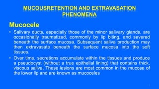
  • 1. Mucocele • Salivary ducts, especially those of the minor salivary glands, are occasionally traumatized, commonly by lip biting, and severed beneath the surface mucosa. Subsequent saliva production may then extravasate beneath the surface mucosa into the soft tissues. • Over time, secretions accumulate within the tissues and produce a pseudocyst (without a true epithelial lining) that contains thick, viscous saliva. These lesions are most common in the mucosa of the lower lip and are known as mucoceles 1
  • 2. 2
  • 3. • The second most common site of mucocele formation is the buccal mucosa. • Mucocele formation results in an elevated, thinned, stretched overlying mucosa that appears as a vesicle filled with a clear or blue-gray mucus. • The patient frequently relates a history of the lesion filling with fluid, rupture of the fluid collection, and refilling of these lesions. • Some instances of mucocele formation regress spontaneously without surgery. For persistent or recurrent lesions, the preferred treatment consists of excision of the mucocele and the associated minor salivary glands that contributed to its formation 3
  • 4. A, Excision of mucocele of right lower lip. B, Gross specimen of intact mucocele is demonstrated. 4
  • 5. • Usually; local anesthesia is administered via a mental nerve block, and an incision is made through the mucosa. • Careful dissection around the mucocele may permit its complete removal; however, in many cases the thin lining ruptures and decompresses the mucocele before removal. • The regional associated minor salivary glands are removed as well and sent for histopathologic evaluation. The recurrence rates of mucoceles may be as high as 15% to 30% after surgical removal, possibly caused by incomplete removal or repeat trauma to the minor salivary glands. 5
  • 6. 6
  • 7. • The most common lesion of the sublingual gland is the ranula, which may be considered a mucocele of the sublingual salivary gland. • Ranulas result from mucous retention in the sublingual gland ductal system or mucous extravasation as a result of ductal disruption. • The two types of ranulas are the simple ranula and the plunging ranula. • The simple ranula is confined to the area occupied by the sublingual gland in the sublingual space, superior to the mylohyoid muscle. 7
  • 8. Left floor of mouth ranula Bilateral floor of mouth ranula. 8
  • 9. • The progression to a plunging ranula occurs when the lesion extends beyond the level of the mylohyoid muscle into the submandibular space . • Ranulas may reach a larger size than mucoceles because their overlying mucosa is thicker and because trauma that would cause their rupture is less likely in the floor of the mouth. • As a result, a plunging ranula has the potential to extend through the mylohyoid muscle into the neck and to compromise the airway, resulting in a medical emergency. 9
  • 10. Right plunging ranula through the mylohyoid muscle seen on computed tomography scan (arrow). 10
  • 11. • The differential diagnosis of a floor of mouth swelling includes ranula, lymphoepithelial cyst, epidermoid or dermoid cyst, salivary gland tumors (e.g. , mucoepidermoid carcinoma) , and mesenchymal tumors (e.g. , lipoma, neurofibroma, or hemangioma) . • The differential diagnosis of a midline neck mass includes thyroid enlargement (i. e . , goiter or tumor) , thyroglossal duct cyst, dermoid cyst, and plunging ranula. • The differential diagnosis of a lateral neck mass includes lymphadenopathy, epidermoid cyst, lipoma, infectious mononucleosis, metastatic carcinoma, lymphoma, salivary gland tumors (e.g. , submandibular gland or tail of the parotid gland), submandibular gland sialadenitis, lymphoepithelial cyst, sarcoidosis, tuberculosis, cat-scratch disease, cystic hygroma, carotid body tumor, or plunging ranula. • The usual treatment of the ranula is marsupialization, in which a portion of the oral mucosa of the floor of the mouth is excised, along with the superior wall of the ranula. 11
  • 12. A, Ranula in the right floor of mouth caused by accumulation of sublingual gland secretions in soft tissues resulting from rupture of salivary duct. B, Diagram of marsupialization incision. C, Marsupialization of ranula, with excision of oral mucosa and superior wall of ranula. D, Completion of marsupialization of left floor of mouth ranula with placement of circumferential sutures. E, Diagram of completed marsupialization. 12
  • 13. • Subsequently, the ranula wall is sutured to the oral mucosa of the floor of the mouth and allowed to heal by secondary intention. • The preferred treatment for recurrent or persistent ranulas is excision of the ranula and sublingual gland via an intraoral approach; several recent studies have indicated that this might be appropriate for initial therapy. 13
  • 14. 14
  • 15. SALIVARY GLAND INFECTIONS • Infections of the major salivary glands can be acute or chronic and are commonly, but not always, related to obstructive disease, especially in the submandibular gland (obstruction leads to infection) . • The cause of acute suppurative sialadenitis of the parotid gland usually involves a change in fluid balance that is likely to occur in patients who are elderly, debilitated, malnourished, dehydrated, or plagued with chronic illness. • In these cases, gland infections are usually bilateral. The mean age of occurrence of infections is 60 years, with a slight male predilection. 15
  • 16. • Salivary gland infections may be caused by a variety of organisms, including aerobic and anaerobic bacteria, viruses, fungal organisms, and mycobacteria. In most cases, mixed bacterial flora is responsible for sialadenitis. • The Single most common organism implicated in salivary gland infection is Staphylococcus aureus because this organism normally colonizes around ductal orifices. • In addition, during instances of decreased or slowed salivary flow (i.e., obstruction or dehydration) , retrograde influx of S. aureus into the ductal system and gland occurs and results in infection. 16
  • 17. • The clinical characteristics of acute bacterial salivary gland infections include rapid onset of swelling in the preauricular (parotid gland) or submandibular regions, with associated erythema and pain. • Palpation of the involved gland reveals no flow or elicits a thick, purulent discharge from the orifice of the duct. 17
  • 18. A, Left parotid gland infection. B, Right acute bacterial parotitis with erythema. This infection is extremely painful and may indicate another serious illness. Treatment requires hospitalization, intravenously administered antibiotics, and possibly surgical drainage. 18
  • 19. Purulent discharge from left parotid duct is demonstrated in patient with an infection involving parotid gland. 19
  • 20. • Treatment of bacterial salivary gland infections includes symptomatic and supportive care, including intravenous fluid hydration, antibiotics, and analgesics. • Initial empirical antibiotics should be aimed at the most likely causative organism, S. aureus, and should include a cephalosporin (first generation) or antistaphylococcal semisynthetic penicillin (oxacillin or dicloxacillin) . • Culture and sensitivity studies of purulent material should be obtained to aid in selecting the most appropriate antibiotic for each patient. 20
  • 21. • Antibiotics should be administered intravenously in high doses for the majority of these patients, who ordinarily require hospitalization. • On most occasions, surgery consisting of incision and drainage becomes necessary in the management of salivary gland infections. Untreated infections may progress rapidly and can cause respiratory obstruction, septicemia, and eventually, death. • In some instances of recurrent salivary gland infection, the repeated insults result in irreversible functional impairment of gland function, and excision of the gland may be indicated. 21
  • 22. • Viral parotitis, or mumps, is an acute, nonsuppurative communicable disease. Before routine vaccination (e.g., measles, mumps, and rubella vaccine) against the disease began, viral parotitis occurred in epidemics during the winter and spring. • Differentiation of viral from bacterial salivary gland infection is important because viral infections are not the result of obstructive disease and require different treatment, not including antibiotics. 22
  • 23. • Mumps is characterized by a painful, nonerythematous swelling of one or both parotid glands that begins 2 to 3 weeks after exposure to the virus (incubation period) . • This disease occurs most commonly in children between ages 6 and 8. The signs and symptoms of mumps include preauricular pain and swelling, fever, chills, and headache. 23
  • 24. • Viral parotitis usually resolves in 5 to 12 days after its onset. Supportive and symptomatic care for fever, headache, and malaise with antipyretics, analgesics, and adequate hydration treats viral parotitis. • Complications of the disease include meningitis, pancreatitis, nephritis, orchitis, testicular atrophy, and sterility in approximately 20% of young males affected. 24
  • 25. NECROTIZING SIAlOMETAPlASIA • Necrotizing sialometaplasia is a reactive, nonneoplastic inflammatory process that usually affects the minor salivary glands of the palate. However, it may involve minor salivary glands in any location. • Necrotizing sialometaplasia is of unclear origin but is thought to result from vascular infarction of the salivary gland lobules. • Potential causes of diminished blood flow to the affected area include trauma, local anesthetic injection, smoking, diabetes mellitus, vascular disease, and pressure from a denture prosthesis. The usual age range of affected patients is between 23 and 66 years. 25
  • 26. • Lesions usually appear as large ( 1 to 4 cm) , painless or painful, deeply ulcerated areas lateral to the palatal midline and near the junction of the hard and soft palate. • Although lesions are usually unilateral, bilateral involvement may occur. Some patients may report a prodromal flulike illness before the onset of the ulceration. 26
  • 27. A, Necrotizing sialometaplasia of posterior palate with ulceration. 27
  • 28. • This condition is of considerable concern because, clinically and histologically, it resembles a malignant carcinoma (squamous cell or mucoepidermoid carcinoma). • The appropriate diagnosis and management of this disease relies on evaluation by an oral maxillofacial surgeon and pathologist who are familiar with this entity because the result of a misdiagnosis may be extensive, unwarranted surgical resection. 28
  • 29. • Helpful histologic criteria for distinguishing necrotizing sialometaplasia from a malignant process include the maintenance of the overall salivary lobular morphology, the generally nondysplastic appearance of the squamous islands or nests, and evidence of residual ductal lumina within the epithelial nests. • The ulcerations of necrotizing sialometaplasia usually heal spontaneously within 6 to 10 weeks after their onset and require no surgical management. 29
  • 30. SJOGREN'S SYNDROME • Sjogren's syndrome (SS) is a multisystem disease process with a variable presentation. The two types of SS are • (1) primary SS, or sicca syndrome, characterized by xerostomia (dry mouth) and keratoconjunctivitis sicca (dry eyes) ; and • (2) secondary SS, which is composed of primary SS and an associated connective tissue disorder, most commonly rheumatoid arthritis. • Although the cause of SS is unknown, there appears to be a strong autoimmune influence. SS shows a female predilection of 9 : 1 , with more than 80% of affected individuals being females with a mean age of 50 years. 30
  • 31. Sjogren's syndrome showing (A) dry eyes (keratoconjunctivitis sicca) and (B) dry mouth (xerostomia). 31
  • 32. • Generally, the first symptoms to appear are arthritic complaints, followed by ocular symptoms, and late in the disease process, salivary gland symptoms. • The involvement of the salivary and lacrimal glands results from a lymphocytic replacement of the normal glandular elements. The xerostomia results from a decreased function of the major and minor salivary glands, with the parotid gland being the most sensitive. • The diagnosis of SS is suggested by the patient's complaints and by a variety of abnormal immunologic laboratory tests. 32
  • 33. • The oral component of SS may be diagnosed using salivary flow rate studies and sialograms that can show acinar destruction. • The use of a labial minor salivary gland biopsy, as mentioned previously, currently is considered to be highly accurate in aiding the diagnosis. • The histopathologic changes seen in the minor glands are similar to those in the major glands (parotid). • Keratoconjunctivitis sicca is suggested by the patient's complaints and a Schirmer's test for lacrimal flow. 33
  • 34. Schirmer's test for dry eyes in a patient with Sjogren's syndrome. Filter paper is placed in the ocular fornix and observed for "wetting" to a certain distance within a specific time limit. 34
  • 35. • The treatment for SS includes symptomatic care with artificial tears for the dry eyes and salivary substitutes for the dry mouth. • Additionally, the medication pilocarpine (Salagen) or the Biotene products may be useful to stimulate salivary flow from the remaining functional salivary gland tissue 35
  • 36. TRAUMATIC SALIVARY GLAND INJURIES • Traumatic injuries, particularly lacerations, involving the salivary glands and their ducts may accompany a variety of facial injuries, including fractures. • Injuries that occur in proximity to one of the major salivary glands or ducts require careful evaluation. 36
  • 37. • Facial lacerations may involve not only the gland and its ductal system but also branches of the facial nerve and branches of major facial vessels. • These structures require meticulous attention for appropriate diagnosis and prompt repair. • Usually, facial nerve lacerations that are anterior to a vertical line from the lateral canthus of the eye to the mental foramen are not amenable to surgical repair. 37
  • 38. A, Diagram showing the position of Stensen's duct along a line drawn from the tragus to the middle of the upper lip. B, Injuries to the terminal branches of the facial nerve anterior to line do not require repair, and function usually returns. 38
  • 39. • Stensen's ductal repair may include ductal anastomoses, in which the proximal and distal portions of the duct are identified, a plastic or metal catheter is placed as a stent, and the duct is sutured over the stent. • The catheter usually remains in place for 10 to 14 days for epithelialization of the duct to occur. • Additionally, nerve anastomoses may be required and performed by placing epineurial sutures, using magnification, to reapproximate the nerve stumps. • The lacerations are closed in a usual layered fashion, after debridement of the soft tissue wounds to cleanse the site of entrapped particles, such as glass or dirt. • Potential sequelae of trauma involving the major salivary glands include infection, facial paralysis, cutaneous salivary gland fistula, sialocele formation, and duct obstruction as a result of scar formation, with eventual glandular atrophy and decreased function. The involved gland may eventually require surgical removal. 39
  • 40. 40
  • 41. NEOPLASTIC SALIVARY GLAND DISORDERS • Although a comprehensive discussion of salivary gland neoplasms is beyond the scope of this chapter and many other sources are available for this information, a brief review of several important aspects of the more common lesions is warranted. • Salivary gland tumors occur much more commonly in the major glands (80% to 85%) as opposed to the minor glands ( 15% to 20%) . • Additionally, between 75% and 80% of major gland tumors are benign, whereas 50% to 55% of minor gland tumors are benign. The overwhelming majority of salivary tumors occur in the parotid gland, and the majority of those are benign (mostly pleomorphic adenomas) . 41
  • 42. 42
  • 43. Benign Salivary Gland Tumors • The pleomorphic adenoma, or benign mixed tumor, is the most common salivary gland tumor. The mean age of occurrence is 45 years, with a male-to-female ratio of 3:2 . • In the major glands the parotid gland is involved in more than 80% of cases; in the minor glands the most common intraoral site is the palate. • Pleomorphic adenomas are usually slow growing, painless masses. The histopathologic examination shows two cell types: (1) the ductal epithelial cell and (2) the myoepithelial cell, which may differentiate along a variety of cell lines (pleomorphic means many forms) . 43
  • 44. • A connective tissue capsule exists, which may be incomplete. The treatment involves complete surgical excision with a margin of normal uninvolved tissue. • Parotid lesions are treated with removal of the involved lobe along with the tumor. Recurrence is possible in rare occasions, as well as a small risk (5%) of malignant transformation to a carcinoma ex pleomorphic adenoma. 44
  • 45. Pleomorphic adenomas. A, Palate. B to D, Parotid gland. E, Submandibular gland. 45
  • 46. • Warthins tumor, or papillary cystadenoma lymphomatosum, almost exclusively affects the parotid gland, specifically the tail of the parotid gland. • The peak incidence is in the sixth decade of life, with a male-to- female ratio of 7 : 1 . This lesion presents as a slow-growing, soft, painless mass . • Warthin's tumor is believed to be caused by entrapped salivary epithelial rests within developing lymph nodes. • The histopathologic examination shows an epithelial component in a papillary pattern and a lymphoid component with germinal centers. The treatment of this lesion is simple surgical excision, and recurrence is rare. 46
  • 47. Warthin's tumor of the tail of the parotid gland. 47
  • 48. • The monomorphic adenoma is an uncommon solitary lesion composed of one cell type, affecting predominantly the upper lip minor glands (canalicular adenoma) and the parotid gland (basal cell adenoma) . • The mean age of occurrence is 61 years, and the lesion usually presents as an asymptomatic, freely movable mass. The histopathologic examination reveals an encapsulated lesion composed of one type (monomorphic) of salivary ductal epithelial cell. The treatment is simple surgical excision. 48
  • 49. Monomorphic (canalicular) adenoma of the left upper lip/vestibule. 49
  • 50. Malignant Salivary Gland Tumors • The mucoepidermoid carcinoma is the most common malignant salivary gland tumor. • The tumor makes up 10% of major gland tumors (mostly parotid) and 20% of minor gland tumors (mostly palatal) . This lesion may occur at any age, but the mean age is 45 years. The male-to- female ratio is 3 : 2 . • The clinical presentation is a submucosal mass that may be painful or ulcerated. The mass may appear to have a bluish tinge because of the mucous content contained within the lesion. 50
  • 51. A, Mucoepidermoid carcinoma of the palate (note bluish tinge from mucin content). B, Mucoepidermoid carcinoma of the palate with ulceration. 51
  • 52. • An intraosseous form of mucoepidermoid carcinoma may present as a multilocular radiolucency of the posterior mandible. • The histopathologic examination shows three cell types: (1) mucous cells, (2) epidermoid cells, and (3) intermediate (clear) cells. • The proportion of each cell type helps to grade the mucoepidermoid carcinoma as high-, intermediate-, or low-grade lesions. • The higher the grade, the more predominance of epidermoid cells and pleomorphism, lack of mucous cells and cystic areas, and overall more aggressive behavior. 52
  • 53. • The treatment of low-grade lesions is wide surgical excision with a margin of uninvolved normal tissue; high grade lesions require more aggressive surgical removal with margins, and possibly, local radiation therapy. • The low-grade lesions have a 95% 5-year survival rate, whereas the high-grade lesions have less than a 40% 5-year survival rate. 53
  • 54. A, Central mucoepidermoid carcinoma of the right retromolar pad minor salivary glands. B, Panoramic radiograph showing an underlying multilocular radiolucency. 54
  • 55. • The polymorphous low-grade adenocarcinoma is the second most common intraoral salivary gland malignancy. This lesion was first described in 1983; before its identification, many cases were probably misdiagnosed as adenoid cystic carcinoma. • The most common site is the junction of the hard and soft palates. The male- to-female ratio is 3 : 1 , with a mean age of 56 years. These tumors present as slow-growing, asymptomatic masses that may be ulcerated. The histopathologic examination shows many cell shapes and patterns (polymorphous) . • Patients experience an infiltrative proliferation of ductal epithelial cells in an "Indian file" pattern. This lesion shows a predilection for invasion of surrounding nerves. The treatment of this tumor is wide surgical excision, with a relatively high recurrence rate of 14%. 55
  • 57. • The adenoid cystic carcinoma is the third most common intraoral salivary gland malignancy, with a mean age of 53 years and a male-to-female ratio of 3:2 . • Approximately 50% of these tumors occur in the parotid gland, whereas the other 50% occur in the minor glands of the palate. • These tumors present as slow-growing, nonulcerated masses, with an associated chronic dull pain. 57
  • 58. Adenoid cystic carcinoma of the palate. 58
  • 59. • Occasionally, parotid lesions may result in facial paralysis as a result of facial nerve involvement • The histopathologic examination demonstrates an infiltrative proliferation of basaloid cells arranged in a cribriform (Swiss cheese) pattern. As seen in the polymorphous low-grade adenocarcinoma, there may be perineural invasion. • The treatment is wide surgical excision, followed in some cases by radiation therapy The prognosis is poor despite aggressive therapy 59