Ischemia, Infarction, Gangrene and Hemorrhage
•Download as PPT, PDF•
2 likes•306 views

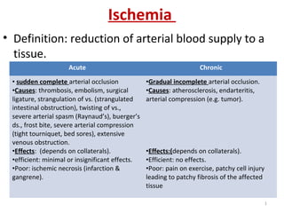
Ischemia is a reduction of arterial blood supply to a tissue. It can be acute or chronic. Acute ischemia occurs with sudden complete arterial occlusion due to various causes like thrombosis or embolism. Chronic ischemia occurs gradually due to incomplete arterial occlusion from atherosclerosis or arterial compression. Effects depend on collateral blood flow. Acute ischemia leads to ischemic necrosis (infarction) if collaterals are poor. Chronic ischemia causes pain on exercise or patchy tissue injury with fibrosis if collaterals are poor.

Recommended
More Related Content
What's hot
What's hot (20)
Similar to Ischemia, Infarction, Gangrene and Hemorrhage
Similar to Ischemia, Infarction, Gangrene and Hemorrhage (20)
More from Lama K Banna
More from Lama K Banna (20)
Recently uploaded
Recently uploaded (20)
Ischemia, Infarction, Gangrene and Hemorrhage
- 1. Ischemia • Definition: reduction of arterial blood supply to a tissue. Acute Chronic • sudden complete arterial occlusion •Causes: thrombosis, embolism, surgical ligature, strangulation of vs. (strangulated intestinal obstruction), twisting of vs., severe arterial spasm (Raynaud’s), buerger’s ds., frost bite, severe arterial compression (tight tourniquet, bed sores), extensive venous obstruction. •Effects: (depends on collaterals). •efficient: minimal or insignificant effects. •Poor: ischemic necrosis (infarction & gangrene). •Gradual incomplete arterial occlusion. •Causes: atherosclerosis, endarteritis, arterial compression (e.g. tumor). •Effects:(depends on collaterals). •Efficient: no effects. •Poor: pain on exercise, patchy cell injury leading to patchy fibrosis of the affected tissue 1
- 2. Infarction • Definition: area of ischemic necrosis (coagulative except CNS) due to sudden ischemia. • Aetiology: – Arterial occlusion (thrombus or embolus) – Extensive venous obstruction. • Types:due to : – Arterial occlusion (thrombus or embolus) – Extensive venous obstruction. • General pathological features. 2
- 3. Infarction • Definition: area of ischemic necrosis (coagulative except CNS) due to sudden ischemia. • Aetiology: – Arterial occlusion (thrombus or embolus) 99%. – Extensive venous obstruction (rare). 3
- 4. Infarction • Types: due to : Due to Arterial occlusion Due to venous occlusion •End artery with poor collateral •immediately : no blood reaching the area (pale). • 24 h. :blood collect in the area (dilatation of the poor collatral (red). •36 h. : swollen necrotic cells (pale anaemic in solid organs or red hrgic in loosly richly vascularised). •red hrgic (+/- arterial occlusion) •e.g: • infarcts of intestine, gonads, brain with JV thrombosis). 4
- 5. Infarction • General pathological features: • Fate: healing +/- dystrophic calcification. • General changes: leukocytosis & fever. gross M/P •Pyramidal or wedge shaped (due to fan shaped distribution of endarteries. •Fibrinous exudate (fibrinous inflamm.) if serosal surface base. •Hyperemic margins (inflamm.). •Early (swollen). •Later contracted (healing). •May be pale or hrgic. •Coagulative necrosis (except CNS liquifactive). •Margins: dilated capillaries & inflam. Cells •Rest of organ appear normal (except lungs due to CVC). 5
- 6. 6
- 7. Examples of infarcts: • Lung infarction: doesn’t occur unless both pulmonary & bronchial (lung congestion & low cardiac output) arteries are affected. Pathogenesis: LSHF followed by RSHF (page 106), C/P: chest pain & hemoptysis. 7
- 8. Gangrene • Massive necrosis followed by putrefaction. • Aetiology: – Causes of necrosis: acute ischemia, bacterial infection. – Putrefaction: saprophytes action on dead tissues hydrogen sulphide iron sulphide (black). • Types: – Dry. – Moist. – Infective – gas gangrene. 8
- 9. 1- Dry gangrene • Pathogenesis: – in limbs. – limb infarction rather than gangrene due to minimal & slow putrefaction in dry media + slight toxemia. – due to occluded arteries & patent veins. – Dry due to: • Limbs not rich in tissue fluid (evaporation). • Distal to occlusion (stop tissue fluid formation & venous drainage of already present tissue fluid. • Examples : senile, arterial embolism, Buerger’s d. (thromboangitis obliterans), raynaud’s d. (a. spasm), early phase of diabetic gangrene. 9
- 10. Senile gangrene • Predisposing factors: in old age, thrombosis is common (spontaneous or with slight injury) due to gradual narrowing of arteries (atherosclerosis) & weak cardiac action (vascular stasis). • Pathological features: start at big toe & progress: – Distal to occlusion: pale & cold (ischemia), red (escape from necrotic capillaries) then black. – Progression of gangrenous process: irritated adjacent tissue further thrombosis of atherosclerotic arteries proximal gangrene progression to reach good collateral. – Line of demarcation:red zone of acute inflammation seen at the margin of the black g. area. – Line of separation: groove near line of demarcation the gradually deepens till separation of dead g. part leaving conical stump (ms & bone separate at lower level 10
- 11. 11
- 12. 2- Moist (wet) gangrene • Aetiology: in circumstances with rich tissue fluid: –Internal organs : as intestine. –Diabetic gangrene: infection fluid exudate. –Occluded artery & vein : limb tight tourniquet. 12
- 13. Dry gangrene • arterial occlusion • Occurs in limbs in cases of: senile g., Buerger’s d., raynaud’s d., early diabetic g. • Does not occur in internal organs. • Very slow putrefaction. • Mild toxemia. • Gangrenous part is dry & mummified. • Prominent line of demarcation. • Line of separation. Moist gangrene • Both artery & vein occlusion. • Occurs in limbs in cases of: crush injuries, tight tourniquet , bed sores & diabetic g. • Occur in internal organs (> serious). • Rapid in limbs & very rapid in intestinal g. • Severe toxemia. • Edematous & swollen. • Poor demarcation. • No line of separation (no self separation). 13
- 14. Oedema • Definition: accumulation of abnormal amounts of fluid in IC tissue spaces &/or body cavities. • May be transudate (> normal), exudate, lymph. Transudate Exudate •Low ptn (< 3 gm/dl). •Mainly albumin. •Doesn’t clot (no fibrinogen). •Low sp. Gravity (<1015) •Poor cellularity. •High ptn (> 3 gm/dl). •Mainly fibrinogen. •Undergoes clotting. •High (>1015). •Ritch in inflammatory cells. 14
- 15. Causes & pathogenesis • capillary H.P.: venous congestion. • Na & water retention: cap. H.P. (renal, heart, hyperaldosteronism). • colloid OP of plasma: hypoproteinemia. • capillary permiability: inflamm. & hypersinsitivity escape of ptn in edema fluid. • tissue OP.: due to capillary permeability. • Lymphatic obstruction: filariasis, lymphatic permiation by malignant cells, surgical removal of lymphatics, post- irradiation, lymphangitis & lymphadenitis, congenital hypoplasia of lymphatics. The lymph is a part of tissue fluid 15
- 16. Classification (types of oedema): • Localized or generalized: • Pitting & non-pitting: – Pitting oedema: in all types of generalized edema & localized congestive edema (fluid is transudate). – Non-pitting oedema: in lymphtic edema & sometimes inflammatory edema. Localized Generalized (anasarca) •Inflammatory: exudate of acute inflam. •Obstructive: -venous: congestion (transudative fluid) Edema of legs, lung, ascities. -lymphatic: edema fluid is lymph. •Cardiac: RSHF •Nutritional: malnutrition, malabsorbtion, chronic liver disease. •Renal: nephrotic, nephretic. 16
- 17. Hemorrhage • Definition: escape of bl. Outside CVS. • Aetiology: apart from physiological loss (menses) • Types: external Internal (into body cavities) interstitial •From skin. •From respiratry tract. •From aleimentary tract. •From urinart t.. •From female genitalia: menorrhagia & metrorrhagia. •Hemothorax. •Hemopericardium. •Hemoperitoneum. •Hematocele (tunica vaginalis) •Hemarthrosis (joint cavities). •According to type of bleeding vs: red (a) or blue (b). 1st green then brown then return normal. •According to it’s extent: petechial, ecchymosis, hematoma (large bl. Swell.) local causes General causes •Traumatic, erosion, aneurysm rupture, local venous congestion, inflammation. •HTN & GVC, blood ds., vit K & C 17
- 18. Effects of hemorrhage: Local effects (hemostasis: natural arrest of hrg) Systemic effects •Michanism: -hypotension. -curling of endothelium (minimize bleeding). - VC by serotonin. -clotting of bl. Around site of bleeding. -gradual fibrous organization of clot lead to permanent arrest of hrge. • causes of failure: - large vascular injury. -coagulation defects & blood ds. - infection before clot organization softning of clot 2ry hrge. • chronic repeated blood loss (Fe anaemia). •Rapid blood loss: depend on amount -very small: no effect. - moderate (<1000ml): compensation: - catecholamines, renins, ADH VC & restore blood pressure. -initial fall of HP withdraw tissue fluid to blood to increase it’s volume & pressure. -decreased renal flow salt & water ret. -bl cells & plasma ptns restored gradually. - massive (> 1000 ml) shock. 18