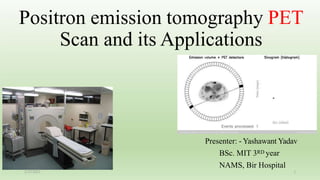
1. Positron emission tomography PET
Scan and its Applications
Presenter: - Yashawant Yadav
BSc. MIT 3RD year
NAMS, Bir Hospital
1
2/25/2021
2. Outlines
• Introduction of PET Scan
• Basic physic behind PET
• Detectors and image acquisition system
• Advancement
• Application of PET
• References
3. Introduction
• Positron Emission Tomography (PET) is a method for measuring biochemical and
physiological processes in vivo in a quantitative way by using
radiopharmaceuticals labelled with positron emitting radionuclides such as 11C,
13N, 15O and 18F.
• PET employs mainly short-lived positron emitting radiopharmaceuticals.
• The most widely used radionuclides are: 11C (t1/2 = 20 min), 13N (t1/2 = 10 min),
15O (t1/2 = 2 min) and 18F (t1/2 = 110 min).
• These radionuclides are produced in a (baby) cyclotron and are then used to label
compounds of biological interest.
4. • In cancerous cells metabolic changes occur much before the cells undergo changes
like dysplasia, metaplasia or anaplasia.
• This is finally followed by structural changes at a later stage.
• PET scan detects the disease at the metabolic level while anatomical imaging
techniques like CT or MRI detect the disease at the structural level.
Why PET ????
5. • Positron
• Gamma ray
• Proton
• Light photons
• Decay Emission Production Detection
What we detect in PET scan ??
6. • Number of proton(z)= atomic number
• Total number of nucleons(A)= mass number
proton+ neutron
Basic atomic physic
12. • The method is based on identifying the increased glycolytic activity in malignant
cells,
• Increase in membrane glucose transporters as well as to an increase in some of
the principal enzymes, such as hexokinase.(at site of tumor )
• Glucose transporter proteins known as GLUT -1 transporters and subsequently
phosphorylated by hexokinase.
Radiopharmaceuticals uptake in PET
13. Decay possibilities for neutron deficient Radionuclides
There is 2 possibilities: -
1. Positron decay
2. Electron capture
For positron decay energy should be at least 1022KeV or more and number of
proton should be lesser (lower atomic number atoms )
For electron capture energy needed lower than 1022KeV and number of protons
should be higher (higher atomic number atoms )
15. Why photons apart at 1800 ??
• With positron decay two conservation laws have to be obeyed:
i) conservation of energy and
ii) conservation of momentum. (m*v)
Conservation of energy is followed by conversion of mass in to gamma ray photons
and momentum is conserved by produced gamma photons moving in opposite
direction to each other and cancel momentum of each other.
The three-quanta annihilation only happens if the formed positronium is in its triplet
state, which is rare, with a half-life of 7 µs.
In the singlet state the positronium decays with a lifetime of 4-8 ns.
16. • Proton range (before annihilation)
• Non collinearity (after annihilation)
Phenomenon
The positron range error is dependent on the energy of the emitted positrons. Non colinearity is
independent of radionuclide, and the error is determined by the separation of the detectors. The deviation
from non colinearity is highly exaggerated in the figure; the average angular deviation from 180° is about
0.25°.
17. Contd..
• The anti-parallel photons are recorded, and the
virtual line connecting the two points is called the
line of response (LoR)
• In a conventional PET system, positron annihilation
is assumed to be localized somewhere along the
LoR without information regarding the exact
interaction point.
18.
19. Contd…
• Coincidence measurements and
ideally measurement of the time
difference, called Time Of Flight
(TOF) measurement.
• It has Depth function
• A major trend in PET
instrumentation is the development
of time-of-flight positron emission
tomography (ToF-PET).
20. Contd…
• ToF-PET leads to better localization of the annihilation event and thus results in
overall improvement in the signal-to-noise ratio (SNR) of the reconstructed image.
• The technique by which the image is reconstructed without using ToF information
is called conventional PET and that incorporating ToF information is abbreviated
as ToF-PET
21.
22. Confounding's needed to be estimated
Patient related confounding’s are attenuation , scattering and random coincidence,
patients motion could be the main confounding Factor
23. Detector of PET system
The scintillator characteristics:-
a) stopping power,
b) light output (yield and wave length) and
c) decay time
• Cesium fluoride (CsF) and barium fluoride (BaF2) detectors were used in the first-
generation TOF-PET.
• CsF needs careful packaging as it is highly hygroscopic. In addition, limited
sensitivity (stopping power), low light output.
24. Contd..
• During the 1970s and early 1980s, bismuth germinate (BGO) was used as the
standard scintillator for PET detectors because of its high detection efficiency and
acceptable light output in commercial PET systems.
1.Newer scintillators such as lutetium orthosilicate (Lu2SiO2) (LSO)
(traditional LSO:Ce scintillato0rs) (In recent years, LSO co-doped with Ca)
(LYSO:Ce) (lutetium yttrium oxyorthosilicate)
2.GSO (gadolinium oxyorthosilicate), LaBr3:Ce (cerium-doped lanthanum
bromide), Ce:GAGG (cerium-doped gadolinium aluminum gallium garnet)
(Gd3Al2Ga3O12:Ce or GAGG:Ce).
25. Contd…
• Detectors based on CZT (cadmium zinc telluride) do not use scintillators because
they directly convert ionizing radiation to charge production, providing higher
energy and spatial resolutions than scintillator-based PET detectors.
• CdTe (cadmium telluride) is another stable crystalline compound
• In terms of detector design, recent PET scanners do not have septa and therefore
images are acquired only in three-dimensional (3D) mode.
26. • There are generally four types of sensor technologies employed:
• photomultiplier tubes (PMTs),
• Avalanche photodiodes (APDs),
• silicon photomultipliers (SiPMs), and
• cadmium zinc telluride (CZT) detectors.
• Spatial resolution of the conventional human PET system was usually 4.5–6 mm
due to the limitation of the sensing technology
Contd..
27.
28. • New-generation PET detectors have silicon photomultipliers (SiPM) instead of
photomultipliers tube (PMT).
• The main benefits of SiPM comprise compact and rugged, high gain , good
intrinsic timing resolution, and higher value of photon detection efficiency than
PMTs.
• In addition, the SiPM detector is insensitive to the electromagnetic interference
and this is the most important feature of the PET/magnetic resonance (MR)
system.
Contd…
33. Clinical application
• The clinical impact and use of PET remained restricted until availability of
Medical/Baby cyclotrons,
• In the late 1990s 18F- fluorodeoxyglucose (FDG) as the radiopharmaceutical
began to be used widely in evaluation of oncology patients.
• The clinical use of PET received a major boost in 1998, when PET scanning was
approved by health care agencies in USA.
• The basis of PET imaging is the detection of altered metabolism in biological
tissues.
34. Contd…
• Tumor Proliferation
Carbon-11 thymidine and F-18 Fluorothymidine (FLT) an analog of thymidine are
markers of cellular proliferation. Analog to FDG (to predict tumor grade in lung
cancers, evaluate brain tumors)
11C methionine and amino acid, has shown great promise in evaluating brain
tumors and other cancers too.
11C-choline and 11C-acetate have been used in prostate cancer to evaluate the
primary and metastatic disease
35. Contd…
• Myocardial Perfusion Imaging
• Rubidium-82 is a potassium analog agent to assess myocardial perfusion in the
same way as Thallium 201 or Technetium -99 labelled compounds.
• Nitrogen-13 labelled ammonia is another PET tracer used for myocardial
perfusion studies.
• Skeletal Imaging
• F-18 Sodium fluoride has shown great promise as a bone scan agent, comparable
to or even superior to Technetium -99 labelled MDP.
•Brain Imaging
Listed above in table
36. Contd…
The goals of oncologic imaging remain lesion detection, lesion characterization,
staging of malignant lesions and assessment of the therapeutic response.
• Brain
• Head and Neck
• Lungs
• Esophageal Cancer
• Colorectal Carcinoma
• Lymphoma
• Carcinoma Breast
• Cervical and Ovarian Carcinoma
• Renal, Prostate and Bladder Cancers
• Testicular Cancers
• Melanoma
• Musculoskeletal Tumors
• Dementias
• Epilepsy
• Movement Disorders
• Stroke and Cerebrovascular Disease (CVD)
• Myocardial Viability
• Coronary Artery Evaluation
• Coronary Perfusion Reserve
37.
38. • Anterior projection images from a PET
scan of a patient undergoing staging of
lymphoma. (a) The initial study was
performed after the patient had eaten a
candy bar 30 minutes prior to FDG
injection. Note the extensive myocardial
and muscle uptake due to high insulin
levels. Diminished activity is seen in the
brain and in tumor sites in the neck and
chest (arrows).
• (b) A repeat study after the patient
complied with routine fasting preparation
shows more normal biodistribution of
tracer and better visualization of tumor
deposits
FINDINGS
39. • A patient being evaluated for metastatic colon cancer. (a) An anterior
projection PET image shows known hepatic metastases, as well as an
indeterminate focus in the left face (arrow). (b) Trans axial PET through the
face shows a distinct hypermetabolic focus (arrow). The
corresponding (c) CT and (d) fusion images show this focus to be a
periodontal abscess in the maxillary alveolar ridge
40. • A patient being restaged for colon cancer. (a) PET image shows focal
uptake consistent with recurrence (dashed circle) but does not allow
localization. (b) CT and (c) fusion images show intraabdominal
recurrence (arrow), as well as lesions involving the psoas and iliacus
muscles (arrowheads). Lesser activity elsewhere is physiologic bowel
uptake.
43. • In any PET/CT study there are three discrete image sets that require display. These
are the stand-alone PET data, the CT and the fused PET/CT images.
Contd….
45. • PET–CT imaging is primarily used in oncology, it has also been used to identify brown fat.
• Murine model of disease (pre clinical PET imaging)
• Primary tumor , nodal and distant metastases. ‘(TNM)
• Staging
46.
47.
48. What about bone Mets in MRI PET SCAN ???
• Zero echo time (ZTE) MR imaging provides enhanced bone contrast in MR
imaging and may obviate concomitant CT and its attendant ionizing radiation.
• (MR-based pseudo-CT image conversion.)
• ZTE differs from UTE in the timing of read-out gradients relative to the
excitation radiofrequency (RF) pulse and its acquisition of a single echo time data
set.
• Signal is acquired immediately after application of the RF pulse (echo time = 8
msec). Furthermore, ZTE uses radial k-space filling strategies,
55. • How positron emitter / neutron deficient nucleoid are made?
• What is momentum and how is it conservation?
• What is proton decay and electron capture ?
• Types of detector with example??
Questions