Chylothorax

A chylothorax is an abnormal accumulation of chyle, a type of lipid-rich lymph, in the space surrounding the lung. The lymphatics of the digestive system normally returns lipids absorbed from the small bowel via the thoracic duct, which ascends behind the esophagus to drain into the left brachiocephalic vein. If normal thoracic duct drainage is disrupted, either due to obstruction or rupture, chyle can leak and accumulate within the negative-pressured pleural space. In people on a normal diet, this fluid collection can sometimes be identified by its turbid, milky white appearance, since chyle contains emulsified triglycerides. Chylothorax is a rare but serious condition, as it signals leakage of the thoracic duct or one of its tributaries. There are many treatments, both surgical and conservative.[1] About 2–3% of all fluid collections surrounding the lungs (pleural effusions) are chylothoraces.[2] It is important to distinguish a chylothorax from a pseudochylothorax (a pleural effusion that happens to be high in cholesterol), which has a similar appearance visually but is caused by more chronic inflammatory processes and requires a different treatment

Recommended
More Related Content
What's hot
What's hot (20)
Similar to Chylothorax
Similar to Chylothorax (20)
More from KararSurgery
More from KararSurgery (11)
Recently uploaded
Recently uploaded (20)
Chylothorax
- 1. CHYLOTHORAX
- 3. • Chylothorax is the collection of an excessive amount of chyle in the pleural space. The continued loss of chyle, which can add up to 2 to 3 liters a day after a thoracic duct injury,1 leads to significant depletion of fats (up to 70% of dietary intake), proteins, and T lymphocytes.2 As a consequence, marked disturbances in the immunologic and nutritional profile are the rule in these patients along with a mass effect created by dislocation of intrathoracic structures by the enlarging fluid collection. Indeed, the flow rate of chyle within the thoracic duct can be as high as 110 mL/hr.1 If left untreated, chylothorax may yield an overall 50% mortality rate.3
- 4. •
- 6. •
- 7. • CLASSIFICATION AND CAUSE The term traumatic is often used to include both iatrogenic and postinjury chylothoraces,2 which usually represent the most common causes of significant chyle accumulation in the chest. Neoplastic causes can account to up to 20% of chylothoraces.2 In a recent report from the Mayo Clinic, the cause was surgery or trauma in 50% of the patients, medical conditions in 44%, and unknown in 6%.4 This unusual distribution, compared to the commonly reported series,5 was explained by the high volume of surgical cases being performed each year at that institution
- 8. • In the pediatric group, congenital chylothorax manifests itself early after birth possibly as a result of a combination of thoracic duct malformation and sudden elevation of venous pressure.6 Neonatal chylothorax has been reported in conjunction with several syndromes, such as Noonan and Down.2,6 In addition, the incidence of chylothorax after cardiothoracic procedures in children is reported to be as high as 3.8%.7 The detection of a pleural effusion in this age group should immediately raise the suspicion of chylothorax.6
- 9. • Tuberculosis with significant mediastinal adenopathy may still be responsible for “spontaneous” bilateral chylothoraces in children because of the obstruction to centripetal flow.8 Excessive chyle collection in the pleural space may occur in association with benign and malignant tumors. Reportedly, almost 50% of the patients with chylothorax have cancer. Of these, 70% have lymphoma.5 Conversely, chylothorax was reported in 10% of the patients with lymphangioleiomyomatosis (LAM) treated at the Mayo Clinic over a 24-year period
- 10. •
- 11. •
- 12. •
- 13. •
- 14. • The centripetal flow toward the left subclavian vein is regulated by three factors17: (1) the vis a tergo created by the continuous enteral absorption of chyle constituents, which pushes the chyle from the cisterna chyli to the left subclavian vein; (2) the aspiration effect given by the negative effect of the intrathoracic pressure facilitating the cephalad flow; and (3) lymphatic vessel contractions, generated by smooth musculature, aiming at emptying the duct into the subclavian vein.
- 15. • PATHOPHYSIOLOGY A chylothorax can result from a chyle leak (direct injury or obstruction of the major lymphatic vessel), generalized oozing, or transdiaphragmatic flow from chylous ascites.9 The distinction between idiopathic (sometimes called spontaneous) and secondary causes of chylothorax depend on the presence of an identified etiology. Secondary causes include neoplastic and inflammatory conditions because chylothorax may ensue from the obstruction to centripetal flow or increased flow rate with extreme dilation of lymphatic vessels leading to extravasation of lymph in a space that changes from virtual to actual despite the presence of an expanded lung. As a consequence, lymphatic vessels are thought to preserve, at the beginning, their structural integrity at the expense of an increased permeability into the pleural space.2,9 Constrictive pericarditis, superior vena cava obstruction, and mediastinal fibrosis resulting from cancer treatment can generate chylothorax according to this pathophysiologic model
- 16. • In this setting, patients with liver cirrhosis can develop spontaneous chylothorax as a result of a twofold or threefold increase in diameter of the thoracic duct from an unusual backflow and pressure in the thoracic duct.16 Another factor implied in the onset of chylothorax is valve competency in the lymphatic vessels as demonstrated by the rarity of such complication following pulmonary resection and extensive mediastinal nodal dissection. Valve insufficiency–induced backflow from the thoracic duct into the areas of nodal dissection and injury of the lymphatic network may explain the chylous effusion11 after pulmonary resections with concurrent nodal dissection.
- 17. • From a clinical standpoint, chylothorax resulting from pleural carcinomatosis or from tubercular involvement may denote a gradual onset and development. This observation can be also explained by progressive lung trapping caused by the visceral pleura thickened by the persistent chemical irritation by chyle components.12 In time, fibrotic visceral pleura may impose a restrictive physiology on the affected lung by significantly reducing its compliance. This scenario is more often the case with the so-called pseudochylothorax
- 18. • Induced or secondary causes of chylothorax include traumatic and iatrogenic causes, which have in common the interruption of the vessel caused by direct injury, inadvertent division, or blunt trauma to the thoracic duct or its tributaries. Rupture of the thoracic duct may also occur following sudden hyperextension of the spine (seat belt injury),18 vertebral fractures or dislocation, or after protracted and vigorous vomiting or coughing
- 19. • SYMPTOMS AND DIAGNOSIS Biochemically, chyle is usually characterized by a content of triglycerides in the pleural fluid greater than the one detected in the plasma (>110 mg/dL1), a cholesterol/ triglycerides ratio less than 1, and, the presence of chylomicrons.2,6,19-21 A detailed description of the chemical components of chyle is already available in the surgical literature.1,6 A differential diagnosis with pseudochylothorax sometimes has to be made.2 Pseudochylothorax refers to those chronic effusions that may resemble chylothorax but do not have its biochemical composition.2 The pleural effusions in patients with tuberculosis or rheumatoid arthritis may have these features (i.e., yellowish or milky color, sterile, high cholesterol levels).
- 20. • One interesting observation is that chyle does not contain fibrinogen, which could seal a small leak.3 Chyle is bacteriostatic and can be a chemical irritant for the pleural surfaces,12 even though this finding is disputed.2,17 In addition, chylothorax can affect the bioavailability of drugs like amiodarone, digoxin, and, cyclosporine.2
- 21. • The acute onset of a significant chyle leak may be characterized by dyspnea and cough associated with a sense of pressure in the chest.2 Hemodynamic disturbance is also common with high-flow chylous fistulas.2 Conversely, severe malnourishment and cachexia may ensue as a result of persistent chylothorax.2 Symptoms related to chyle accumulation were described in a report by the Mayo Clinic.4 Over a 21-year observation period, 203 patients developed chylothorax (male-female ratio, 1.21; median age, 54 years). Of these patients, 57% presented with dyspnea, whereas 37% were asymptomatic. More than 7 weeks elapsed from symptom development to diagnosis.4
- 22. • A milky pleural effusion accumulating at a rate of more than 400 to 700 mL per day is suggestive of chylothorax.12 An empty pleural space after pneumonectomy may accommodate a greater chyle flow.12 The administration of a high-fat meal 3 hours before surgery, with or without dye, or the subcutaneous injection of dye 1 hour before surgical exploration can facilitate the identification of the site of leakage when the decision for surgery to ligate the duct is made
- 23. •
- 25. •
- 26. •
- 27. •
- 28. • the lymphographic finding of a leak from small tributaries may predict the success of conservative management.
- 29. • Three approaches to managing chylothorax are: 1. Conservative (nonsurgical) 2. Surgical, aimed at identifying and isolating the lymphatic duct causing the leak so that it can be closed 3. Surgical, aimed at obliterating the space otherwise to be filled by chyle
- 30. • Conservative management represents a mainstay of treatment of postoperative chylothorax developing in children following cardiothoracic interventions.7 The principles of conservative treatment include (1) nothingby mouth (NPO), (2) medium-chain triglyceride diet, or (3) parenteral nutrition
- 31. • In adults, it has been suggested that the detection of recurrent chylothorax greater than 1 liter/day after 1 week or between 0.1 and 1 liter chyle loss during the first two weeks16 is evidence of failure of conservative management. Others12 suggest opting for a surgical intervention if the leak is greater than 1 liter/day for 5 days, if the chylothorax does not subside after 2 weeks, or if there is severe nutritional or metabolic imbalance. Advocates of early surgical intervention following esophagectomy support the idea of reintervention if the leak is consistently greater than 2 liters during 2 consecutive days.3 An interesting perspective for chylothorax treatment has been outlined by Cope, who suggested percutaneously injecting a sclerosing agent into the cisterna chyli.24 Surgical intervention or reintervention can be performed via an open or a video-assisted thoracic surgery
- 32. •
- 34. •
- 35. • The increasingly frequent resort to safe minimally invasive techniques to achieve control of the thoracic duct may change the balance between conservative and operative approaches. Indeed, the therapeutic possibilities revolve around the identification of the leak, closure of the fistula, and obliteration of the pleural space, especially if the chylothorax complicates an esophagectomy (see Table 29-1). On principle, the side of the identified leak should be preferentially approached and surgically treated.3 In the event of a bilateral chylothorax, the right side should be preferentially entered to ligate the supradiaphragmatic thoracic duct. If the duct cannot be isolated, mass ligation of the fibrotic area around the duct should be performed
- 36. • Chylothorax after Pulmonary Resection Despite the fact that a well-expanded residual lung after lobectomy and postpneumonectomy chylothorax have similar etiologies and pathophysiologies, the former usually responds better to a conservative approach.21 In the latter, sudden accumulation of chyle in the empty hemithorax can cause a contralateral shift of the mediastinum with the attendant cardiorespiratory functional impairment. This rare clinical scenario, reported in almost half of the pneumonectomy patients who develop chylothorax,26 mandates a more aggressive protocol to ensure early ligation of the thoracic duct—within 3 to 5 days of onset if the chyle loss is greater than 400 mL after an observation period lasting two consecutive 8-hour shifts.21 Reportedly, chylothorax occurs more frequently on the right side, possibly because of a more radical mediastinal nodal dissection following resection for bronchial carcinoma
- 37. • Chylothorax after Esophagectomy Patients undergoing esophagectomy are often older adults with significant cardiorespiratory comorbidities and malnourishment.13 In addition, multiple operative fields are necessary to complete the esophageal resection and restore the gastrointestinal continuity. The resort to VATS has reduced the impact of this operation on patients’ overall condition. In a series of 1787 patients operated on during an 18-year period, 26% underwent a transhiatal esophagectomy as a result of their substantial cardiorespiratory comorbidities.13 However, no significant difference in the prevalence of chylothorax was noted between transthoracic and transhiatal approaches.13 In a Mayo Clinic series, the incidence of this complication following esophagectomy was reported to be 2.9%.1 Investigators renowned for their long-standing experience with transhiatal esophagectomy (reaching almost 2000 patients subjected to this procedure) have reported an incidence of chylothorax of 1.5%, favorably comparing with the 2% to 4% range reported in the literature.
- 38. • Early reoperation with mass ligation of the thoracic duct is advocated to limit the immunologic and nutritional imbalances that can heavily affect the postoperative course of these patients.13 The mortality rates for reoperation reach 16% compared with more than 80% after conservative treatment,3 making an early surgery option to control chylothorax a preferable option. However, the prophylactic ligation of the immediately supradiaphragmatic azygos vein along with the thoracic duct during esophagectomy has been recommended to drastically reduce the incidence of chylothorax.2,1
- 39. • CONCLUSION Among different types of chylothoraces, postoperative chylothorax represents a serious challenge, especially when esophagectomy is performed after induction therapy (Table 29-2). Current conservative treatment, in addition to time-honored strategies, tends to include diagnostic lymphangiography37 and midodrine, an α-adrenergic drug available for oral administration.38 Conservative treatment, combined with surgical treatment when necessary, leads to resolution in most patients.
- 41. • Hepatic hydrothorax is an uncommon complication of cirrhotic patients with ascites. It causes serious respiratory distress in these patients whose ventilation is usually already compromised by the elevated diaphragm caused by ascites. Denver pleurovenous shunt insertion is a minimally invasive alternative to other surgical procedures such as repeated thoracentesis, tube thoracostomy, chemical and mechanical pleurodesis, and Transjugular Intrahepatic Portosystemic Shunts (TIPS). Placement of Denver pleurovenous shunt with a pump chamber allows unidirectional flow of fluid from the pleural cavity to the central venous system. It helps replace the lost plasma volume in the central venous system by shunting the pleural fluid to the superior vena cava. The pump chamber is placed subcutaneously within the chest wall and allows daily manual compression and movement of fluid across a less favorable pressure gradient created between the pleural cavity and the central venous system. Potential complications of the procedure are bleeding, air embolism, infection and occlusion, and leakage. Here, we present a complication of the Denver shunt in the form of a leak presenting as a neck mass.
- 42. • Complications of pleurovenous shunts • Pulmonary edema and development of respiratory distress syndrome is reported as an early complication due to fluid overload in the early post shunt replacement period.[6] • Post shunt coagulopathy is reported in up to 5% of patients following shunt placement. Coagulopathy is presumed to be initiated by a large amount of tissue plasminogen factors being introduced to the blood from the peritoneal or the pleural fluid.[6] • Infection is reported to have a lower incidence due to use of prophylactic antibiotics and wound irrigation with aminoglycosides.[6] • Deep vein thrombosis involving the upper extremity, ipsilateral to the shunt insertion has also been reported. • Shunt failure is the most common complication and is reported in 10% to 15% of the patients. Failure can be due to mal-positioning of the venous tip of the shunt or mechanical obstruction such as a kink or a thrombus.[6] The main cause of Denver shunt occlusion is the accumulation of fibrin and cellular debris that often get impacted after each manual compression.[10] • Air embolism as a complication is uncommon and has been reported in the literature. To prevent air embolism, pleural or peritoneal fluid should not be aspirated to its fullest extent and a residue amount should always be left in pleural or peritoneal cavity.[9] • Although catheter leakage is a known entity, the leakage encountered in our patient led to the formation of a collection within the soft tissue that presented as a neck mass.
- 43. • Post-Operative Chylothorax in Children Undergoing Congenital Heart Surgery
- 44. • Abstract • Chylothorax is a rare postoperative complication of congenital heart surgery. It has high morbidity with increased hospital stay and cost of treatment. Damage to the thoracic duct, disruption of accessory lymphatic vessels, and increased venous pressure exceeding that in the thoracic duct have been proposed as the possible causes of chylothorax after surgery for congenital heart disease. Prompt diagnose with early initiation of treatment will reduce the duration of drainage. Staged treatment is the general principle in managing this serious complication. Loss of chyle leads to volume, nutritional and electrolyte depletion, immunological deficiencies and hematological complications. Identifying the underlying cause and addressing it is crucial to definitive management.
- 45. Congenital chylothorax Traumatic High venous pressures Associated with tumors Miscellaneous (A) Congenital lymphatic malformation (A) Surgical Thrombosis of superior vena cava Neurogenic tumors (A) Granulomatous diseases Lymphangiomatosis Excision of lymph nodes Deep vein thrombosis of upper extremity Lymphoma Tuberculosis Lymphangiectasia Congenital heart surgery Post-operative Fontan Surgery Teratoma Histoplasmosis Atresia of thoracic duct Scoliosis operations Wilm’s tumor Sarcoidosis (B) Associated with syndromes Excision of vascular rings Ovarian tumor (B) Others Down syndrome Diaphragmatic hernia Kaposi sarcoma Staphyloccocal discitis Turner’s syndrome (B) Invasive diagnostic procedures Henoch Schonlein purpura Noonan’s syndrome Subclavian vein catheterization Gorham-Stout syndrome (C) Other trauma Yellow nail syndrome Blunt or penetrating chest injury Hydrops fetalis Thoracic spine surgery
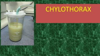
- 47. • Clinical features • The first sign of development of post-operative chylothorax is pleural fluid turning milky white in the chest tube. Sometimes chylothorax is serous, sanguineous or bloody [12]. It can develop from the 1st day up to 24th day after surgery [4] and can be unilateral or bilateral. However, in patients who are fasting post-operatively, effusions may appear serious. Pleural empyema can also produce opaque pleural fluid, as can pseudo- chylothorax (long-standing pleural effusion in which transudate becomes turbid due to accumulation of cholesterol and lecithin). The latter two can be distinguished by clinical features and laboratory investigations (see below) [8,14]. • Without the chest tube, low volume chylothorax can be clinically silent. High volume collections can lead to dyspnea, cough, hypovolemic symptoms, and rarely, with rapid accumulation of fluid, may cause tension chylothorax. Since the accumulation is non-inflammatory, fever and pleuritic chest pain are not present
- 48. • Investigations • Investigations essentially focus on confirmation of chylothorax by fluid analysis and diagnosis of the cause. A persistent chest tube drainage of >5 ml/kg/day on 4th post- operative day or a milky nature of the fluid warrants investigation and management. Chest X-Ray or ultrasound may show unilateral or bilateral pleural effusion. Examination of fluid obtained by pleurocentesis will differentiate between chylothorax, pseudo-chylothorax and pleural empyema. Chyle will have high levels of triglycerides (>110 mg/dl or higher than serum triglycerides), proteins (>20 g/l), and a cholesterol content <200 mg/dl, absolute white cell count of >1,000/cumm with >80% of cells being lymphocytes [1,2,8,13,15]. A triglyceride content <50 mg/dl almost rules out chyle. Ambiguity exists when the level is between 50 mg/dl and 100 mg/dl. Lipoprotein electrophoresis which is considered to be a gold standard in diagnosing chylothorax should be considered in such a setting because rarely chylothorax may have low triglyceride levels [14]. Typical composition of chyle is given in Table 2 [8]. Pseudo- chylothorax, which is also milky, is characterized by a cholesterol concentration of >200 mg/dL, lower triglyceride composition (<110mg/dl), cholesterol/triglyceride ratio >1 and a pleural/serum cholesterol ratio >1 [8,16,17]. For prognostication certain laboratory investigations have been used by clinicians. These include serum C-reactive protein/pre- albumin ratio or their levels and transferrin as an acute phase reactant
- 49. Components Amount pH 7.4-7.8 Absolute cell count 1,000 cells/L Lymphocytes 400-6,800/cumm Erythrocytes 50-600/cumm Calories 200 Kcal/L Total fat 0.4-0.8 g/dl Cholesterol 65-200 mg/dl Triglycerides 110 mg/dl (1.1 mmol/L) Chylomicrons present Total protein 2-6 g/dl Albumin 1.2-4.1 g/dl Globulin 1.1-3.1 g/dl Glucose 2.7-3.1 g/dl Sodium 104-108 mmol/L Potassium 3.4-5.0 mmol/L Chloride 85-130 mmol/L Calcium 3.4-6 mmol/L Phosphate 0.8-4.2 mmol/L Lactate dehydrogenase <160 IU/L
- 50. • Management of post-surgical chylothorax • Treatment of post-surgical chylothorax has two primary goals: relief of respiratory symptoms by drainage of fluid and prevent or reduce chyle collection in pleural space [12]. Management strategies for the second goal will depend upon the cause, volume and rate of accumulation of effusion, underlying disease and co-morbidities. The initial treatment in all cases is conservative and interventional therapy is reserved for refractory cases.
- 51. • 1. Conservative treatment • The goals of conservative management are to reduce chyle production by nutritional measures and relieve symptoms by image-guided chest tube drainage. This helps in re-expansion of lung, optimizes lung function and also guides treatment strategies. Sometimes drainage approximates lung and pleural surfaces, thereby sealing the leak. • Nutritional management should be aggressive with advice from a nutrition expert. Chylothorax diet aims at providing low long-chain triglycerides (because they undergo second esterification and enter lymphatic duct in the form of chylomicrons), and high medium-chain triglycerides (MCT, because they get coupled to albumin and directly enter portal circulation) either as oral or nasogastric tube feeding. A 10-day treatment with long-chain fatty acid-free MCT diet was found to be effective in 71% of patients in one study [2]. In case of oral intolerance best approach would be total parenteral nutrition (TPN). TPN is also recommended for high output of chyle or if central venous pressures are >15mmHg [2]. Fat-soluble vitamins, albumin or protein diet, electrolytes and calcium may be added as required.
- 52. • Drugs may be added as indicated, like diuretics, sildenafil, angiotensin-converting enzyme (ACE) inhibitors, or heparin for thrombosis. Cardiac catheterization may be needed to document increased venous pressures and address stenosis with balloon dilatation or placement of stents [2]. • Figure 2 is a guiding algorithm to an overall management approach. Management is in phases and escalation of treatment is decided by the chyle output and number of days of treatment. First phase is nutritional management for five to seven days. If chyle production exceeds 15 ml/kg/day (or 20 ml/kg/day) [18], the 2nd phase of treatment would be to stop oral feeds and provide TPN for five to seven days. If TPN fails to reduce chylous output then the 3rd phase of a five to seven days trial of drugs is initiated. Previously steroids were used but recent protocols have not included it. All protocols use intravenous infusion or subcutaneous boluses of octreotide, with a starting dose of 0.5-4 mcg/kg/hr or 10 mcg/kg/day in three divided doses, increasing 5-10 mcg/kg/day every 72-96 hours, maximum of 40 mcg/kg/day. Indication for starting octreotide is a chylous drainage for >2 weeks or drain output of >15 ml/kg/day [4]. Octreotide, a somatostatin analogue, reduces lymph formation by directly acting on vascular somatostatin receptors and indirectly by reducing intestinal blood flow and motility. Adverse reactions are mild and include abdominal distension, hypoalbuminemia and rarely may contribute to septicemia through its inhibitory role on immune responses [4]. Duration of treatment with octreotide is generally for five to seven days and then weaning off over four days
- 53. • Success of any conservative regimen is when the drainage output becomes <2 ml/kg/day. Throughout the treatment, chylothorax nutritional management should be continued. Even after reduction in chyle output, dietary management with MCT diet should be continued for 6-8 weeks and with low-fat diet for another six weeks [15]. • Majority of patients, up to 80%-85%, respond to conservative treatment [9,20]. Treatment failure with octreotide warrants further investigations and interventional treatment with weaning off of the drug at 25% dose daily in four days [2].
- 55. • Interventional treatment • Patients who fail to respond to conservative treatment have the option of surgical or interventional treatment. Surgical treatment reduces mortality from 50% to 10%. Indication for surgical treatment include chyle production exceeding 100 ml/kg/day or 100 ml/year of age for five days, persistent chyle drainage of >100 ml/day for >2 weeks despite conservative management, unchanged drainage output for one to two weeks [14,15], or clinical deterioration (hemodynamic, nutritional, immunological or metabolic). • Early reoperation for chylothorax may put anastomosis at risk and conservative treatment for two to four2-4 weeks is therefore recommended. However early surgical treatment is recommended in small children with high volume losses due to their delicate fluid and electrolyte balance.
- 56. • a: Surgical Treatment • Direct surgical ligation of thoracic duct is done from above the diaphragm between T8 and T12. After ligation, the lymph drains via lymphatic collaterals and lympho-venous anastomoses. The challenge is identification of the thoracic duct or the leakage site which can be made prominent by giving cream intra-operatively by nasogastric tube. Thoracoscopic ligation of the thoracic duct has also been done. If leakage site is not identified, then mass ligation of the thoracic duct and tissue around it, aorta, azygous vein and esophagus is done or ligation of cisterna chyli may also be helpful [8,14]. Thoracic duct ligation is successful in 90% of cases [15]. • Pleurodesis, a procedure involving chemical obliteration of pleural space using talc, tetracyclin, bleomycin or povidone-iodine, may be successful in patients who continue to produce chyle in large amounts after surgery [8]. Pleuro-peritoneal shunt or external intermittent drainage are other options for refractory patients whose thoracic duct ligation has failed.
- 57. • b: Interventional Radiological Treatment • Expertise in this field is very limited and therefore it is recommended in refractory cases of chylothorax. Lymphangiography (conventional or magnetic resonance) outlines the thoracic duct and the leakage site. Embolization is done through micro-catheters using ethiodized oil (lipiodol), endovascular coils and n-butyl cyanoacrylate glue, alone or in combination [10,21]. After successful thoracic duct embolization, short-term complications noted are hypotension, systemic inflammatory response syndrome, pulmonary edema and rarely procedure-related stroke [2]. Delayed complications may be seen like chronic diarrhea and lymphedema of lower extremities [19,22,23].
- 58. • Morbidity from post-surgical chylothorax • The impact of chylothorax is considerable because it increases morbidity and puts patients twice at risk of dying as compared to patients who do not develop chylothorax [6]. Delayed diagnosis correlates with longer duration of chest tube drainage [13]. Chyle leak, proportional to its volume, leads to volume depletion, lymphopenia, hypoalbuminemia, loss of lipids, electrolytes which would lead to a catabolic state and malnutrition, immunologic deficiencies, metabolic and hematological complications, all having a detrimental effect on an already compromised post-operative state [3,4]. Lymphopenia is an absolute peripheral lymphocyte count of <1500/dl and directly correlates with duration of chylothorax [15]. • There is a reported increased risk of sepsis due to the bacteriostatic properties of lecithin and fatty acids in the chyle as well as decrease in cellular and humoral immunity (hypogammaglobulinemia). There is an increased loss of anti-thrombin and fibrinogen, the former causing increased risk of thrombosis and the latter bleeding diathesis [15]. Electrolyte loss leads to hyponatremia, hypocalcemia and metabolic acidosis. • Large effusions compromise lung function, which is relevant in patients with single ventricle physiology. In patients with Fontan surgery, plastic bronchitis is a frequent comorbidity associated with chylothorax, both related to abnormal pulmonary lymphatic perfusion [10]. Long-term complications of chylothorax in neonates and children have not been reported [20,21].
- 59. • Conclusions • Chylothorax is a rare complication of congenital heart surgery. It significantly increases morbidity and mortality, cost of treatment and length of hospital stay. Early diagnosis and prompt initiation of treatment may lead to early resolution. There is no overall consensus on best management protocol or therapeutic strategies. Different algorithms reported reflect Institutions or physicians experience and preference. Mainstay of treatment is conservative with MCT diet and dietary supplements. Other treatment modalities if needed also have a high success rate. Prevention may be important by careful patient selection in single ventricular physiology, creating fenestration in Fontan circulation and foramen ovale in anticipated post-operative right atrial hypertension and aggressive medical management of restrictive right ventricular physiology in tetralogy of Fallot.
- 62. • The goal of operative therapy is to ligate the thoracic duct and to also perform a pleurodesis. We prefer a mechanical pleurodesis, but some use chemical pleurodesis. Others have used fibrin glue over the duct as well. Another option is to use a pleural shunt, which we believe does not address the problem, but merely reroutes it. The best way to identify the chylous fistula at the time of surgery is to give the patient a fatty meal, either through the feeding tube (that many surgeons place at the time of esophageal resection), or though the nasogastric tube. Cream, milk, or olive oil can be given approximately 1 hour before the operation. This maneuver is done so the surgeon can see the duct actively spurting the milky discharge. Once identified, the duct can be clipped or ligated.
- 63. •
- 64. •
- 65. •
- 67. • Congenital Although they represent the minority of chylothoraces in children, congenital chylous effusions can represent a significant management challenge. Multiple etiologies exist for congenital chylothorax, including abnormalities of the lymphatic system (including lymphangiectasia and less severe intrathoracic lymphatic malformations), genetic syndromes such as Noonan’s syndrome, and various chromosomal abnormalities, congenital heart disease, and mediastinal tumors (seeTable 46.1).6,7A higher proportion of children with congenital chylothorax fail conservative therapy than those whose effusions occur after cardiothoracic surgery or trauma.
- 68. • Acquired Traumatic chylothorax, either postoperative from thoracic surgery or by accidental or nonaccidental trauma,8,9 represents the majority of acquired cases in infants and children. Most studies place the incidence of postcardiac surgery chylothorax in children around 3–7%,10 but these cases represent 65–80% of all pediatric chylous effusions. In addition to direct injury to the duct, thrombosis of the superior vena cava leads to high central venous pressure transmitted back to the thoracic duct. This form of acquired chylothorax accounted for 27% of the cases of neonatal and pediatric chylothorax in one large study.
- 69. •
- 70. • Antenatal Primary Fetal Hydrothorax
- 71. • Treatment of PFHT includes expectant management with a single drainage in the immediate prenatal period, serial thoracocenteses with each reaccumulation of fluid in recurrent PFHT, and pleuroamniotic shunting with placement of a double-pigtail catheter between the fetal chest cavity and the amniotic sac.11 The authors of the meta-analysis offer a management algorithm, which begins with emergency thoracocentesis for fetuses who initially present with fetal distress. Once the emergency has resolved, a full workup to rule out causes of secondary hydrothorax (the same as for congenital chylothorax – see Table 46.1) is needed. Once PFHT has been diagnosed ultrasound scans should be done semimonthly. Regression of the PFHT portends near 100% survival with observation alone. In fetuses whose PFHT stabilizes, monitoring with a single thoracocentesis immediately before delivery is recommended. For those whose chylothorax worsens, the preferred approach depends on the gestational age of the fetus. Results are best with pleuroamniotic shunting in fetuses less than 32 weeks and with serial thoracocentesis in those greater than 32-weeks gestational age.
- 72. • Diagnosis The presence of milky fluid in the pleural space raises the suspicion of chylothorax although in children who are not being fed the fluid can appear completely clear. An elevated level of triglyceride within the fluid is necessary to confirm the diagnosis. Triglyceride level greater than 110 mg/dl in the pleural drainage is the generally accepted level for diagnosis. In addition, the high chylomicron (in enterally fed patients) and lymphocyte counts in normal lymphatic fluid make these markers useful diagnostic tools in differentiating chylothorax from other types of pleural effusions.
- 73. • Medical Therapy Dietary The mainstay of treatment for chylothorax has been drainage of the pleural space and complete bowel rest with total parenteral nutrition. More recently medium chain triglyceride (MCT) milk formulas have been used as initial therapy. Unlike long-chain fatty acids, MCT lipids are taken up directly into the venous circulation without first being packaged in the lymph. The high-MCT feeds therefore decrease overall lymphatic flow. Most reports indicate little difference in outcome between parenteral nutrition and high-MCT enteral nutrition, with about three-fourths of patients responding to these measures and chest drainage alone.14,15 In several pediatric studies time to cessation of drainage generally ranges from 1 to 4 weeks although more recent authors have recommended more aggressive treatment if drainage does not decrease to less than 250 cc per day after 2 weeks
- 74. • Adjunctive Medications In addition to bowel rest or restriction of dietary fat several drugs have been used for the treatment of chylothorax in children. The most extensively studied and widely used of these is the synthetic somatostatin analogue, octreotide. Although the mechanism of action is unclear several studies have demonstrated the efficacy of somatostatin in children with congenital and acquired chylothorax resistant to dietary measures.16–18 Octreotide may reduce lymph fluid production by acting directly on somatostatin receptors in the gut circulation. It is also thought to decrease gastric, pancreatic, and biliary secretions, thus decreasing the overall volume and protein content of fluid going to the thoracic duct
- 75. • Cannizaro and colleagues studied 85 neonatal and pediatric patients with chylous pleural effusion over a 5-year period. They used a staged protocol with increasing therapeutic invasiveness ranging from fat-free diet to total parenteral nutrition to the addition of octreotide to surgical intervention. Once patients were deemed to have failed a level of therapy they moved to the next level in the protocol. Eighty-five percent of their patients were cured with fat-free diet or total parenteral nutrition alone. Only 15% required the addition of octreotide. Approximately half of these failed to resolve with octreotide therapy with three deaths (one of whom responded to octreotide but died of sepsis 2 weeks later). The other four patients went on to surgical treatment. This represents the largest reported cohort of pediatric patients treated with octreotide for chylous effusions
- 76. • Surgical Approaches Drainage Simple drainage, along with dietary modification, has been the mainstay of therapy for many years in pediatric patients with chylothorax who fail medical management or for those in whom the effusion is symptomatic. In one of the largest pediatric series of chylous effusions Chan et al. reported a median drainage period of 5 days for chylothoraces resulting from cardiothoracic surgery.21 More than one-half of their patients required chest tube reinsertion after drainage had apparently stopped. Longer duration of drainage, and increased failure rates after drainage and dietary modification, are generally seen in patients with congenital chylothorax or vena caval obstruction than in those with direct trauma to the thoracic duct. Few reports mention the use of pleurodesis with the authors that do indicating that it has usually failed as a therapeutic option
- 77. • Thoracic Duct Ligation Continued significant drainage after a lengthy course of dietary modification and bowel rest represents the main indication for surgery in children with chylothorax. Historically, the most common surgical approach to this problem has been ligation of the thoracic duct. Surgeons have attempted to ligate the duct at the site of injury,1 or perform mass ligation of the duct at its origin just above the right hemidiaphragm.22 More recently, case reports and small series have highlighted the safety and efficacy of a thoracoscopic approach to thoracic duct ligation. In both open and thoracoscopic approaches, the introduction of high-fat liquids (e.g., cream) into the stomach has anecdotally been found to increase lymphatic flow and turn the chyle white, improving the chances of identifying the lymphatic leak at the time of operation
- 78. • Although widely cited as the most common surgical approach to recalcitrant pediatric chylothorax, thoracic duct ligation has generally yielded equivocal results. In one large series of postcardiac surgery chylothorax, all four of the patients treated with duct ligation failed and three of them died.21 Other authors have reported success rates between 25 and 100%, but no series includes more than five patients treated with this procedure.6,22,23 It is likely that the equivocal results are related to selection bias toward sicker patients.21 As such, some authors have suggested that a more aggressive approach earlier in the course of the chylothorax might improve both surgical and overall outcomes
- 79. • Pleuroperitoneal Shunt While traditional medical and surgical approaches are usually successful, a subset of patients remains refractory to dietary manipulation, medications, thoracic duct ligation, and external drainage. Approximately 25 years ago, Azizkahn introduced the technique of pleuroperitoneal shunting for such patients.3 Several series since then have demonstrated greater than 75–85% success rates in treating refractory pediatric chylothorax.2,3,23 The procedure involves creation of subcutaneous tunnels in both the chest wall (mid-axillary line of the affected side) and abdominal wall. The two cuffed ends of the shunt are passed through the tunnels and into the pleural and peritoneal cavities. The middle of the shunt tube contains a 1.5–2.5-cc pumping chamber that can be positioned either externally24 or subcutaneously.3 The chamber includes a one-way valve, and when manually pumped pushes fluid
- 80. • from the chest cavity into the abdomen. The fluid is then absorbed by peritoneal vessels. The chest cavity is emptied several times daily until the pumping sessions can be weaned and ultimately discontinued.24 Wolff et al. reported successful clearance of 16 out of 19 refractory chylothoraces in patients aged 1 month to 11 years (median 3 months) who had failed at least 2 weeks (and up to 2 years) of parenteral nutrition and external drainage.24 The patients required from 12 to 365 days of pleuroperitoneal shunting for their effusions to disappear (median 14 days of therapy). Complications included six shunt malfunctions requiring further surgical intervention and two shunt infections requiring removal.
- 81. •