conducting system of heart.pdf
•
0 likes•10 views

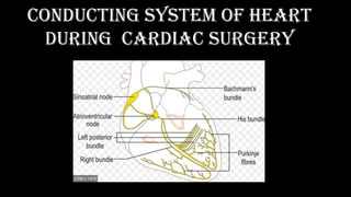
surgical techniques aimed at avoiding damage to the conduction system in manipulations mainly on the cardiac surgery

Recommended
More Related Content
Similar to conducting system of heart.pdf
Similar to conducting system of heart.pdf (20)
More from KararSurgery
More from KararSurgery (12)
Recently uploaded
Recently uploaded (20)
conducting system of heart.pdf
- 1. conducting system of heart during cardiac surgery
- 2. Directed by DR.Karar .A.Ali department of cardiothoracic surgery kararbenign@gmail.com
- 3. •
- 4. •
- 5. •
- 6. •
- 7. •
- 8. •
- 9. •
- 10. •
- 11. •
- 13. •
- 14. •
- 16. • Figure 2: Representative angiographic images of the SNA (arrows) demonstrating the general feasibility of its visualization. • (A) a normal SNA; (B) SNA with a focal stenosis (arrowhead); (C) diffuse atherosclerotic disease involving also the SNA; (D) stenosis of the main left coronary artery upstream the SNA (arrowhead).
- 17. •
- 18. •
- 20. •
- 21. •
- 22. •
- 23. •
- 24. • This heart with tetralogy of Fallot shows the typical devia- tion of the outlet septum and the overriding aorta. The yellow line depicts the position of the atrioventricular conduction tissues on the posteroinferior border of the ventricular septal defect.
- 25. • Schematic representation of the atrioventricular conduction tissues in the presence of a perimembranous ventricular septal defect. The bundle usually lies on the left ventricular aspect of the posteroinferior border of the defect. The star marks the position of the atrioventricular node. Modified, adapted, and used with permission from Davies et al. 9
- 26. • Right ventricular view of the atrioventricular transition, showing a perimembranous ventricular septal defect (VSD) extending slightly to the outlet. The yellow line shows the location of the atrioventricular conduction axis.
- 27. •
- 28. • These two hearts opened through the right ventricle show muscular ventricular septal defects (VSD) positioned in different parts of the septum (trabecular and inlet). The yellow lines indicate the location of the conduction axis, running posteroinferiorly or anterocephalad relative to the VSD.
- 29. •
- 30. • These two hearts with atrioventricular septal defect were dissected differently. The upper panel shows a view of the common atrioventricular junction and the aorta (Ao) displaced anterosuperiorly. The lower panel demonstrates a heart opened through the right ventricular chambers. The atrioventricular node is displaced posteroinferiorly due to the deficiency of the atrioventricular septation. The initial part of the nonbranching bundle is usually covered by the inferior bridging leaflet but the anterior part lies on the bare surface of the crest of the ventricular septum.
- 31. •
- 32. • The left-sided panel shows a heart with atrioventricular septal defect with two valvar orifices due to the fusion of the bridging leaflets. Such tissue fusion protects the long atrioventricular conduction axis from surgical damage during defect correction. The right panel shows in a histological section the presence of a bundle branch hidden by the leaflet tissues.
- 33. •
- 34. • Opened right atrium in a case of absent right atrioventricular connection. The star shows the anticipated site of the atrioventricular node on the floor of the atrium. ICV indicates inferior caval vein.
- 35. • Opened right atrium showing the tricuspid valvar orifice overriding the inlet ventricular septum. The anomalous node is located at the site where the ventricular septum reaches the inferior right atrial wall. The atrioventricular bundle runs on the bare surface of the ventricular crest.
- 36. •
- 37. •
- 38. •
- 39. • Different surgical techniques for correction of an AVSD. Single patch correction (A), double patch correction (B) and modi fi ed single patch (C) where the valve is attached to the ventricular septum are shown. In all techniques closure of the so-called cleft in the left atrioventricular valve (LAVV) is performed (1) and often approximation of the septal parts of the right atrioventricular valve (RAVV) (2) and approximation of the 2 left lateral commissures of the LAVV (3) are performed. RA = right atrium, LA = left atrium, RV = right ventricle, LV = left ventricle.
- 55. triangle of Koch
- 62. (A) Main panel shows a heart cut in a plane simulating transesophageal view with the superior vena cava (SVC) at 11 O'clock position. The circles mark the leaflet commissures and the white lines trace the segments of the hinge line. Note the smooth wall vestibule of atrial wall leading to the valve orifice. The smaller panels taken from random specimens demonstrate the thinness of the vestibular wall and variations in relationship of the hinge line (arrows), depth of fat- filled atrioventricular groove, and location of right coronary artery(*) at the numbered segments.