
Recommended
More Related Content
What's hot
What's hot (20)
Similar to RDT-112-PRELIM-LESSON-1.docx
Similar to RDT-112-PRELIM-LESSON-1.docx (20)
Recently uploaded
Recently uploaded (20)
RDT-112-PRELIM-LESSON-1.docx
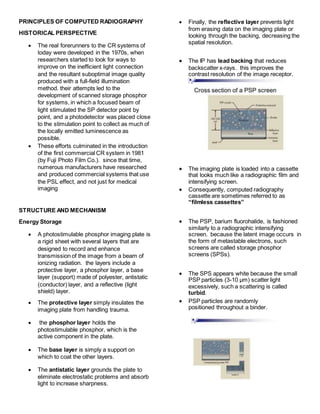
- 1. PRINCIPLES OF COMPUTED RADIOGRAPHY HISTORICAL PERSPECTIVE The real forerunners to the CR systems of today were developed in the 1970s, when researchers started to look for ways to improve on the inefficient light connection and the resultant suboptimal image quality produced with a full-field illumination method. their attempts led to the development of scanned storage phosphor for systems, in which a focused beam of light stimulated the SP detector point by point, and a photodetector was placed close to the stimulation point to collect as much of the locally emitted luminescence as possible. These efforts culminated in the introduction of the first commercial CR system in 1981 (by Fuji Photo Film Co.). since that time, numerous manufacturers have researched and produced commercial systems that use the PSL effect, and not just for medical imaging STRUCTURE AND MECHANISM Energy Storage A photostimulable phosphor imaging plate is a rigid sheet with several layers that are designed to record and enhance transmission of the image from a beam of ionizing radiation. the layers include a protective layer, a phosphor layer, a base layer (support) made of polyester, antistatic (conductor) layer, and a reflective (light shield) layer. The protective layer simply insulates the imaging plate from handling trauma. the phosphor layer holds the photostimulable phosphor, which is the active component in the plate. The base layer is simply a support on which to coat the other layers. The antistatic layer grounds the plate to eliminate electrostatic problems and absorb light to increase sharpness. Finally, the reflective layer prevents light from erasing data on the imaging plate or looking through the backing, decreasing the spatial resolution. The IP has lead backing that reduces backscatter x-rays. this improves the contrast resolution of the image receptor. The imaging plate is loaded into a cassette that looks much like a radiographic film and intensifying screen. Consequently, computed radiography cassette are sometimes referred to as “filmless cassettes” The PSP, barium fluorohalide, is fashioned similarly to a radiographic intensifying screen. because the latent image occurs in the form of metastable electrons, such screens are called storage phosphor screens (SPSs). The SPS appears white because the small PSP particles (3-10 µm) scatter light excessively, such a scattering is called turbid. PSP particles are randomly positioned throughout a binder.
- 2. SPSs are mechanically stable, electrostatically protected, and fashioned to optimize the intensity of stimulated light. SOme SPSs incorporate phosphors grown as linear filaments that enhance the absorption of x-rays and limit the spread of stimulated emission. In order for CR to function, the imaging plate material must have the ability to store and release the image information in a usable form. The most common phosphors with characteristics favorable for CR are barium fluorohalide bromides and iodides with europhium activators, (BaFBr:Eu and BaFi:Eu). The halides are approximately 85 percent bromide and 15 percent iodide. Some other compounds that may be used in the PSP are BaSrFBr:Eu, RbBr:T1, RbCI, and CsBr:Eu Photostimulable Luminescence (PSL) When these electrons return to the ground state, visible light is emitted. Over time, these metastable electrons return to the ground state on their own. However, this return to the ground state can be accelerated or stimulated by exposing the phosphor to intense infrared lightfrom a laser. Hence the term photostimulable luminescence from a photostimulable phosphor (PSP). Notes: During photostimulable luminescence: o X-rays interacts with the material in the imaging plate most specificallythe phosphor. o Once it interacts the electrons within the phosphor will go to an excited metastable state. o In order for them to be accelerated or stimulated, we use the laser light in order for the electrons to go back to their ground state. Thus, emitting the light needed for the image. Energy Release The sequence of events engaged in producing a PSL signal begins when an x-ray beam exposes a PSP, the energy transfers results in excitation of electrons intoa metastable state. Approximately 50% of these electrons return to their ground state immediately,resulting in prompt emission of light.
- 3. The remaining metastable electrons return to the ground state over time. This causes the latent image to fade and requires that the Imaging Plate must be read soon after exposure. CR signal loss is objectionable after approximately 8 hours. The next step in CR imaging is stimulation. The finely focused beam of infrared light with a beam diameter of 5O to 100 µm is directed at the PSP. o As laser beam intensity increases, so does the intensity of the emitted signal. The diameter of the laser beam determines the spatial resolution of the CR imaging system. Note: That as the laser beam penetrates, it spreads. The amount of spread increases with PSP thickness. The thicker the phosphor is, the more your laser beam penetrates and spread. The third step in this imaging process, which is detecting or reading the stimulated emission. The laser beam causes metastable electrons to return to the ground state withthe emissionof a shorter wavelengthlightin the blue regionof the visible spectrum. Through this process, the latent image is made visible. o Some signal is lost as the result of: 1) Scattering of the emitted light 2) The collection efficiency of the photodetector o Photodiodes (PDs) are the light detectors of choice for CR. The stimulation cycle of PSL signal acquisition does not completely transition all metastable electrons to the ground state. Some excited electrons remain. (Final stage - erase) If residual latent image remained, ghosting could appear on subsequent use of the IP. Any residual latent image is removed by flooding the phosphor with very intense white light from a bank of specially designed fluorescent lamps.
- 4. Notes: Crystals has both: VALENCE BAND and CONDUCTION BAND Electrons within the valence band will be transformed and pump and become exciteddue to introduction of energy through x-rays. These excitedelectrons will be trapped inthe color center of the phosphor crystals. Once they are trapped here and if they are exposed to the laser light, they are transported into the luminescence center. The PSP is sufficiently sensitive that it can become fogged by background radiation. The laser light used to stimulate the PSP is monochromatic. A solid-state laser is the stimulating source of choice. The resulting emission has a polychromatic spectrum. The emitted light intensity is many orders of magnitude lower than that of the stimulating light. Solid-state lasers produce longer wavelength light and therefore are less likely to interfere with emitted light. Even so, optical filters are necessary to allow only emitted light to reach the photodetector while blocking the intense stimulated light.
- 5. Digitalization -emitted light from the IP (Imaging Phosphor) is channeled into a funnel-like fiber-optic collection assembly and is directed at the photodetector, PMT (Photomultiplier tube), PD (Photo Diode), or charge-coupled device (CCD). Note: How does this x-ray become a digital signal? From x-rays, it becomes light and it becomes a digital signal. In Order for the x- rays to become a signal, it must undergo through the wires. Note: After the laser light is produced, there is an emitted light which will be filtered through by your optical filter ands photodetector. Before Photodetection occurs, the light is filtered so that none of the long- wavelength stimulation light reaches the photodetector and swamps emitted light. (Note: short wavelength lang ang ginadawat sa photodetector that is why mayroon tayong tinatawag na optical filter) In this case, emitted light is the signal and stimulating light is the noise; therefore, proper filtering improves the signal-to-noise ratio. (Note: Two results of the film that was exposed in laser light --- emitted light and stimulating light. Optical filter only allows the emitted light. Dapat mas madami ang signal compared to the noise.) The output of the photodetector is a time-varying analog signal that is transmitted to a computer system that has multiple functions. The time-varying analog signal from the photodetector is processed for amplitude, scale, and compression. This shapes the signal before the final image is formed. (Note: From x-rays, it becomes light and then it becomes an analog signal) Then the analog signal is digitized, with attention paid to proper sampling (time between samples) and quantization (the value of each sample). The image buffer is usually a hard disc. This is the place where a completed image can be stored temporarily until it is transferred to a workstation for interpretation or to an archival computer. The computer of the CR readers is in control of the slow scan and the fast scan. This control works off the computer clock in gigahertz (GHz). Note: From x-rays, it becomes light, it becomes a metastable energy within the electrons. The electrons are emitted in the form of light. Light is passed through the optical filter and detected by the photo detector. It begins to be an analog signal and then, it becomes a digital signal through analog-to-digital converter (ADC).
- 6. Note: Because of the sampling and quantization and shipping of the signal, it becomes a digital signal and then, computer compliment. Energy is neither created nor destroyed. Kung unsa tong energy na naa sa x- rays or the value of the energy or the strength of the x-rays, it is the same pag mahimo na siyang light. When x-rays interact with the imaging platespecifically sa phosphor, it becomes a metastable energy. Gihimo niya na excited ang electrons. It puts the electrons into a metastable state. After that, your electrons will emit that energy in the form of emitted light. The light is passed through the optical filter which filters both emitted and stimulated light, but ang ginatanggap niya lang talaga mostly is the emitted light. It passes through the photodetector, after it becomes detected by the photodetector, it becomes an analog signal, then after quantization and sampling, shipping, scaling, amplitude, compression, it becomes a digital signal. And then, imaged is displayed in the monitor. The heart of the computed radiography is the imaging plate.
- 7. Film-Screen vs Computed Radiography Film-Screen CR Exposure Medium Film and intensifying screen Phosphor and light (both emitted and the laser light) Processing Involves the use of an automatic processor with the use of chemistry Uses an automatic reader. Processing Time Longer processing time Shorter reading times Evaluation A viewbox or negatoscope is needed to access the images. The images pop up in the monitor after reading the image plate. Archiving Requires a huge space for storing and ordering of films. Hard copy Only needs a small space for the device storage. Uses PACS Soft copy Availability Only few medical facilities utilize this. Widely available