Methods For Improving The Cellular Uptake Of Dna Origami...
Science_2005
1. standing the biology of obesity and how best
to treat it. The propensity of obese persons to
sit more than lean individuals has several
potential explanations. Rodent studies sup-
port the concept that there are central and
humoral mediators of NEAT (10, 11). For
example, we have shown that a neuropeptide
associated with arousal, orexin (12), in-
creases NEAT in rats when injected into
the paraventricular nucleus (PVN) of the
hypothalamus. Preliminary data suggest that
PVN injections of orexin also cause dose-
dependent increases in standing posture
allocation in rats (13). Thus, there may be
central and humoral mediators that drive the
sedentary behavior of obese individuals. The
negative relationship between fat mass and
movement (Fig. 1B) raises the intriguing
possibility that body fat releases a factor that
slows physical activity in obesity. However,
these data also demonstrate that posture
allocation is not the mechanism by which
NEAT is modulated with short-term over-
feeding. One hypothesis is that this occurs
through modulation of energy efficiency;
this is an area worthy of future investigation.
These data may also have implications
for obesity intervention. One could argue
that obese individuals have a biologically
determined posture allocation and therefore
are destined to become obese. If this were
true, obesity would have been as common 50
years ago as it is today. However, obesity
rates have increased and continue to do so
(14). We speculate that obese and lean
individuals respond differently to the envi-
ronmental cues that promote sedentary be-
havior. If the obese volunteers adopted the
NEAT-enhanced behavior of their lean
counterparts, they could expend an addition-
al 350 kcal per day. Over a year, this alone
could result in a weight loss of È15 kg, if
energy intake remained unchanged. Herein
lies the rationale behind nationwide ap-
proaches to promote NEAT in small incre-
ments (15). For example, in Rochester,
Minnesota, in 1920 before car use was com-
monplace the average walk to and from work
was 1.6 miles (16). If walking this distance
to work were reinstituted by our obese
subjects, all of whom currently drive to
work, an extra 150 kcal per day could be
expended. We will need to use similar
measures to promote NEAT as an impetus
to create an active and dynamic environment
in which, for example, dancing supersedes
television as a leisure activity. Approaches
that succeed in getting people out of their
chairs and moving could have substantial
impact on the obesity epidemic.
References and Notes
1. A. H. Mokdad, J. S. Marks, D. F. Stroup, J. L.
Gerberding, JAMA 291, 1238 (2004).
2. E. A. Finkelstein, I. C. Fiebelkorn, G. Wang, Obes. Res.
12, 18 (2004).
3. World Health Organization, Obesity: Preventing and
Managing the Global Epidemic (Geneva, Switzerland,
1997).
4. J. A. Levine, N. L. Eberhardt, M. D. Jensen, Science
283, 212 (1999).
5. J. A. Levine, P. A. Baukol, K. R. Westerterp, Med. Sci.
Sports Exerc. 33, 1593 (2001).
6. J. A. Levine, E. L. Melanson, K. R. Westerterp, J. O.
Hill, Eur. J. Clin. Nutr. 57, 1176 (2003).
7. J. A. Levine, E. L. Melanson, K. R. Westerterp, J. O. Hill,
Am. J. Physiol. Endocrinol. Metab. 281, E670 (2001).
8. Materials and methods are available as supporting
material on Science Online.
9. D. A. Schoeller, C. A. Leitch, C. Brown, Am. J. Physiol.
251, R1137 (1986).
10. J. A. Levine, J. Nygren, K. R. Short, K. S. Nair, J. Appl.
Physiol. 94, 165 (2003).
11. K. Kiwaki, C. M. Kotz, C. Wang, L. Lanningham-Foster,
J. A. Levine, Am. J. Physiol. 286, E551 (2004).
12. J. G. Sutcliffe, L. de Lecea, Nature Med. 10, 673
(2004).
13. C. M. Kotz, personal communication.
14. J. O. Hill, H. R. Wyatt, G. W. Reed, J. C. Peters,
Science 299, 853 (2003).
15. More information about promoting NEAT in small
increments can be found at www.smallstep.gov and
www.americaonthemove.org.
16. L. Lanningham-Foster, L. J. Nysse, J. A. Levine, Obes.
Res. 11, 1178 (2003).
17. We thank the volunteers, dietitians, food technicians,
nursing staff, and the Mass Spectrometer Core at the
General Clinical Research Center, A. Oberg for
assistance with statistics, and P. Baukol for techni-
cal support. Supported by NIH grants DK56650,
DK63226, DK66270, and M01 RR00585, by T. S. and
D. B. Ward, and by the Mayo Foundation.
Supporting Online Material
www.sciencemag.org/cgi/content/full/307/5709/584/
DC1
Materials and Methods
Figs. S1 to S4
Tables S1 and S2
References
20 October 2004; accepted 7 December 2004
10.1126/science.1106561
Sequence-Directed DNA
Translocation by Purified FtsK
Paul J. Pease,1
* Oren Levy,2
* Gregory J. Cost,1
Jeff Gore,3
Jerod L. Ptacin,1
David Sherratt,5
Carlos Bustamante,1,3,4
.
Nicholas R. Cozzarelli1,2
.
DNA translocases are molecular motors that move rapidly along DNA using
adenosine triphosphate as the source of energy. We directly observed the
movement of purified FtsK, an Escherichia coli translocase, on single DNA
molecules. The protein moves at 5 kilobases per second and against forces up
to 60 piconewtons, and locally reverses direction without dissociation. On
three natural substrates, independent of its initial binding position, FtsK
efficiently translocates over long distances to the terminal region of the
E. coli chromosome, as it does in vivo. Our results imply that FtsK is a
bidirectional motor that changes direction in response to short, asymmetric
directing DNA sequences.
DNA translocases are adenosine triphosphate
(ATP)–driven machines required for DNA
replication, recombination, and transfer
within and between cells (1–6). FtsK is a
membrane-bound and septum-localized E.
coli translocase that coordinates cell division
with chromosome segregation (7). At times,
the product of chromosome replication is a
circular dimer rather than two monomers.
These dimers are resolved by the XerCD
site-specific recombinase at a site near the
terminus of replication, termed dif (8).
Recombination between the distant dif sites
requires FtsK. In addition, at the time of cell
division some DNA may remain in the septal
region, and FtsK appears to act as a pump to
clear this region of DNA (9, 10). FtsK may
also promote disentanglement of chromo-
some termini by means of an interaction with
topoisomerase IV (11).
Translocases move along DNA or move
the DNA if the translocase is anchored. In
either case, translocases must be able to move
in a specific direction relative to the DNA.
Translocase directionality could be determined
by strand polarity for single-strand tracking
enzymes, nonrandom orientation of trans-
locase binding through localized accessory
factors or binding sites, or DNA sequences
that affect the enzyme during translocation. In
E. coli, provocative genetic experiments
showed that lambda-phage DNA inserted
near dif disrupted chromosome dimer reso-
lution in a manner dependent on insertion
orientation (9). Thus, DNA sequences could
be directing FtsK toward dif so that it can
activate XerCD recombination.
1
Department of Molecular and Cell Biology, 2
Biophysics
Graduate Group, 3
Department of Physics, 4
Howard
Hughes Medical Institute, University of California,
Berkeley, CA 94720–3204, USA. 5
Division of Molecular
Genetics, Department of Biochemistry, University of
Oxford, OX1 3QU, UK.
*These authors contributed equally to this paper.
.To whom correspondence should be addressed.
E-mail: ncozzare@socrates.berkeley.edu (N.R.C.);
carlos@alice.berkeley.edu (C.B.)
R E P O R T S
28 JANUARY 2005 VOL 307 SCIENCE www.sciencemag.org586
onFebruary20,2017http://science.sciencemag.org/Downloadedfrom
2. Here, we measured FtsK translocation on
single DNA molecules and showed that FtsK
moves fast (5 kb/s) and can work against a
heavy load (960 pN) in an ATP-dependent
manner. We directly observed its movement
along DNA and found that purified FtsK
moves toward dif, demonstrating that DNA
sequence alone is sufficient to direct FtsK
toward its target. Our results are consonant
with a model of FtsK in which bidirectional
motors alternate activity in response to
directing DNA sequences.
Our experiments used the C-terminal
motor domain of FtsK (FtsK50C) (12, 13).
We first asked whether FtsK50C is able to pull
DNA against a load, as its role in vivo
suggests. To this end, we tethered a single
molecule of lambda-phage DNA between two
beads, one of which was held on a micro-
pipette (Fig. 1A). The DNA was then ex-
tended under constant tension by buffer flow.
When FtsK50C and ATP were introduced, the
DNA tether rapidly shortened against the ap-
plied force, visualized as a decrease in the
bead-to-bead distance (Fig. 1A and Movie S1).
We conclude that tether shortening results
from the formation of an expanding DNA
loop closed off by two points of contact with
FtsK50C. This loop is probably topologically
constrained by the translocase, because bulk
assays indicate that FtsK generates twin super-
coiled regions (13). Topological domain for-
mation by translocases may be widespread, as
another translocase, human Rad54, has been
shown by scanning force microscopy to form
supercoiled DNA loops (14).
Activity occurred in bursts that were
identified by an algorithm that recognizes
continuous changes in interbead distance, thus
excluding periods of enzyme inactivity (12).
A plot of DNA extension versus time for a
typical experimental run is shown in Fig. 1B.
At a constant force from 10 to 15 pN, the
velocity of individual bursts from four exper-
imental runs (totaling 108 bursts) averaged
5.0 T 0.9 kb/s at 25-C (fig. S1), a rate com-
parable with those of a recent study (15). The
kinetics of FtsK50C are considerably faster
than other DNA translocases assayed in
single-molecule experiments (16–20). Not
only is FtsK50C exceptionally fast, it is also
unusually powerful. In the same assay, against
high loads of 35 to 40 pN, FtsK50C velocity
only drops by about half to 2.8 T 0.5 kb/s.
Although decreasing bead-to-bead distance
results from an expanding DNA loop, we also
observed that the bead-to-bead distance could
increase, as if the DNA loop were contracted.
The rate was nearly the same during loop
expansion as during loop contraction (Fig.
1B inset), which suggests that contraction
too is an active process of FtsK50C. This bi-
directionality of FtsK50C suggests that it
consists of more than a single unidirectional
motor (21). Occasionally, we saw an abrupt
collapse of the loop, consistent with com-
plete release of one of the contact points
between the DNA and FtsK50C. Trans-
location and loop formation then immediate-
ly resumed, which indicates that FtsK50C
remained firmly bound to the DNA at at least
one contact point during these events. These
rapid reversals along with the small standard
deviation of the observed rates make it very
unlikely that there are multiple FtsK com-
plexes acting simultaneously.
In a complementary assay for DNA
translocation, a molecule of lambda DNA
was tethered between two beads as in the
reeling assay, but instead of being left free,
the second bead was caught in an optical
trap, allowing direct measurement of the
force exerted on the DNA by FtsK50C. As
FtsK50C translocated, it pulled the trapped
bead toward the micropipette with a con-
comitant increase in force, indicative of loop
formation. As the DNA loop enlarged,
FtsK50C experienced an increasing load that
was released by reverse translocation or, less
frequently, by a dissociation of the protein
from one of the DNA contact points.
Visible aggregates of the enzyme, re-
ferred to here as particles, were observed at
higher protein concentrations, particularly
above 100 nM. These particles bound indi-
vidually to the DNA at random positions as
protein was introduced to the flow cell
during the optical-trap assay (Fig. 1C and
Movie S2). As soon as an FtsK50C particle
bound to the DNA, DNA looping was
initiated, as evidenced by the shortening of
the tether. These particles traveled along the
DNA at 4.9 T 0.9 kb/s, the same rate measured
in the reeling assay, thus making it possible to
track a single active FtsK50C complex.
Strikingly, the FtsK50C particles con-
sistently translocated along the lambda
DNA in the same overall direction (75 out
of 75 observations), even though occasion-
ally some would temporarily reverse direc-
tion, as in the reeling assay. To be certain
that unidirectionality was in fact a property
of the translocase, we inverted the DNA
molecule with respect to either the optical
trap or the two types of beads used. The
particles always moved in the same direction
relative to the lambda DNA sequence. It is
notable that they move in the direction
predicted by genetic experiments (9). The
occasional reversals show that FtsK50C
directionality is dynamic; it can change on
2 4 6 8 10 12 14 16 18
6.0
12.0
18.0
24.0
Time (sec)
Extension(kb)
A
1 sec
13 sec
B
C
Flow
FtsK reels bead
against flow
14.2 14.8 15.4
0 sec 6 sec
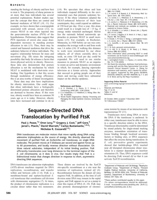
Fig. 1. Measurement of FtsK translocation with two assays. (A) In the reeling assay shown, each
end of a lambda DNA molecule is attached to a bead. One bead is held by suction through a
micropipette. Translocation by FtsK forms a loop in the DNA tether, which reels in the free bead
against a flow force of 10 to 15 pN. The shortening of the tether with 100 nM FtsK50C and 3 mM
ATP is seen in the video frames spaced at 1-s intervals. (B) Tracking of free-bead position as a
function of time shows bursts reeling in (green) and reeling out (blue). Reeling in and reeling out
velocities are often similar (insert). (C) FtsK50C translocation can be directly visualized on single
lambda DNA molecules. The DNA was tethered between two beads with one held by a
micropipette and the other in an optical trap. The tether was maintained at a constant tension by
moving the micropipette in response to DNA looping with an automated force-feedback program.
Video frames at 1-s intervals are shown with the FtsK particle surrounded by a red circle.
R E P O R T S
www.sciencemag.org SCIENCE VOL 307 28 JANUARY 2005 587
onFebruary20,2017http://science.sciencemag.org/Downloadedfrom
3. small scales but is ultimately unidirectional
over large distances.
To test the directionality of FtsK50C on its
natural substrate, we constructed three DNA
tethers from the dif region of the E. coli
chromosome (Fig. 2A). These 27- to 30-kb
substrates contain the DNA immediately to
the left (L) of dif on the chromosome, the
right (R) of dif, or centered (C) on dif. If
DNA sequences neighboring dif are suffi-
cient to direct FtsK, then the net movement
of FtsK50C particles should always be toward
dif. The particle-tracking assay described
above was used to test this hypothesis.
Every FtsK50C particle observed on the L
and R substrate (15 of 15) translocated to the
dif proximal end of the DNA tether. Traces
of FtsK50C particles moving on L and R are
shown in Fig. 2B. The rate of movement was
4.8 T 0.9 kb/s for the bursts and 3.2 kb/s
overall, the same rate as on lambda DNA.
Also, as on lambda DNA, FtsK50C occasion-
ally reversed direction before turning back
toward the dif proximal end.
In vivo, FtsK must bind to a long DNA
molecule (the chromosome) and efficiently
find its way to dif. The behavior of FtsK50C
on our C tether mimics the in vivo situation.
Here, the FtsK50C particles moved rapidly
toward dif from either direction. Upon
reaching dif, the particles oscillated for a
number of minutes around dif (Fig. 2B and
Movie S3), and their average position
coincided with dif. These oscillations were
only observed on the C substrate and indicate
that FtsK50C constantly samples DNA se-
quence to ensure proper directionality.
Our findings contradict the conclusion of
a recent study that FtsK is a sequence-
independent translocase (15). That study,
however, used magnetic tweezers in which
DNA is viewed down its axis. Therefore,
the position or movement of FtsK could
not be detected. In our assay with optical
tweezers, the direction of view is perpen-
dicular to the helix axis, and we can readily
see FtsK particles. The data in the other
study is readily explained by the model we
present below.
We tested whether FtsK50C pulls DNA
into a loop from two directions at once, as
does T antigen (22), by measuring simulta-
neously the position of the FtsK50C particle
and the two beads. Analysis of 25 trans-
location events on the C substrate showed
that the DNA shortening occurred only from
one direction at any given time. For exam-
ple, during FtsK50C translocation toward the
micropipette, as diagrammed in Fig. 3A, the
tether length below the particle decreased,
whereas that above it increased slightly as a
result of stretching caused by the high forces
generated by FtsK50C translocation (Fig. 3, C
and D) (23, 24). We conclude that the
FtsK50C complex is coupled in such a way
that only one motor is active at any given
time (25).
After observing that DNA enters the loop
from only one side of the FtsK50C complex,
we used the bead-particle correlation analy-
sis to observe loop release. The DNA in the
loop could be released from either side of the
complex. However, there would only be net
BA
dif E. coli
chromosome
dif
L R
C
0 10 20 240 250 260 270
18
12
6
6Kb
12Kb
18Kb
Time (sec)
FtsKposition(kb)
12
6
18
L
R
C
dif
Fig. 2. FtsK translocates toward dif in vitro.
(A) To test FtsK directionality on its natural
substrate, the three DNA regions of the E. coli
chromosome shown were amplified to give
products spanning 29.5 kb to the left of dif (L),
28.7 kb to the right of dif (R), and 27.9 kb
centered on dif (C). (B) FtsK50C quickly translocated toward the dif proximal end with both the
L (red) and R (green) substrates (15 out of 15 observations). Once the FtsK particle reached the end
of the tether, it was obscured by the bead and no longer visible. The particles did not translocate
back in the opposite direction even after several minutes. With the C substrate (blue), FtsK50C
oscillated about dif for 5 to 15 min. The location of dif is shown as a black horizontal dot-dash line.
Fig. 3. Simultaneous tracking of FtsK, force on the tether, and
flanking DNA lengths demonstrates that DNA is reeled from
only one side at a time. (A) Diagram of the experiment
showing a DNA molecule held between two beads. The
portions of the tether above and below FtsK are colored black
and orange, respectively, to illustrate that only the DNA
below FtsK is reeled into the loop. The upper bead is in an
optical trap (not shown), and the lower bead is on a
micropipette. The FtsK complex is shown as two coupled
bidirectional motors, with the active motor depicted as a blue
triangle pointing toward the direction of movement and the
inactive one as a green square. Translocation can generate a
loop and displace the bead in the optical trap and also release
the loop, returning the bead to the trap. (B) Correlated
movement of the bead in the trap (red) and FtsK particle
(blue), corresponding to the diagram in (A). The bead trace is
offset to facilitate comparison. (C) As FtsK50C translocates
down toward the micropipette, only the tether below the
FtsK50C complex (orange) decreases in length (25 of 25
observations, including the opposite case in which the FtsK50C
complex moves up and the upper tether length decreases),
which demonstrates that DNA is reeled from only one side at
any one time. The tether lengths are corrected for stretching
because of the high forces on the tether. (D) The force
generated by FtsK50C during this burst.
4.2
4.4
4.6
4.8
5
Position(µm)
9 9.5 10 10.5
10
20
30
40
50
Time (sec)
Force(pN)
11.8
12.4
13.0
13.6
14.2
TopTetherlength(kb)
A
C
B
D
Bead
FtsK
BottomTetherlength(kb)
R E P O R T S
28 JANUARY 2005 VOL 307 SCIENCE www.sciencemag.org588
onFebruary20,2017http://science.sciencemag.org/Downloadedfrom
4. translocation if the loop were released from
the opposite side from which it was formed.
We observed all four possible movements,
i.e., DNA entering the loop from either side
and releasing from either side (fig. S2, A to
D). The correlated movement of the bead in
the optical trap and the FtsK50C particle
provides clear evidence that the only site of
looping is at the visible FtsK50C particle. The
symmetry of movements seen here supports
a model of two coordinated, bidirectional
motors, which we propose are rectified by
short, asymmetric DNA sequences.
We also observed translocation in the
absence of detectable looping, especially at
forces above 35 pN (fig. S2, E and F). This
type of movement presumably occurs when
there is no stable, static FtsK-DNA contact
point. Therefore, even though translocation
at low forces typically proceeds with two
points of contact between the motors and
DNA, only one may be needed.
We estimated the strength of the FtsK50C
motor using the C tether in the optical-trap
assay (fig. S3). The tension in the DNA tether
increases as FtsK50C decreases the DNA end-
to-end distance by looping and pulls the bead
from the center of the trap. We measured the
force at which the bead suddenly reverses
direction, because the DNA loop is presum-
ably released as a result of a slip at the con-
tact point with the active motor. DNA loops
were released at forces ranging from 15 pN to
more than 60 pN, with a mean of 35 pN (fig.
S3). The upper bound of this force range is
close to that which causes a phase transition
in DNA (26, 27).
Our working model for FtsK translo-
cation (Fig. 4) depicts the FtsK complex as
two bidirectional motors coupled in such a
way that only one is active at any given
time. The inactive motor remains firmly bound
to the DNA, thus creating the second con-
tact point necessary for loop formation. An
expanding supercoiled domain is generated
between the two motors as one binds the
DNA tightly while the other tracks the DNA
helix. We propose that each motor is bi-
directional to account for all of the patterns
of movement we observed in our correlation
analyses (fig. S2): The FtsK complex must
be able to pull DNA through either motor
and also release it through either motor. Net
translocation of FtsK occurs, however, only
when DNA is taken in through one motor
and released through the opposite motor
(Fig. 4). It is unclear how FtsK reverses di-
rection, but if it tracks one strand of the
DNA duplex, a switch to the complemen-
tary strand would cause translocation rever-
sal. Although it is not shown explicitly in
Fig. 4, we believe that the net directionality
of FtsK is imprinted when it moves past a
short, asymmetric sequence whose polarity
inverts at dif. Directionality is then main-
tained over a distance long enough to give
overall unidirectionality but with occasion-
al switches.
The high speed at which FtsK translocates
along DNA (5 kb/s at 25-C) is about the rate
expected for this family of translocases in
vivo (2, 15). Thus, the simple purified system
we studied recapitulates the rate as well as
the directionality of translocation in vivo.
The ability of FtsK to work against a heavy
load is essential for its role in vivo. FtsK is
rendered stationary by its insertion into the
septum and must move a considerable
section of the chromosome burdened with
large transcription and translation complexes.
Moreover, during translocation FtsK will
encounter proteins and RNA bound to the
DNA, so the ability to apply force during
translocation would allow FtsK to clear these
potential roadblocks without slowing down
appreciably. The proposed annular structure
of FtsK with DNA in the lumen would make
FtsK and similar translocases effective
Bwire-strippers,[ clearing DNA (28) to reset
epigenetic and cell-cycle–specific nucleo-
protein structures at the apt time of replica-
tion termination.
We observe that FtsK changes direction
during translocation at several places on the
DNA around dif, which suggests that the
DNA sequences responsible for directionality
are widespread. In fact, many short sequences
with a skewed orientation flip abruptly at dif
(29), and these have been proposed to bias
the direction of FtsK translocation toward dif
(9). The mechanism of FtsK movement could
be analogous to the bipolar helicase
RecBCD, which switches active motors when
it encounters the skewed octamer called Chi
in the correct orientation (30).
Bulk experiments with several trans-
locases showed that these enzymes efficiently
generated (þ) and (–) supercoils in a circular
DNA (13, 31). Our finding that FtsK forms a
stable DNA loop provides a ready explana-
tion for why these opposing supercoils did
not cancel: They are segregated into separate
domains delimited by the protein. FtsK and
other translocases could contribute to do-
main formation in vivo; the mobility of the
contact by FtsK is consistent with recent
findings of random placement of domain
boundaries in the cell (32). The rapid forma-
tion of a large loop at the terminus could
concentrate catenane links, making decatena-
tion by Topo IV more efficient (11, 33).
References and Notes
1. M. R. Singleton, D. B. Wigley, EMBO J. 22, 4579 (2003).
2. J. Errington, J. Bath, L. J. Wu, Nature Rev. Mol. Cell
Biol. 2, 538 (2001).
3. G. D. Recchia, M. Aroyo, D. Wolf, G. Blakely, D. J.
Sherratt, EMBO J. 18, 5724 (1999).
4. F. X. Barre et al., Proc. Natl. Acad. Sci. U.S.A. 98,
8189 (2001).
5. E. C. Hendricks, H. Szerlong, T. Hill, P. Kuempel,
Mol. Microbiol. 36, 973 (2000).
6. T. L. Raoul Tan, R. Kanaar, C. Wyman, DNA Repair
(Amst.) 2, 787 (2003).
7. G. Liu, G. C. Draper, W. D. Donachie, Mol. Microbiol.
29, 893 (1998).
8. G. Blakely, S. Colloms, G. May, M. Burke, D. J. Sherratt,
New Biol. 3, 789 (1991).
9. J. Corre, J. M. Louarn, J. Bacteriol. 184, 3801 (2002).
10. I. F. Lau et al., Mol. Microbiol. 49, 731 (2003).
11. O. Espeli, C. Lee, K. J. Marians, J. Biol. Chem. 278,
44639 (2003).
12. Materials and methods are available as supporting
material on Science Online.
13. L. Aussel et al., Cell 108, 195 (2002).
14. D. Ristic, C. Wyman, C. Paulusma, R. Kanaar, Proc.
Natl. Acad. Sci. U.S.A. 98, 8454 (2001).
15. O. A. Saleh, C. Perals, F. X. Barre, J. F. Allemand,
EMBO J. 23, 2430 (2004).
16. M. D. Wang et al., Science 282, 902 (1998).
17. N. R. Forde, D. Izhaky, G. R. Woodcock, G. J. Wuite,
C. Bustamante, Proc. Natl. Acad. Sci. U.S.A. 99, 11682
(2002).
18. D. E. Smith et al., Nature 413, 748 (2001).
19. P. R. Bianco et al., Nature 409, 374 (2001).
20. G. J. Wuite, S. B. Smith, M. Young, D. Keller, C.
Bustamante, Nature 404, 103 (2000).
21. We use the term ‘‘motor’’ to mean an enzyme that
translocates or binds DNA at one point, with a
molecular structure yet to be established.
22. R. Wessel, J. Schweizer, H. Stahl, J. Virol. 66, 804 (1992).
23. FtsK stopped translocating above È63 pN, presum-
ably because of DNA distortion, but remained bound
and continued translocating if the force was lowered
below the over-stretch transition.
Fig. 4. Model for FtsK translocation along a
single molecule of DNA. A nicked DNA molecule
is held between two beads. One bead is held
with a micropipette, and the other is held in an
optical trap (not shown). The two coupled
motors in an FtsK complex (green and blue)
are shown as a triangle when active and as a
rectangle when providing a static contact point.
(A) The complex binds to the DNA, and the
lower motor begins moving downward. (B) The
DNA is bound statically to the upper motor so
that translocation by the lower motor creates a supercoiled DNA loop between the two motors,
decreasing the end-to-end distance of the DNA, thus displacing the bead in the optical trap. (C)
The lower motor continues enlarging the loop, further displacing the optically trapped bead. (D)
The motors switch activity so that the lower motor becomes static while the upper motor begins
pulling out the DNA loop, relaxing the force on the trapped bead. (E) The upper motor finishes
pulling out the loop, returning the DNA tether to its full extension. The net transfer of DNA from
one side of the FtsK complex to the other results in FtsK complex translocation. No net trans-
location is seen when the bottom motor reverses direction. This and other alternatives are
illustrated in fig. S2. The reversibility of the two motors makes it possible for asymmetric DNA
sequences (not shown) to dictate the overall direction of FtsK translocation by switching bi-
directional motor activity.
R E P O R T S
www.sciencemag.org SCIENCE VOL 307 28 JANUARY 2005 589
onFebruary20,2017http://science.sciencemag.org/Downloadedfrom
5. 24. S. B. Smith, Y. Cui, C. Bustamante, Science 271, 795
(1996).
25. This experiment also supports our conclusion that we
are observing the activity of a single FtsK complex. If
multiple complexes were bound to the DNA, we
would expect to see by chance occasional reeling in
from above and below the observed particle.
26. It is difficult to measure a conventional stall force,
because in almost all cases, the DNA loop releases
before the motor velocity falls to zero.
27. Because processive motion is observed against a force
of È63 pN, we can place an upper limit of 1.6 nm
(100 pN nm/63 pN) on the step size of the motor. This
upper bound was calculated by assuming that a single
ATP molecule is hydrolyzed per step and by recogniz-
ing that the efficiency must be less than 100%.
28. D. L. Kaplan, M. O’Donnell, Mol. Cell 15, 453 (2004).
29. S. L. Salzberg, A. J. Salzberg, A. R. Kerlavage, J. F. Tomb,
Gene 217, 57 (1998).
30. M. Spies et al., Cell 114, 647 (2003).
31. J. Bath, L. J. Wu, J. Errington, J. C. Wang, Science 290,
995 (2000).
32. L. Postow, C. D. Hardy, J. Arsuaga, N. R. Cozzarelli,
Genes Dev. 18, 1766 (2004).
33. J. Louarn, F. Cornet, V. Francois, J. Patte, J. M. Louarn,
J. Bacteriol. 176, 7524 (1994).
34. We thank T. H. Massey for providing the FtsK50C His-
tagged construct and S. B. Smith for expert assist-
ance. This work was supported by NIH grants
GM31657 (N.R.C.), GM32543 (C.B.), GM07232-27
(J.L.P.), Wellcome Trust and the Royal Society (D.S.),
Ruth L. Kirschstein National Research Service Award,
grant GM08295-15 (P.J.P.), Fannie and John Hertz
Foundation (J.G.), Damon Runyon Cancer Research
Foundation grant 1702-02 (G.J.C.), and U.S. Depart-
ment of Energy grants DE-AC03-76DF00098,
GTL2BN ‘‘Microscopies of MolecularMachines,’’ and
SNANOB ‘‘Design of Autonomous Nanobots’’ (C.B.).
Supporting Online Material
www.sciencemag.org/cgi/content/full/307/5709/586/
DC1
Materials and Methods
Figs. S1 to S3
Movies S1 to S3
7 September 2004; accepted 1 December 2004
10.1126/science.1104885
Restoration of Tolerance in Lupus
by Targeted Inhibitory
Receptor Expression
Tracy L. McGaha,1
Brian Sorrentino,2
Jeffrey V. Ravetch1
*
Lupus, a multigenic autoimmune condition in which a breakdown of tolerance
results in the development of autoantibodies, leads to a variety of pathologic
outcomes. Despite the heterogeneity of factors influencing disease suscepti-
bility, we demonstrate that the partial restoration of inhibitory Fc receptor
(FcgRIIB) levels on B cells in lupus-prone mouse strains is sufficient to restore
tolerance and prevent autoimmunity. FcgRIIB regulates a common B cell
checkpoint in genetically diverse lupus-prone mouse strains, and modest
changes in its expression can result in either tolerance or autoimmunity.
Therefore, increasing FcgRIIB levels on B cells may be an effective way to treat
autoimmune diseases.
The ability of the immune system to dis-
tinguish self from nonself is central to its
ability to protect against pathogens and, at the
same time, maintain nonresponsiveness to
self. This property is established at discrete
checkpoints both during development and
in the adult. To date, several early develop-
mental checkpoint mechanisms have been
identified. These include the deletion of
autoreactive lymphocytes during early devel-
opment of the immune system (1–3); anergy,
which converts autoreactive cells to a state
that precludes them from becoming activated
(4, 5); and editing, a mechanism for modifying
autoantibodies that renders them nonauto-
reactive (6, 7). Although these developmental
checkpoints purge the immune repertoire of
autoreactive cells, the processes of central
tolerance remain incomplete, allowing self-
reactive cells that express antigen receptors
to escape into the periphery (8, 9). In ad-
dition, mechanisms that enhance antibody
diversity, such as somatic mutation, can
generate potentially autoreactive antigen
receptors in the adult (10). Thus, checkpoints
that operate in the periphery of mature
individuals are critical for maintaining toler-
ance and for establishing tolerance to self-
antigens that only appear after maturity. Less
is known about these peripheral checkpoints,
although a principal element has emerged
whereby the balance between stimulatory
and inhibitory signals regulates the activa-
tion and expansion of lymphoid cells. Inhib-
itory signaling, in particular, is a critical
feature of peripheral tolerance, providing a
means for establishing thresholds for stimu-
lation and for active deletion of autoreactive
cells from the peripheral repertoire. Pertur-
bations in inhibitory signaling pathways
have been shown to be genetically associated
with autoimmunity (11, 12).
Genetic studies have associated a large
number of loci and candidate genes, in ad-
dition to inhibitory signaling pathways, with
susceptibility to the development of autoim-
mune diseases (11, 13). In the context of mul-
tifactorial and multigenic diseases such as
lupus, it is possible that single overriding fac-
tors may ultimately dictate whether the disease
progresses or not. The selection and prolifer-
ation of immunoglobulin G (IgG)–producing
B cells represents one such overriding periph-
eral checkpoint that is under the potential
control of inhibitory signaling pathways.
Our previous work demonstrated that the
expression of the inhibitory Fc receptor
FcgRIIB was required for the maintenance
of tolerance (14). C57BL/6 mice that are
deficient in this receptor develop spontane-
ous lupus-like autoimmunity and progress to
fulminate glomerulonephritis and premature
mortality (14). Studies of bone marrow trans-
fer into recombinase-activating gene (RAG)–
deficient mice suggested that FcgRIIB defi-
ciency in the B cell compartment is most
likely responsible for the loss of tolerance
seen in these mice. In support of this idea,
several strains of mice that develop sponta-
neous autoimmune disease, such as NZB,
NOD, BXSB, and MRL/lpr, have also been
shown to express reduced levels of FcgRIIB
on activated or germinal-center B cells. This
reduced expression results from a polymor-
phism in the promoter of this gene (15–18).
These results suggest that the absolute level
of FcgRIIB expressed on some B cells may
regulate the ability of these cells to maintain
tolerance and that relatively small changes in
the expression of this inhibitory receptor may
permit the survival and expansion of auto-
reactive cells. To test this hypothesis, we
developed retroviral vectors that are capable
of expressing FcgRIIB upon transduction of
bone marrow cells, which can restore the
wild-type level of FcgRIIB to B cells derived
from autoimmune-prone strains. Bone mar-
row was derived from the autoimmune-
susceptible strains NZM 2410, BXSB, and
B6.Fcgr2bj/j and transduced with either
FcgRIIB-expressing retrovirus or parental
(mock) virus lacking FcgRIIB. The bone
marrow of irradiated recipients was recon-
stituted with autologous retroviral-transduced
bone marrow, and the mice were followed for
the development of autoimmunity and autoim-
mune disease. Mice that received autologous
bone marrow transduced with the parent virus
developed autoimmune disease and had re-
duced viability comparable to that of unma-
nipulated autoimmune-prone strains (Fig. 1A).
In contrast, mice that received autologous bone
marrow transduced with FcgRIIB-expressing
retrovirus showed improved survival.
The basis for this protection was inves-
tigated by examination of the immune status of
1
Laboratory of Molecular Genetics and Immunology,
The Rockefeller University, 1230 York Avenue, New
York, NY 10021, USA. 2
St. Jude Children’s Research
Hospital, 332 North Lauderdale, Memphis, TN 38105,
USA.
*To whom correspondence should be addressed.
E-mail: ravetch@rockefeller.edu
R E P O R T S
28 JANUARY 2005 VOL 307 SCIENCE www.sciencemag.org590
onFebruary20,2017http://science.sciencemag.org/Downloadedfrom
6. (5709), 586-590. [doi: 10.1126/science.1104885]307Science
Cozzarelli (January 27, 2005)
Ptacin, David Sherratt, Carlos Bustamante and Nicholas R.
Paul J. Pease, Oren Levy, Gregory J. Cost, Jeff Gore, Jerod L.
Sequence-Directed DNA Translocation by Purified FtsK
Editor's Summary
This copy is for your personal, non-commercial use only.
Article Tools
http://science.sciencemag.org/content/307/5709/586
article tools:
Visit the online version of this article to access the personalization and
Permissions
http://www.sciencemag.org/about/permissions.dtl
Obtain information about reproducing this article:
is a registered trademark of AAAS.ScienceAdvancement of Science; all rights reserved. The title
Avenue NW, Washington, DC 20005. Copyright 2016 by the American Association for the
in December, by the American Association for the Advancement of Science, 1200 New York
(print ISSN 0036-8075; online ISSN 1095-9203) is published weekly, except the last weekScience
onFebruary20,2017http://science.sciencemag.org/Downloadedfrom