1. Bacterial scaffold directs pole-specific
centromere segregation
Jerod L. Ptacina
, Andreas Gahlmannb
, Grant R. Bowmana,1
, Adam M. Pereza
, Alexander R. S. von Diezmannb
,
Michael R. Eckartc
, W. E. Moernerb
, and Lucy Shapiroa,2
a
Department of Developmental Biology and c
Stanford Protein and Nucleic Acid Facility, Stanford University School of Medicine, Stanford, CA 94305;
and b
Department of Chemistry, Stanford University, Stanford, CA 94305
Contributed by Lucy Shapiro, March 21, 2014 (sent for review January 14, 2014)
Bacteria use partitioning systems based on the ParA ATPase to
actively mobilize and spatially organize molecular cargoes through-
out the cytoplasm. The bacterium Caulobacter crescentus uses a
ParA-based partitioning system to segregate newly replicated chro-
mosomal centromeres to opposite cell poles. Here we demonstrate
that the Caulobacter PopZ scaffold creates an organizing center at
the cell pole that actively regulates polar centromere transport by
the ParA partition system. As segregation proceeds, the ParB-bound
centromere complex is moved by progressively disassembling ParA
from a nucleoid-bound structure. Using superresolution microscopy,
we show that released ParA is recruited directly to binding sites
within a 3D ultrastructure composed of PopZ at the cell pole,
whereas the ParB-centromere complex remains at the periphery
of the PopZ structure. PopZ recruitment of ParA stimulates ParA
to assemble on the nucleoid near the PopZ-proximal cell pole. We
identify mutations in PopZ that allow scaffold assembly but spe-
cifically abrogate interactions with ParA and demonstrate that
PopZ/ParA interactions are required for proper chromosome seg-
regation in vivo. We propose that during segregation PopZ seques-
ters free ParA and induces target-proximal regeneration of ParA
DNA binding activity to enforce processive and pole-directed cen-
tromere segregation, preventing segregation reversals. PopZ there-
fore functions as a polar hub complex at the cell pole to directly
regulate the directionality and destination of transfer of the mitotic
segregation machine.
soj | spo0J | parAB | prokaryotic | replication
The bacterial cytoplasm is a complex mixture of dynamic
macromolecules densely packed into a tiny compartment.
Recent studies have revealed unexpected levels of organization
of bacterial cytoplasmic components, including hundreds of
proteins, specific lipids, mRNA molecules, and even the nucleoid
itself (1). One strategy used by bacteria to generate subcellular
organization of specific macromolecular complexes is active
segregation by ParA-mediated molecular partitioning machines.
ParA-based partitioning systems are found throughout bacteria
and have been shown to spatially organize diverse macromo-
lecular complexes to facilitate their equal distribution to progeny
during cell division (2). An important question is how direc-
tionality is provided to ParA partitioning machines.
One family of highly conserved ParA-based partitioning sys-
tems segregates plasmid or chromosomal centromeres to daughter
cells during cell division. ParA-mediated DNA partitioning sys-
tems (Par systems) are composed of three core components: a
centromeric DNA sequence parS, a site-specific DNA binding
protein ParB that binds to the centromere parS sequence, and the
ATPase ParA. Structural studies demonstrate that the activity of
ParA is regulated by a molecular switch in which ATP-bound
ParA forms dimers that bind tightly to DNA, and ParB stimulates
ATP hydrolysis and release of ADP-bound ParA as monomers (3).
During centromere partitioning in vivo, ATP-bound ParA as-
sembles into a multimeric nucleoid-bound structure (4). At the
centromere, ParB binds to the parS locus and nearby DNA to
create a compact nucleoprotein complex (5). This ParB/parS
complex binds to ParA subunits within the ParA/nucleoid
structure, stimulating ATP hydrolysis and release of ParA-ADP
(6–8). The multivalent ParB/parS complex has thus been pro-
posed to bind to and shorten the ParA superstructure on the
nucleoid, moving along a receding track via a dynamic disas-
sembly mechanism (6, 8–10). The result of this process is the
movement of the chromosomal centromere (parS) relative to the
nucleoid bulk, and therefore to the cell itself.
Whereas the fundamental operating principles of ParA-
mediated movement seem conserved, how these machines target
transfer to specific subcellular destinations is unknown. Many
chromosomal Par systems maintain a single origin-proximal ParB/
parS complex at the old cell pole and, after replication, move one
newly replicated parS locus to the opposite pole (9, 11, 12). Polar
protein complexes that interact with chromosome segregation
factors have been identified in various bacteria, but the mecha-
nistic consequences of these interactions have not been estab-
lished (13–15). In Caulobacter, two distinct polar protein factors
affect ParA-mediated centromere segregation: the new pole-
specific protein TipN (16, 17) and the polar organizing protein
PopZ (18, 19). TipN is a large, membrane-anchored, coiled-
coil rich protein that localizes to the new pole throughout the
cell cycle and, in addition to roles in localization of flagellar
synthesis (16, 17), affects processive parS segregation via an
unknown mechanism (6, 20).
In contrast, PopZ is a small, acidic protein that forms a poly-
meric network at the cell pole (18, 19). In the prereplicative cell,
PopZ localizes exclusively at the old cell pole, where it anchors
Significance
Bacteria use molecular partitioning systems based on the
ATPase ParA to segregate chromosome centromeres before cell
division, but how these machines target centromeres to spe-
cific locations is unclear. This study shows that, in Caulobacter
crescentus, a multimeric complex composed of the PopZ pro-
tein directs the ParA machine to transfer centromeres to the
cell pole. Spent ParA subunits released from the mitotic appa-
ratus during segregation are recruited throughout a 3D PopZ
matrix at the pole. ParA recruitment and sequestration by PopZ
stimulates the cell-pole proximal recycling of ParA into a nu-
cleoid-bound complex to ensure pole-specific centromere
transfer. PopZ therefore utilizes a 3D scaffolding strategy to
create a subcellular microdomain that directly regulates the
function of the bacterial centromere segregation machine.
Author contributions: J.L.P., A.G., G.R.B., and L.S. designed research; J.L.P., A.G., G.R.B.,
A.M.P., A.R.S.v.D., and M.R.E. performed research; W.E.M. and L.S. supervised the study;
J.L.P., A.G., G.R.B., A.M.P., A.R.S.v.D., and W.E.M. contributed new reagents/analytic tools;
J.L.P., A.G., A.M.P., and A.R.S.v.D. analyzed data; and J.L.P. and L.S. wrote the paper.
The authors declare no conflict of interest.
1
Present address: Department of Molecular Biology, University of Wyoming, Laramie,
WY 82071.
2
To whom correspondence should be addressed. E-mail: shapiro@stanford.edu.
This article contains supporting information online at www.pnas.org/lookup/suppl/doi:10.
1073/pnas.1405188111/-/DCSupplemental.
E2046–E2055 | PNAS | Published online April 28, 2014 www.pnas.org/cgi/doi/10.1073/pnas.1405188111
2. the ParB-bound parS locus via direct interactions with ParB
(18, 19). During chromosome replication initiation, PopZ releases
ParB from the old pole and adopts a bipolar PopZ distribution
that seems to capture ParB/parS complexes during the segregation
process (18, 19). Whereas cells lacking tipN are only mildly elon-
gated, popZ deletion causes severe filamentation (16–19), sug-
gesting that PopZ plays a more important role in the regulation of
segregation. However, the molecular mechanism by which PopZ
affects segregation has remained elusive.
Here we demonstrate that the multifunctional PopZ complex
plays a crucial role in pole-directed movement of ParA-mediated
chromosome segregation by interacting directly with ParA. We
show that PopZ, but not TipN, is required for robust polar re-
cruitment of ParA and demonstrate that a polar PopZ scaffold
recruits and concentrates free ParA released during segregation.
Recruitment of ParA within the PopZ matrix sequesters free
ParA and locally regenerates ParA DNA binding activity. Active
ParA complexes are released for recycling into nucleoid-bound
structures near the cell pole, which we propose drives centro-
mere segregation toward pole-localized PopZ. Thus, PopZ
orchestrates a positive feedback mechanism that forces ParA-
mediated centromere transfer to the cell pole. The polar PopZ
scaffold complex creates a unique 3D microenvironment at the
pole that spatially separates distinct centromere tethering and
ParA-modulation activities, enabling coupling between chromo-
some segregation with the initiation of cell division.
Results
PopZ Is Required for ParA Recruitment to the Caulobacter Cell Pole.
Caulobacter ParA accumulates at cell poles during and after
chromosome segregation (6, 20, 21). To examine the roles of
PopZ and TipN in the polar recruitment of ParA, we used
a previously characterized monomeric variant of ParA (the di-
merization-deficient ParAG16V) that exhibits preferential lo-
calization at the cell pole rather than the nucleoid DNA in
Caulobacter (6, 20). We created merodiploid strains that ex-
pressed ParAG16V-enhanced YFP (eYFP) in Caulobacter strains
deficient in popZ or tipN. When expressed in a wild-type Cau-
lobacter background, ParAG16V-eYFP localized as foci at the cell
poles (Fig. 1A) as described (6, 20). In a ΔtipN background,
ParAG16V-eYFP also efficiently formed foci at cell poles,
whereas in the ΔpopZ background ParAG16V–eYFP was diffuse
throughout the cell (Fig. 1A). In cells overexpressing TipN-
mCherry in the ΔpopZ background, ParAG16V-eYFP foci colo-
calized with TipN-mCherry foci at the cell poles (Fig. S1A).
These results suggest that TipN overexpression can rescue the
recruitment of ParA to the cell pole in the ΔpopZ background,
implying functional redundancy between these proteins. How-
ever, under physiological expression levels, only PopZ is required
for ParA focus formation at the cell pole.
ParA and PopZ Interact Directly in Vitro and in Vivo. Because PopZ
is required for recruitment of ParA to the Caulobacter cell poles,
we tested whether ParA and PopZ proteins interact in vivo. We
created Caulobacter strains that replaced the native parA gene on
the chromosome with an M2 epitope-tagged version of parA
(parA-M2) and immunoprecipitated ParA-M2 from lysates of
this strain or wild type using resin-immobilized anti-M2 antibody.
Western blotting of these samples using anti-PopZ sera showed
that PopZ specifically coimmunoprecipitates with ParA-M2 (Fig.
S1B, Upper). We blotted these samples with antisera against the
abundant nonspecific DNA binding protein HU2 and detected
signal in the lysates but not in the pulldown eluates, showing that
ParA does not coimmunoprecipitate other nonspecific DNA-
binding proteins (Fig. S1B, Lower). These results confirm that
ParA and PopZ are in close proximity in vivo in Caulobacter.
To test whether PopZ and ParA interact directly, we purified
PopZ and ParA proteins and examined their interactions in vitro
using surface plasmon resonance (SPR). His-tagged PopZ was
immobilized directly to the sensor chip, and this surface was
probed for interaction with ParA-6His in the presence or ab-
sence of nucleotide. When we added ParA-6His in the presence
of ATP, we observed a robust, ParA concentration-dependent
signal (Fig. 1 B and C). In contrast, ParA-6His injected in the
presence of ADP or no nucleotide displayed reduced or no re-
sponse, respectively (Fig. 1C), suggesting that ParA and PopZ
interact directly in vitro and that this interaction depends on the
nucleotide-bound state of ParA.
To further investigate the biochemical states of ParA neces-
sary for PopZ interaction, we coexpressed ParA-eYFP protein
variants and mCherry-PopZ in the heterologous Escherichia coli
strain BL21 (which lacks a Par system), which allows facile vi-
sualization of protein/protein and protein/DNA interactions us-
ing fluorescence microscopy. We used a set of characterized
ParA mutant proteins that disrupt specific steps in the ParA
ATPase cycle, including ATP binding [ParAK20Q (6, 9, 22, 23)],
ParA dimerization [ParAG16V (6, 20, 24, 25)], ATP hydrolysis
[ParAD44A (3, 6, 20, 25)], and DNA binding [ParAR195E (6, 8, 20,
26, 27)] (Fig. 1D). In the presence or absence of mCherry-PopZ,
the ATP-binding-deficient ParAK20Q localized diffusely through-
out the cytoplasm (Fig. 1D), confirming that ATP binding is
requisite for ParA recruitment by PopZ. The ATP-binding-pro-
ficient but ParA dimerization-deficient ParAG16V mutant protein
localized diffusely in the absence of PopZ but in the presence of
PopZ was recruited to polar PopZ foci with some diffuse signal
(Fig. 1D), demonstrating that PopZ can recruit ParA monomers.
Interestingly, both the wild-type ParA and ATP-hydrolysis-
deficient ParAD44A localized exclusively to the nucleoid in the
presence or absence of PopZ (Fig. 1D). In contrast, the DNA
binding-deficient ParAR195E alone was diffuse but when
expressed with PopZ was strongly colocalized with PopZ foci
(Fig. 1D), suggesting that nucleoid DNA outcompetes PopZ
for ParA interaction. Finally, when we expressed a combination
mutant protein defective in ATP hydrolysis and DNA binding
(ParAD44A/R195E) we observed diffuse localization in the ab-
sence of PopZ but robust polar recruitment to PopZ foci (Fig.
1D), suggesting that PopZ can recruit dimeric ParA when not
bound to the nucleoid. Together, these results suggest that ParA
monomers and dimers are recruited to PopZ but that nucleoid
interactions outcompete PopZ for dimeric ParA-ATP recruitment.
Amino Acid Substitutions in the Conserved N-Terminal Region of PopZ
Differentially Disrupt Interactions with ParA and/or ParB. To identify
PopZ mutant variants defective in interaction with ParA and
ParB, we mutated residues in the highly conserved N-terminal
domain of PopZ, which has been previously implicated in
chromosome segregation (Fig. 2A (28, 29). We screened these
mutants in E. coli for polar recruitment of the monomeric mu-
tant ParAG16V-eYFP variant, which readily interacts with PopZ
rather than the nucleoid, and CFP-ParB using fluorescence mi-
croscopy. Using this approach, we identified alleles of popZ that
form foci at the cell pole but fail to interact with ParA, ParB, or
ParA/B (Fig. 2 A and B). In the absence of PopZ expression,
ParAG16V-eYFP and CFP-ParB were diffusely localized (Fig.
2B). In the presence of wild-type PopZ, ParAG16V-eYFP and
CFP-ParB both were recruited into foci at the cell pole (Fig. 2B).
In the presence of the PopZ mutant variant E12K/R19E (here-
after referred to PopZ-KE), ParAG16V-eYFP was recruited to
the cell pole, whereas CFP-ParB was diffuse (Fig. 2B). Con-
versely, in the presence of the mutant PopZ-S22P derivative
(hereafter referred to as PopZ-SP), ParAG16V-eYFP was diffuse,
whereas CFP-ParB formed foci at the pole (Fig. 2B). As ex-
pected, in the presence of a PopZ variant in which these muta-
tions were combined (E12K/R19E/S22P, or PopZ-KEP), both
ParAG16V-eYFP and CFP-ParB were diffusely localized through-
out the cell (Fig. 2B). Therefore, this set of popZ alleles specifically
Ptacin et al. PNAS | Published online April 28, 2014 | E2047
MICROBIOLOGYPNASPLUS
3. abrogates PopZ interactions with ParA and/or ParB in the E. coli
expression/localization assay.
To biochemically characterize the interactions of ParA and
ParB with each PopZ variant, we used SPR. When we immobi-
lized PopZ on the surface of the sensor chip and injected ParA
with ATP, we observed a rapid, concentration-dependent re-
sponse (Fig. 2C) as shown above (Fig. 1B). Similarly, when we
flowed ParB, we observed a robust response, confirming that
wild-type PopZ interacts with ParB in vitro (Fig. 2C) (18).
Immobilized PopZ-KE displayed a moderately reduced inter-
action with ParA-ATP compared with wild type but strongly
abrogated ParB recruitment (Fig. 2C). Immobilized PopZ-SP,
however, displayed the opposite characteristics, with ParA-ATP
signals severely diminished compared with wild type, yet robust
for ParB (Fig. 2C). Finally, the combination mutant variant
PopZ-KEP was severely deficient in both ParA and ParB inter-
actions (Fig. S1C). These results confirm the specific defects of
these PopZ mutant proteins in ParA and/or ParB interactions
and thus comprise a set of alleles to test the effects of blocking
ParA and/or ParB interactions with PopZ in vivo.
A
B
mCHY-PopZParAeYFPoverlay
C
D
0 100 200 300 400 500
time (seconds)
response(R.U.)
0
200
800
400
600
ParA-ATP
ParA-ADP
ParA only
no ParA
ParAeYFP
no PopZ
ParA eYFPG16V
0 100 200 300 400 500
0
200
400
600
800
1000
1200
ParA-ATP
1500 nM
1000 nM
500 nM
250 nM
125 nM
62 nM
0 nM
response(R.U.)
time (seconds)
ParA wt
PopZ
ParA R195E
DNA binding
ParA G16V
dimerization
ParA K20Q
ATP-binding
ParA D44A
ATP hydrolysis
ParA RE/DA
ATP hydrolysis/
DNA binding
PopZ
PopZ
PopZ
PopZ
PopZ
ParA
ParA
ParA
ParA
ATP binding
dimerization
ATP hydrolysis
ParB
ParB
DNA
ATP
ADP
wild type tipN popZ
Proposed ParA
biochemical cycle
Fig. 1. PopZ interacts with ParA in vitro and in vivo. (A) PopZ is required for ParA recruitment to the Caulobacter cell pole. Images of Caulobacter cells
expressing ParAG16V-eYFP protein (a monomeric form of ParA that is recruited to the cell pole) in the indicated genetic backgrounds are shown with phase-
contrast and eYFP (green) fluorescence overlaid. (Scale bar, 1 μm.) (B) Purified ParA and PopZ interact directly in a concentration-dependent manner. SPR
analysis using immobilized PopZ. ParA-ATP was injected at the indicated concentrations at t = 150 s, followed by buffer only (t = 300 s). Response units (R.U.s)
are plotted versus time (seconds). (C) Purified ParA and PopZ interact robustly in the presence of ATP, and more weakly in the presence of ADP. SPR analysis
using immobilized PopZ. ParA (1 μM) injected with ATP (green), ADP (red), no nucleotide (blue), or no ParA (black) at t = 150 s, followed by buffer only
(t = 300 s). R.U.s are plotted versus time (seconds). (D) Schematic depicting a proposed ParA biochemical cycle (adapted from ref. 6). Upon ATP binding,
monomeric ParA adopts a conformation that favors ParA dimerization. Dimeric ParA may interact with DNA or bind to ParB. ParB interaction stimulates ATP
hydrolysis by ParA, resetting the cycle. (Left) E. coli expression/colocalization assay for mCherry-PopZ recruitment of mutant ParA-eYFP variants. Images of
E. coli BL21 (DE3) cells expressing wild-type or mutant Caulobacter ParA-eYFP variant proteins (green, as indicated) in the presence of mCherry-PopZ (red) or
no PopZ (right column). Colocalized red and green foci appear yellow. (Scale bar, 1 μm.)
E2048 | www.pnas.org/cgi/doi/10.1073/pnas.1405188111 Ptacin et al.
4. PopZ Interaction with ParA Is Required for Proper Chromosome
Segregation and Cell Division in Caulobacter. To visualize the
effects of mutant popZ alleles on cell morphology and chro-
mosome segregation in Caulobacter, we replaced the native
chromosomal popZ gene with mcherry-popZ variants in strains
containing the centromere-marking cfp-parB allele (Fig. 3A).
Strains bearing mcherry-popZ and cfp-parB alleles displayed
doubling times similar to the cfp-parB control strain (Fig. S2).
Furthermore, these strains showed similar distributions of cell
lengths (Fig. 3B), demonstrating that the mcherry-popZ allele
supports normal cell growth. In contrast, strains that contained
mutant mcherry-popZ alleles showed moderate to severe growth
and cell division defects. The ParB binding-deficient mcherry-
popZ-KE strain doubled more slowly than wild type in rich media
and were 12% elongated on average (Fig. 3B and Fig. S2). The
ParA-deficient mcherry-popZ-SP strain displayed more severe
growth and morphological defects, doubled more slowly, and
were on average 80% longer in length compared with wild type
(Fig. 3B and Fig. S2). Strains that contained the mcherry-popZ-
KEP allele were similar to the ΔpopZ strain, with comparable
doubling times and mean cell lengths 90% longer than wild type
(Fig. 3B and Fig. S2). Importantly, the percent of mCherry-PopZ
that was localized into foci and the percent of cells that con-
tained bipolar positioned mCherry-PopZ complexes in these
strains were comparable to wild-type mCherry-PopZ (Fig. S3 A
and B), demonstrating that the observed growth and morpho-
logical defects in these mutant strains did not result from PopZ
mislocalization. Together, these results suggest that normal PopZ
interactions with ParA are essential for proper growth and cell
division, whereas robust interactions with ParB are not.
We directly observed the effects of mutant popZ alleles on
centromere segregation in vivo using fluorescence microscopy
to assess the localization of CFP-ParB foci in mcherry-popZ
mutant strains. In asynchronous populations, wild-type cells con-
tained predominantly cell-pole-localized centromere complexes,
as CFP-ParB localization showed pronounced peaks at the
cell poles (Fig. 3C). The popZ-KE cells also displayed polar ParB
localization peaks, but the peaks were broader and showed
a higher frequency of ParB localization in nonpolar regions of
the cell (Fig. 3C), consistent with defects in polar centromere
anchoring caused by lack of ParB interactions. In contrast,
popZ-SP, KEP, and ΔpopZ strains exhibited virtually no polar
preference for CFP-ParB localization, because ParB foci were
distributed evenly along the cell length (Fig. 3C). Consistent with
the observed cell length elongation, strains that contain mutant
popZ alleles affecting ParA interactions contained more CFP-
ParB foci than wild type (Fig. S4A). However, the distributions
of ParB foci per normalized cell length in these strains were
virtually identical (Fig. S4B), suggesting that ongoing rounds of
DNA replication accompany cell growth without proper cell di-
vision in these cells. Together, these results show that ParA in-
teraction with PopZ is required for centromere partitioning and
positioning in vivo, whereas ParB interaction defects result in
“loose” centromere tethering at the cell pole.
We observed the effects of mutant popZ alleles on ParB cen-
tromere segregation dynamics by tracking CFP-ParB foci in syn-
chronized mcherry-popZ mutant strains over time. In wild-type cells,
ParB foci were localized to one cell pole before duplication, and
after duplication one ParB focus was rapidly and unidirectionally
transferred to the opposite cell pole, where it became immobilized
(Fig. 3D and Fig. S5) (11). In cells carrying the popZ-KE allele,
single ParB foci rapidly fluctuated between cell poles before du-
plication (Fig. 3D and Fig. S5) and pole-proximal ParB foci showed
a greater degree of motion than in wild-type popZ cells (Fig. 3D
and Fig. S5), suggesting defects in polar anchoring of ParB com-
plexes before and after segregation. Despite anchoring defects,
popZ-KE cells demonstrated processive ParB motions toward cell
poles and completion of bipolar separation (Fig. 3D and Fig. S5),
implying a generally functional segregation machine. In contrast,
ParB foci movements in popZ-SP and ΔpopZ cells were erratic,
characterized by frequent reversals in the direction of movement
and no preference for centromere segregation toward the cell pole
(Fig. 3D and Fig. S5). These results explain the severe defects in
polar ParB positioning observed in population averages of popZ-SP
and ΔpopZ strains (Fig. 3C), because ParB foci are not efficiently
mobilized to the cell poles in these strains. Together, these results
are consistent with a model in which robust interactions between
PopZ and ParA are required for proper functioning of the ParA
centromere segregation machine.
Free ParA, but Not ParB, Is Recruited and Concentrated Within a 3D
PopZ Matrix at the Cell Pole. Previously it was shown that PopZ
forms matrices of uniform density at the cell pole (30) that
A
B
ParA/ParB overlay
ParA eYFP
CFP-ParB
no
PopZ
PopZ
wt
PopZ
KE
PopZ
SP
PopZ
KEP
ParA-ATP ParB
wt
KE
SP
response(R.U.)response(R.U.)response(R.U.)
0
200
400
600
800
0
200
400
600
800
0
200
400
600
800
0
100
200
300
0
100
200
300
0
100
200
300
time (s)
0 100 200 300 400
time (s)
0 100 200 300 400
time (s)
0 100 200 300 400
time (s)
0 100 200 300 400
time (s)
0 100 200 300 400
time (s)
0 100 200 300 400
G16V
R2 R3
residue 1-23 24-101 102-177
* **
R1 R3
oligomerizationlinker regiondynamic localization
ParAB recruitment
C
E12 R19 S22
1000nM
500nM
250nM
0nM
ParA or ParB
concentration
Fig. 2. Mutant PopZ variants are specifically defective in interacting with
ParA and ParB. (A) Schematic depicting the domain structure of the PopZ
protein and its functional domains R1, R2, and R3 (28). The R1 region (red) is
composed of the N-terminal 24 residues, which are required for dynamic
PopZ localization during the Caulobacter cell cycle and recruitment of
chromosome partitioning proteins in E. coli (28, 29). The amino acid positions
mutated in subsequent sections are indicated. The R2 region (white) has
been shown to be a required linker between R1 and R3. The R3 region (blue)
is necessary and sufficient for oligomerization (28, 29). (B) Heterologous
E. coli expression/colocalization assay showing that the indicated PopZ mu-
tant proteins [PopZ wt, PopZ-KE (E12K/R19E), PopZ-SP (S22P), and PopZ KEP
(E12K/R19E/S22P)] are specifically defective in recruiting ParA or ParB in
E. coli. Overlaid phase contrast and fluorescence micrographs of E. coli BL21
(DE3) cells expressing Caulobacter ParAG16V-eYFP proteins (green, middle
row) and CFP-ParB (red, bottom row) in the presence of the indicated PopZ
mutant protein (untagged). Foci that recruit ParA or ParB are indicated by
white arrows. (Scale bar, 1 μm.) (C) PopZ variants are defective in interaction
with ParA and/or ParB in vitro. SPR analysis using immobilized PopZ variants
indicated. ParA-ATP (Left) or ParB (Right) at the indicated concentrations
(legend) were injected at t = 150 s, followed by buffer only at t = 300 s. R.U.s
are plotted versus time (seconds).
Ptacin et al. PNAS | Published online April 28, 2014 | E2049
MICROBIOLOGYPNASPLUS
5. displace large objects such as ribosomes and nucleoid DNA but
allow smaller molecules to enter (18, 19, 29, 31). This suggests
that the filamentous PopZ structure forms a 3D microenviron-
ment near the pole that is distinct from the general cytoplasm.
Because this porous matrix contains a multitude of ParA in-
teraction sites, we reasoned that these microdomains could
capture and concentrate free ParA at the cell pole, locally al-
tering the kinetics of ParA biochemical interactions and, on
a larger scale, the pool of available ParA. To test the ability of
PopZ to recruit and concentrate ParA in vivo, we used a PopZ
overexpression/colocalization assay in Caulobacter (29, 31). We
overexpressed untagged PopZ from a high-copy plasmid in
Caulobacter and observed the ability of the resulting polar PopZ
complexes to recruit monomeric ParAG16V-eYFP (Fig. 4A).
When we overexpressed PopZ, we observed extended phase-
light regions at the cell pole that correspond to large PopZ polar
matrices (Fig. 4A), as previously reported (29, 31). Through-
out extended PopZ polar zones, ParAG16V-eYFP was robustly
A
0 0.2 0.4 0.6 0.8 1
0
0.04
0.08
0.12
0.16
0.20
normalized cell length
normalizedfrequency
B
normalizedfrequency
cell length (microns)
0 2 4 6 8 10 12 14
0
0.1
0.2
0.3
0.4
0.5
0.6
196 popZ
468 mchy-popZ
502 mchy-popZ KE
470 mchy-popZ SP
451 mchy-popZ KEP
446 popZ
overlay
CFP-ParB
mChy-PopZ
variants
cfp-parB
popZ
JP446
cfp-parB
mchy-popZ
SP
JP470
cfp-parB
mchy-popZ
KEP
JP451
cfp-parB
popZ
JP196
cfp-parB
mchy-popZ
wt
JP468
C
cfp-parB
mchy-popZ
KE
JP502
Cell lengths
ParB focus position along
normalized cell length
D
0 20 40 60 80 100 120 140 160 180 200
0
0.2
0.4
0.6
0.8
1
ParBpositionalong
normalizedcelllength
Time (min)
0 20 40 60 80 100 120 140 160 180 200
0
0.2
0.4
0.6
0.8
1
Time (min)
0 20 40 60 80 100 120 140 160 180 200
0
0.2
0.4
0.6
0.8
1
Time (min)
0 20 40 60 80 100 120 140 160 180 200
0
0.2
0.4
0.6
0.8
1
Time (min)
popZ wt popZ-KE
popZ-SP popZ-
ParBpositionalong
normalizedcelllength
196 popZ
468 mchy-popZ
502 mchy-popZ KE
470 mchy-popZ SP
451 mchy-popZ KEP
446 popZ
Fig. 3. PopZ interaction with ParA is required for proper cell division and centromere segregation in Caulobacter. (A) Mutant popZ alleles that disrupt PopZ/
ParA interaction cause cell morphology and centromere positioning defects in Caulobacter. Fluorescence micrographs of representative cells from strains that
contain the centromere-marking cfp-parB chromosomal replacements and the indicated popZ or mcherry-popZ allele. Phase images are overlaid with CFP
(green) and mCherry (red) channels (Upper) or fluorescence overlays only (Lower). Example minicells containing mCherry signal are shown for popZ-SP and
popZ-KEP strains (Insets). (Scale bars, 1 μm.) (B) Mutant popZ alleles cause filamentous cell growth and cell length variability in Caulobacter. Histograms of cell
lengths for the indicated strains are shown, with frequency plotted versus cell length (micrometers)(n >639 cells). (C) Mutant popZ alleles cause defective
polar centromere positioning in Caulobacter. The positions of ParB foci in mixed populations of the indicated strains were quantitated and plotted versus
normalized cell length (n >1,591 foci per strain). (D) Mutant popZ alleles that prevent PopZ/ParA interactions cause erratic and nonproductive ParB segre-
gation dynamics in Caulobacter. Synchronized populations of the indicated mcherry-popZ mutant strains were subjected to time-lapse fluorescence mi-
croscopy. The positions of CFP-ParB foci along normalized cell length were determined computationally (Materials and Methods) and plotted versus time
(imaging interval 5 min). ParB foci position tracks from three representative cells (red, blue, and green lines) are shown for popZ-wt, -KE, -SP, and Δ strains.
Additional tracks for each strain are found in Fig. S5.
E2050 | www.pnas.org/cgi/doi/10.1073/pnas.1405188111 Ptacin et al.
6. recruited, creating large regions of colocalization with ParA
fluorescence (Fig. 4A). When we expressed PopZ-KE, similar
robust ParAG16V-eYFP recruitment was observed, whereas
PopZ-SP structures showed diminished ParAG16V-eYFP con-
centration and higher background fluorescence throughout the
cell (Fig. S6). These results show that PopZ structures specifi-
cally recruit ParA to binding sites in the PopZ matrix.
To observe PopZ recruitment of ParA under near-physiolog-
ical PopZ levels, we expressed PAmCherry-PopZ and mono-
meric ParAG16V-eYFP or eYFP-ParB and used quantitative
two-color 3D superresolution microscopy (30) to measure the
colocalization of single ParA and ParB molecules relative to PopZ
at the Caulobacter cell pole (Fig. 4B). When ParAG16V-eYFP
and PAmCherry1-PopZ were coexpressed, we found that Par-
AG16V-eYFP molecules were localized throughout the polar
PopZ complex (Fig. 4 B and C). We quantitated the spatial
separation between the ParAG16V and PopZ distributions along
the cell axis and found that the distances were normally dis-
tributed with an average distance of 0.02 ± 28.9 nm (Fig. 4C),
demonstrating that the ParA and PopZ distributions tightly
overlap in space. In contrast, when eYFP-ParB and PAmCherry1-
PopZ were coexpressed, we found that eYFP-ParB molecules were
clustered and localized on the cytoplasmic periphery of the PopZ
complex at the new pole (Fig. 4 B and C). The spatial separation
between the ParB and PopZ distributions along the cell axis were
normally distributed with an average distance of 53.1 ± 34 nm (Fig.
4C), demonstrating that, in contrast to ParA, the ParB distributions
were spatially distinct from PopZ and offset toward the cytoplasmic
side of the PopZ complex. These results show that the PopZ
scaffold can spatially separate distinct ParA recruitment and ParB
tethering activities at the cell pole.
In the Presence of Both ParB and PopZ, ParA Assembles a Gradient-
Like Localization Pattern That Peaks Near PopZ Foci. We have shown
that PopZ recruits ParA throughout a 3D matrix (above) and
that DNA outcompetes PopZ for ParA-ATP recruitment (Fig.
1D). We thus hypothesized that, during segregation, ParB might
stimulate ParA to adopt a conformation or biochemical state
that releases it from the nucleoid and allows PopZ interaction.
Using the heterologous E. coli expression assay, we coexpressed
ParA-eYFP in the presence or absence of mCherry-PopZ and
ParB. Strikingly, in the presence of both ParB and PopZ, ParA-
eYFP formed a pronounced gradient-like distribution that peaked
near the PopZ focus (Fig. 5 A and B and Fig. S7A). In the absence
of ParB, or in the presence of a variant of ParB that does not
interact with ParA [ParBL12A (6)], ParA-eYFP localization was
uniformly distributed across the nucleoid with no preference for
the PopZ polar region (Fig. 5 A and B and Fig. S7B). When the
ParB-binding-deficient PopZ-KE was expressed with ParA and
ParB, asymmetric ParA gradients were observed similar to wild
type, whereas PopZ-SP expression produced symmetric and uni-
form ParA-eYFP localization (Fig. 5A). Thus, ParA, but not ParB,
interaction with PopZ is required to drive ParA localization into
asymmetric structures near PopZ foci, implying a stepwise path-
way during segregation in which ParB stimulates ParA conversion
to a biochemical state that is incompetent for DNA interaction (6)
but competent for interaction with PopZ. Subsequent recruitment
of ParA to the PopZ matrix reactivates the DNA binding activity
of ParA, releasing active ParA to associate with the nearby nu-
cleoid in a manner strikingly similar to the localized activation
model recently proposed for the ParA ortholog MipZ (32).
Discussion
Our results demonstrate that the polar PopZ matrix directly
modulates the dynamics of ParA-mediated centromere segre-
gation. During segregation, ParB interacts with nucleoid-bound
ParA assemblies, stimulating ParA ATPase activity and the re-
lease of ParA monomers. ParB complexes then move along
phase overlayA
B
ParA eYFPG16V
C
PopZ
ParB
Distance (nm)
Counts
3D single-molecule localizations
separation of distributions along cell axis
PopZ
ParA
0100200300
0
100
200
300
0
100
200
300
400
y (nm)
x (nm)
z(nm)
Distance (nm)
Counts
0100200300
0
100
200
300
400
0
100
200
300
400
y (nm)
x (nm)
z(nm)
-50 0 50 100 150
0
4
8
12
16
20
-50 0 50 100 150
0
2
8
10
4
6
G16V
PopZ / ParAG16V PopZ / ParB
3D single-molecule reconstruction
PopZ / ParAG16V
PopZ / ParB
400
Fig. 4. Free ParA is recruited and concentrated into a 3D PopZ matrix at the
cell pole, whereas ParB is clustered on the cytoplasmic side of the PopZ com-
plex. (A) Overexpressed PopZ forms large polar structures at the Caulobacter
cell pole that recruit ParA throughout the PopZ matrix. Untagged PopZ is
overexpressed in Caulobacter ΔpopZ to form extended complexes at the cell
pole (black arrow indicating polar phase-bright region (18, 19) that recruit
ParAG16V-eYFP (green). (Scale bar, 1 μm.) (B) Superresolution imaging reveals
that ParAG16V-eYFP is recruited throughout the PopZ matrix at the cell pole,
whereas ParB is clustered and offset from PopZ along the long axis of the cell.
Two-color 3D superresolution reconstruction image of ParAG16V-eYFP (green)
and PAmCherry-PopZ (red) (Left) and eYFP-ParB (green) and PAmCherry-PopZ
(Right) localizations with respect to the estimated cell outline [white line
showing the cell boundary is shown to guide the eye (Inset, viewing angle is
indicated by the red arrow)]. Fluorescent molecule localizations are plotted as 3D
Gaussian distributions corresponding to the localization precision of the in-
dividual emitters (Materials and Methods). Spatially overlapping red and green
distributions appear yellow. Blue gridlines (500-nm squares) are included for
scale and perspective. Below each reconstruction are plots of 3D localizations of
ParAG16V-eYFP or eYFP-ParB (Left and Right, respectively) (green) and PAm-
Cherry-PopZ (red) from the representative cells displayed above, showing the
centroid of the distributions (blue). (C) Histograms plotting the frequency of
interdistribution distances along the long cell axis for the ParA/PopZ (Left) and
ParB/PopZ (Right) localization distributions obtained from 3D image cross-
correlation analysis. The ParA/PopZ distributions displayed a mean difference
of 0.02 ± 28.9 nm (SD, n = 83 individual cell poles), versus a mean difference of
53.1 ± 34 nm (SD, n = 57 individual cell poles) for the ParB/PopZ distributions.
Ptacin et al. PNAS | Published online April 28, 2014 | E2051
MICROBIOLOGYPNASPLUS
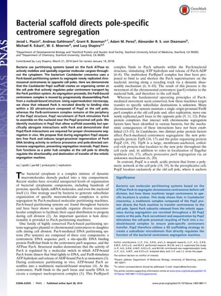
7. a shortening nucleoid-bound ParA structure toward the opposite
cell pole via dynamic and directional disassembly of the ParA/
DNA complex. We propose that ParA molecules that have been
released from the nucleoid by ParB during segregation are
recruited to the cell pole by the polar PopZ scaffold (Fig. 6 A and
B). Polar recruitment of inactive ParA would then sequester free
ParA molecules, preventing ParA reassembly on other nucleoid
regions and allowing processive ParB movement toward the
new pole.
In addition to sequestration, our E. coli reconstitution ex-
periments revealed another unexpected function of PopZ in
ParA dynamics: the modulation of ParA DNA binding activity.
In the presence of ParB, PopZ directly stimulated ParA reloc-
alization into an asymmetric gradient-like distribution along the
nucleoid with the highest concentration of ParA near PopZ foci,
suggesting that PopZ may affect the enzymatic activity of ParA.
Our epifluorescence and superresolution microscopy experiments
show that whereas PopZ recruits the ParB-centromere complex
to the cytoplasmic side of the scaffold, ParA monomers are
recruited throughout the 3D PopZ matrix. This recruitment may
locally increase the concentration of ParA and drive dime-
rization, or PopZ may activate ParA allosterically. We hypoth-
esize that the resulting activated ParA-ATP dimers are released
into the cytoplasm and encounter abundant nucleoid DNA near
the cell pole, binding with high affinity (Fig. 6 A and B). This
model is similar in principle to the orthologous ParA-family
protein MipZ, which was demonstrated to dimerize in response
to ParB interactions, causing MipZ to assemble a localized gra-
dient on nearby nucleoid DNA (32). The architecture of the re-
sulting ParA/nucleoid assembly might thus be similar to the
gradient-like distribution formed by MipZ. In a previous publica-
tion, we published single-molecule superresolution reconstruction
images that showed a high density of ParA localizations along the
long axis of the cell (6). However, the actual active structure, and
A ParAeYFP
mCHY-PopZ
ParAeYFP
mCHY-PopZ
ParB
mCHY-PopZ
ParAeYFP
overlay
no ParB
ParAeYFP
mCHY-PopZ-KE
ParB
ParAeYFP
mCHY-PopZ-SP
ParB
0 20 40 60 80 100
position along normalized cell axis
0 20 40 60 80 1000 20 40 60 80 100
0.02
0.04
0.06
0.08
0
ParA PopZ + ParB ParA PopZ no ParB
ParA + ParB
0 20 40 60 80 100
ParA only
meannormalizedPopZintensity
B
ParAeYFP
only
ParAeYFP
ParB
0.02
0.04
0.06
0.08
0
0.01
0.02
0.03
0.04
0
0.01
0.02
0.03
0.04
0
meannormalizedParAintensity
Fig. 5. ParB stimulates large-scale ParA localization into asymmetric struc-
tures near PopZ foci in E. coli. (A) ParB and PopZ direct the formation of an
asymmetric ParA structure in E. coli. Images of E. coli cells expressing wild-
type ParA-eYFP (green) in the presence or absence of mCherry-PopZ variants
(red) and ParB expression (untagged). Fluorescence micrographs are overlaid
as shown. (Scale bar, 1 μm.) Asymmetric ParA-eYFP localization (white
arrowheads) occurs only when coexpressed with ParB and requires a ParA
interaction-proficient PopZ variant. (B) Quantitation of mean fluorescence
intensity profiles for ParA-eYFP (green) and mCherry-PopZ (red) when
expressed in the presence or absence of ParB in the E. coli expression/
colocalization assay. Images of representative cells were oriented with re-
spect to the position of the polar mCherry-PopZ focus (where applicable),
and the fluorescence profiles were averaged and plotted (red scale corre-
sponds to PopZ signal (Left) and green scale corresponds to ParA signal
(Right) versus normalized cell length (n >18 cells). The double hump pattern
adopted by ParA-eYFP reflects accumulation on the nucleoid regions (6).
Horizontal dashed lines indicate eYFP signal maxima, and vertical dashed
lines indicate centers of mCherry-PopZ (red) and ParA-eYFP (green) peaks.
A
B
chromosome replication/ segregation
Polar PopZ matrix recruitment and concentration of
inactive ParA subunits
ParA activation/release
and nucleoid association
dimeric ParA-ATP
inactive ParA
ParB polymeric PopZ
nucleoid FtsZ ring
Fig. 6. Model for PopZ-catalyzed ParA reassembly, a feedback mechanism
to drive segregation toward the cell pole. (A) Molecular schematic for PopZ
recruitment and modulation of ParA activity. A 3D matrix of PopZ (structure
unknown, shown here as green lattice for clarity) recruits released/inacti-
vated ParA molecules (purple spheres) throughout the complex. Interactions
with, or increased local concentrations of, inactive ParA within the matrix
facilitates localized ParA activation, and resulting activated dimeric ParA-
ATP (yellow spheres) is released to encounter nearby nucleoid DNA (blue),
binding with high affinity. (B) Model for PopZ modulation of ParA activity in
the context of the Caulobacter cell during replication and centromere seg-
regation. In a swarmer cell, PopZ (green) anchors ParB/parS complexes (red
spheres) at the old cell pole (18, 19) and ParA-ATP (yellow spheres) localizes
along the nucleoid (blue oval). Upon replication initiation, the ParB/parS
complex is released from the pole (21, 31) and duplicated. Entropic forces
resulting from accumulating newly replicated DNA between ParB/parS may
drive centromeres apart (39), moving one ParB/parS complex away from the
pole. Upon encountering the ParA/nucleoid structure, the ParB complex
binds to nucleoid-bound ParA, stimulating ATP hydrolysis, releasing ParA
molecules (purple spheres) from the structure and tracking along the re-
ceding edge of the shortening ParA assembly (6, 20, 21). Released ParA
molecules are recruited to the cell pole by PopZ. PopZ recruitment concen-
trates and may allosterically stimulate ParA activation and release active
molecules to bind neighboring DNA. This ParA sequestering/feedback
mechanism may facilitate efficient centromere segregation and subsequent
anchoring of ParB/parS to the cell pole.
E2052 | www.pnas.org/cgi/doi/10.1073/pnas.1405188111 Ptacin et al.
8. the involvement of polymerization or cooperativity in DNA
binding by ParA, is poorly understood and awaits further study.
After segregation, PopZ was proposed to capture the segre-
gated ParB/parS complexes at the cell poles, terminating segre-
gation and preventing reversals (18, 19). Surprisingly, strains
bearing popZ alleles that significantly disrupt PopZ interactions
with ParB did not display severe growth or cell division defects
but showed marked defects in polar ParB anchoring before and
after segregation. In contrast, strains containing popZ alleles
defective in ParA interactions were severely defective in cell
division and centromere segregation, suggesting the primary role
of PopZ may be to modulate ParA segregation dynamics, with
polar centromere anchoring providing additional robustness to
the process. However, our ParB tracking experiments demon-
strate that the new pole ParB/parS complex seems immobilized
to the pole, even in the popZ-KE strain, whereas the old pole
ParB/parS complex seems dynamic (Fig. 3D and Fig. S5), sug-
gesting that ParA may facilitate attachment of the ParB/parS
complex specifically to the new pole.
The subcellular localization of PopZ scaffold complexes
changes dynamically during chromosome segregation. PopZ
initially forms a unipolar matrix at the old pole of the swarmer
cell and, during the process of chromosome replication and seg-
regation, assembles an additional network at the new pole
(Fig. 6B) (18, 19). It was proposed that ParA accumulation at the
new pole during segregation could stimulate the formation of
a new complex of PopZ at this position (29). Here we observe
that popZ alleles defective for ParA binding are not significantly
defective in bipolar PopZ complex formation. Instead, our results
suggest that once established a polar PopZ complex regulates
ParA dynamics during segregation, as PopZ modulates the re-
localization of ParA to neighboring regions of the nucleoid in the
presence of ParB. Thus, a PopZ-directed ParA recycling mecha-
nism may function once the newly formed PopZ complex is po-
sitioned to recruit free ParA to the pole. Assembly of the new
PopZ network could prevent reversals by ensuring ParA relocal-
ization to the nucleoid between the segregating centromere and
the destination of transfer, which may also facilitate anchoring of
the ParB complex at the new pole. Similarly, these mechanisms
may function at the old cell pole to continuously maintain the
nonmobilized ParB/parS complex near the pole (21), facilitating
bidirectional segregation.
An important question that remains to be addressed is the role
of TipN in regulating ParA dynamics. Previous studies demon-
strated that new pole localized TipN prevents ParA assembly
into aberrant structures that cause reverse segregation (6, 20).
However, a tipN deletion leads to relatively mild cell division
defects compared with a popZ deletion, suggesting a more
prominent role for PopZ in regulating segregation. Over-
expression of TipN can partially rescue cell division defects in
cells lacking popZ (20). Our data suggest that this rescue results
from recruitment of ParA monomers to the cell pole by over-
expressed TipN during segregation and imply that a shared
mechanism for TipN and PopZ regulators may include seques-
tration of free ParA. However, in wild-type cells after segrega-
tion, significantly more ParA accumulates at the new pole than at
the old (20), which may suggest a synergistic effect of PopZ and
TipN on ParA recruitment or modulation, conceivably via
a handoff of free ParA from TipN to PopZ. Recently, Laloux
and Jacobs-Wagner (29) showed that PopZ recruitment to the
new pole is significantly delayed in cells lacking TipN. In light of
our data, we hypothesize that segregation defects in ΔtipN strains
could result in part from such a delay in positioning a PopZ
complex at the new pole, which would prevent PopZ from
directing ParA reassembly to terminate segregation.
Overall, our results demonstrate that a major function of the
polar PopZ network is to modulate ParA activity during cen-
tromere segregation and ensure termination of segregation at
the cell pole. PopZ scaffolds, therefore, function not only as
polar docking stations for cell cycle regulatory complexes (18, 19,
31), but also as local activation centers that direct the centro-
mere-positioning machine. PopZ thus comprises a polar nexus
that enables the spatial and temporal coupling of chromosome
replication and segregation to ensure proper cell cycle pro-
gression. The filamentous network-like properties of PopZ cre-
ate a specialized 3D microenvironment within the bacterial
cytoplasm. In the absence of membrane-bounded cytoplasmic
compartments, bacteria may use a similar 3D scaffolding strategy
to create other subcellular microdomains, allowing spatial par-
titioning and organization of important molecular transactions
within the cytoplasm.
Materials and Methods
Bacterial Strains and Culture Conditions. The Caulobacter crescentus strains,
E. coli strains, and plasmids used in this study are listed in Tables S1–S3, re-
spectively. All Caulobacter strains used were derived from the synchroniz-
able strain CB15N (33) and were cultured as described (34). Generalized
transduction was performed with phage ФCr30 as described (35). Caulobacter
synchronies were performed as described (36). Relevant details of culture
conditions for each experiment can be found below, and details of plasmid
and strain construction can be found in Supporting Information.
Epifluorescence Microscopy and Image Analysis. Sample preparation and
epifluorescence image acquisition was performed as described (28). Briefly,
Caulobacter and E. coli strains were cultured as indicated before deposition
onto M2G/1.5% agarose pads for imaging. Image acquisition and analysis
was performed using the MetaMorph package. Quantitative image analysis
of cell length distributions and fluorescent ParB foci was performed using
custom software written in MATLAB (The MathWorks, Inc.) using modules of
the MicrobeTracker software suite (37). Cell outlines were computed from
phase images using MicrobeTracker. To ensure correct cellular coordinate
positions of ParB foci, fluorescence images were aligned to one or several
cell outlines by maximizing the image cross-correlation between fluores-
cence images and binary mask images constructed from the cell outlines. The
resulting shift vectors were used as control point pairs to register the entire
set of cell outlines in a given camera frame to the corresponding fluores-
cence image using a 2D affine transformation (cp2tform function in
MATLAB). Fluorescent foci were then fit with asymmetric 2D Gaussian func-
tions and the fitted center positions were converted into the registered cel-
lular coordinates using the projectToMesh function of MicrobeTracker.
Coimmunoprecipitation and Western Blotting. Wild-type (JP1) and parA-M2
(JP88) strains were cultured in peptone-yeast extract (PYE) at 28 °C to A at
600 nm = 0.5 before pelleting by centrifugation at 8,000 rpm in a JA10 rotor
for 15 min at 4 °C. Pellets were washed twice with ice-cold Co-IP buffer
[20 mM Hepes (pH 7.5), 100 mM NaCl, and 20% (vol/vol) glycerol] before the
addition of formaldehyde (Sigma) to 1% and incubation at room tempera-
ture for 30 min. Reactions were quenched using 125 mM glycine. Cells were
washed in Co-IP buffer II [50 mM Hepes, 500 mM NaCl, and 20% (vol/vol)
glycerol, pH 7.5], supplemented with complete protease inhibitors (Roche)
and 1μL benzonase nuclease (Sigma). Cells were then lysed by passing three
times through a French pressure cell at 16,000 psi. Lysates were supple-
mented with 0.1% Triton X-100 and centrifuged 30 min 4 °C at 20,000 × g,
after which the supernatants were normalized for protein concentration
and incubated with Dynabeads anti-M2 magnetic particles (Invitrogen).
Particles were washed five times in wash buffer [50 mM Hepes, 500 mM
NaCl, 20% (vol/vol) glycerol, and 0.05% Nonidet P-40] supplemented with
complete protease inhibitors (Roche) before elution with 3× FLAG peptide
for 1 h at 4 °C according to the manufacturer’s instructions (Invitrogen).
Samples were then subjected to SDS/PAGE electrophoresis and transferred
to a PVDF membrane (Millipore). Immunoblotting was with anti-PopZ sera
(1:10,000) (18) or with HU2 antisera (1: 5,000) followed by goat anti-rabbit
secondary and detection using chemiluminescent substrate (Pierce).
E. coli Expression/Colocalization Assay for Mutant ParA Protein Recruitment by
PopZ. The E. coli BL21(DE3) strains eJP590-595 (mCherry-PopZ and ParA-eYFP
variant expression), eJP146-147, 175–177, and 406 (ParA-eYFP variant only)
(Table S2) were cultured to log phase at 37 °C in LB containing ampicillin
and/or chloramphenicol, respectively. Cultures were induced by the addition
of 100 μM isopropyl β-D-1-thiogalactopyranoside (IPTG) and incubated for
1 h at 37 °C before imaging.
Ptacin et al. PNAS | Published online April 28, 2014 | E2053
MICROBIOLOGYPNASPLUS
9. E. coli Expression/Colocalization Assay for ParA-eYFP Localization in Response
to PopZ and ParB Expression. The E. coli BL21(DE3) strains eJP147 (ParA-eYFP
only), eJP290 (ParA-eYFP and ParB), eJP576 (ParA-eYFP, mCherry-PopZ, and
ParB), and eJP582 (ParA-eYFP and mCherry-PopZ) (Table S2) were cultured to
log phase at 37 °C in LB containing the appropriate combination of ampi-
cillin, chloramphenicol, and spectinomycin. Cultures were induced by the
addition of 100 μM IPTG and incubated for 1 h at 37 °C before imaging.
Recruitment of ParAG16V-eYFP to the Cell Pole in Caulobacter. The Caulobacter
strains JP308, JP258, JP437, and JP443 (Table S1) were cultured to log phase
at 28 °C in PYE containing oxytetracycline (and gentamicin for JP443). Cul-
tures were induced by the addition of 0.5 mM vanillic acid (and 0.06%
D-xylose for JP443) and incubated for 2 h at 28 °C before imaging.
E. coli Expression/Colocalization Assay for PopZ Variant Recruitment of
ParAG16V-eYFP and CFP-ParB. The E. coli BL21(DE3) strains eJP619-622
(Table S2) were cultured to log phase at 37 °C in LB containing ampicillin and
chloramphenicol. Cultures were induced by the addition of 100 μM IPTG and
0.04% D-arabinose and incubated for 1 h at 37 °C before imaging.
PopZ “Plug” Assay for Recruitment of ParAG16V-eYFP. The Caulobacter strains
241, 242, and 255 (Table S1) were cultured to log phase at 28 °C in PYE
containing tetracycline and gentamycin. Cultures were induced by the ad-
dition of 0.3% D-xylose and incubated for 4 h at 28 °C before imaging.
Protein Expression and Purification. For expression and purification of hex-
ahistidine-tagged PopZ and mutant derivatives, E. coli Rosetta/DE3 strains
containing pET28-6his-popZ variants (Table S3) were cultured at 37 °C in LB
containing 30 μg/mL kanamycin and 20 μg/mL chloramphenicol. Upon re-
aching midlog phase, cultures were induced with 1mM IPTG for 2 h. Cell
pellets were collected by centrifugation and stored at −80 °C. Pellets were
resuspended in Buffer 1 [100 mM phosphate (pH 8), 10 mM Tris·Cl, 300 mM
NaCl, 8 M urea, and 20 mM imidazole] supplemented with complete pro-
tease inhibitors (Roche). After resuspension and solubilization, lysates were
applied to Talon metal affinity resin (Clontech) and washed with buffer1
before elution in 100 mM phosphate, 10 mM Tris·Cl, 300 mM NaCl, 8 M urea,
and 250 mM imidazole, pH 8. Purified samples were refolded via dialysis
against 20 mM Tris·Cl, pH 8.5. Wild-type and mutant derivative 6His-PopZ
samples displayed similar oligomerization/ assembly characteristics as mea-
sured by native gel electrophoresis and gel filtration analysis (not shown).
For expression and purification of hexahistidine-tagged ParB, E. coli
Rosetta/DE3 carrying the plasmid pJP137 (containing pET28-6his-parB)
(Table S3) was cultured and expressed as above. Pellets were resuspended
in buffer 1 [50 mM Hepes (pH 7.5), 500 mM NaCl, 5% (vol/vol) glycerol,
10 mM imidazole, 1mM PMSF, and 20 μg/mL RNaseA] supplemented with
complete protease inhibitors (Roche). After resuspension on ice, lysis was
carried out by passing three times through a French pressure cell (16,000 psi)
at 4 °C before centrifuging at 20,000 × g for 30 min. The supernatant was
loaded onto a 1-mL nickel HisTrap column (GE Healthcare), washed with
20 column volumes of wash buffer [50 mM Hepes (pH 7.5), 500 mM NaCl,
10 mM imidazole, and 5% (vol/vol) glycerol], and eluted using a linear
gradient of imidazole from 10 to 500 mM in wash buffer at 1 mL·min−1
. Pure
fractions were dialyzed into 20 mM Hepes (pH 7.5) and 50 mM KCl, applied
to a 1-mL HiTrap heparin column (GE Healthcare), and eluted with a linear
gradient of KCl in 20 mM Hepes, pH 7.5. Pure fractions (by SDS PAGE stained
with Coomassie blue) were pooled and dialyzed into 50 mM Hepes (pH 7.5),
100 mM KCl, and 10% (vol/vol) glycerol.
For expression and purification of hexahistidine-tagged ParA, a E. coli
Rosetta/DE3 strain containing the plasmid pJP325 (pET28-parA-6his) (Table
S3) was cultured and expressed as above, except expression was carried out
at 30 °C. Pellets were resuspended in lysis buffer [20 mM Hepes (pH 7),
500 mM KCl, 1 mM ADP, 20 mM imidazole, 10 mM MgCl2, 0.1 mM EDTA, and
1 mM DTT] supplemented with complete protease inhibitors (Roche). After
resuspension on ice, lysis was carried out by passage through a French
pressure cell (16,000 psi) at 4 °C before centrifuging at 20,000 × g for 30 min.
Lysates were passed over 1 mL of Ni-NTA agarose (5Prime), washed with
WashBuffer [20 mM Hepes (pH 7), 500 mM KCl, 1 mM ADP, 50 mM im-
idazole, 10 mM MgCl2, 0.1 mM EDTA, and 1mM DTT] and eluted with
elution buffer [20 mM Hepes (pH 7), 500 mM KCl, 1 mM ADP, 300 mM
imidazole, 10 mM MgCl2, 0.1 mM EDTA, 1 mM DTT, and 50% (vol/vol)
glycerol]. The resulting eluate containing pure ParA-6His was aliqoted
and frozen. Aliquots were thawed on ice and exchanged into buffer
HMK [20 mM Hepes-NaOH (pH 7.5), 200 mM KCl, and 2 mM MgCl2] and
spun at 80,000 rpm in a TL100.3 rotor at 4 °C for 30 min immediately
before use.
SPR. SPR experiments were performed on a Biacore 3000 system at 25 °C using
a flow rate of 30 μL·min−1
in buffer HMK (20 mM Hepes/KOH, 2 mM MgCl2,
and 100 mM KCl, pH 7.5) and, where indicated, contained 1 mM ATP or ADP
(Sigma). Purified 6His–PopZ and mutant variants were exchanged into
20 mM Hepes (pH 8) and immobilized directly to the carboxymethylated
dextran surface of CM5 biosensor chips through amine coupling. ParA-6His
and 6His-ParB samples were exchanged into buffer HMK before injection.
Data were corrected for nonspecific interactions by subtracting the signal in
a control flow cell that lacked immobilized ligand and analyzed using the
BIAevaluation software 4.1 (G.E. Healthcare).
Superresolution Imaging. Multicolor 3D superresolution images were ac-
quired and reconstructed as described (30). Briefly, Caulobacter strains JP464
and JP465 (Table S1) were cultured to log phase in M2G containing kana-
mycin and tetracycline. JP464 cultures were induced by the addition of be-
tween 0.06–0.09% xylose and 0.1–0.15 mM vanillic acid for 90 min before
imaging. For JP465, cultures were induced with 0.1mM vanillic acid for
45 min before synchrony and maintained at this concentration postsyn-
chrony in M2G. To avoid perturbation of centromere translocation, 0.15%
xylose was added 40 min postsynchrony, followed by sample preparation
(∼30 min) and imaging. Proteins labeled with eYFP and PAmCherry1 were
detected on an inverted fluorescence microscope using an interleaved pulse
sequence of 405 nm (Coherent Cube, 403 nm, <1 W/cm2
: PAmCherry1 acti-
vation), 514 nm [Coherent Sapphire 514–100 continuous wave (CW): eYFP
blinking and detection], and 561 nm (Coherent Sapphire 561–100 CW:
PAmCherry1 detection) illumination. To image strain JP464, 100-ms frame
integration times were used, whereas 200-ms integration was used for strain
JP465 to ensure that stationary molecules of eYFP-ParB were preferentially
detected. Peak intensities for 514-nm illumination were 636 W/cm2
and 281
W/cm2
, respectively, with a 1/e2
width of 63 μm. The corresponding peak
intensities for 561-nm illumination were 758 W/cm2
and 438–634 W/cm2
,
with a 1/e2
width of 60 μm. Fluorescence was split into two color channels
and imaged onto separate sections of the EMCCD camera chip. Three-di-
mensional information was encoded in single-molecule localizations using
the double-helix point spread function (DH-PSF). The DH-PSF was generated
by convolving the image in each channel with the appropriate phase pattern
imprinted on a transmissive phase mask placed in the Fourier plane of a 4f
imaging system. The calibrated single-molecule localization precisions for
eYFP were 28–47 nm (lateral) and 39–57 nm (axial); for PAmCherry, these
values ranged from 42 to 71 nm and 59–86 nm (ranges given as first-third
quartiles) (30). Drift was compensated with fluorescent beads emitting in
both channels. Single-molecule positions were determined using the Easy-
DHPSF software suite (38) and color channels were merged using a locally
weighted transformation function generated by using localizations of fluo-
rescent beads as control points.
Superresolution Image Rendering and Density Correlation Analysis. The dis-
tances between subcellular spatial domains of PAmCherry1-PopZ and
ParAG16V-eYFP were determined using a custom algorithm implemented in
MATLAB as follows. Sets of clustered single-molecule localizations compos-
ing a dense protein domain at the cell poles were manually identified
(minimum of 30 single-molecule localizations). The underlying 3D distribu-
tion of each cluster was estimated by blurring the localizations with a 3D
Gaussian kernel with σ(x/y/z) equal to the calibrated localization precision,
sampled in 4-nm voxels. The relationship between the resulting densities
was analyzed using the cross-correlation function
rði,j,kÞ =
X
x,y,z
fðx,y,zÞgðx + i,y + j,z + kÞ,
where f(x, y, z) and g(x, y, z) are the sampled densities of each protein. The
3D Euclidean distance between the densities f and g was given by the vector
(i, j, k)max, defined as the offset from the initial position to the position
where r(i, j, k) is maximized.
ACKNOWLEDGMENTS. We thank Keren Lasker for helpful comments on the
manuscript and Jordan Harrison for help in screening to identify the popZ-SP
allele. This work was supported by National Institutes of Health (NIH) Grants
R01 GM51426 and R01 GM32506 (to L.S.), NIH/National Institute of General
Medical Sciences (NIGMS) Fellowship F32GM088966-3 (to J.L.P.), NIH/NIGMS
Award R01GM086196 (to W.E.M.), and Swiss National Science Foundation
Postdoctoral Fellowship PA00P2_145310 (to A.G.).
E2054 | www.pnas.org/cgi/doi/10.1073/pnas.1405188111 Ptacin et al.
10. 1. Nevo-Dinur K, Govindarajan S, Amster-Choder O (2012) Subcellular localization of
RNA and proteins in prokaryotes. Trends Genet 28(7):314–322.
2. Lutkenhaus J (2012) The ParA/MinD family puts things in their place. Trends Microbiol
20(9):411–418.
3. Leonard TA, Butler PJ, Löwe J (2005) Bacterial chromosome segregation: Structure
and DNA binding of the Soj dimer—a conserved biological switch. EMBO J 24(2):
270–282.
4. Vecchiarelli AG, Mizuuchi K, Funnell BE (2012) Surfing biological surfaces: Exploiting
the nucleoid for partition and transport in bacteria. Mol Microbiol 86(3):513–523.
5. Murray H, Ferreira H, Errington J (2006) The bacterial chromosome segregation
protein Spo0J spreads along DNA from parS nucleation sites. Mol Microbiol 61(5):
1352–1361.
6. Ptacin JL, et al. (2010) A spindle-like apparatus guides bacterial chromosome segre-
gation. Nat Cell Biol 12(8):791–798.
7. Hwang LC, et al. (2013) ParA-mediated plasmid partition driven by protein pattern
self-organization. EMBO J 32(9):1238–1249.
8. Vecchiarelli AG, et al. (2010) ATP control of dynamic P1 ParA-DNA interactions: A key
role for the nucleoid in plasmid partition. Mol Microbiol 78(1):78–91.
9. Fogel MA, Waldor MK (2006) A dynamic, mitotic-like mechanism for bacterial chro-
mosome segregation. Genes Dev 20(23):3269–3282.
10. Ringgaard S, van Zon J, Howard M, Gerdes K (2009) Movement and equipositioning
of plasmids by ParA filament disassembly. Proc Natl Acad Sci USA 106(46):
19369–19374.
11. Viollier PH, et al. (2004) Rapid and sequential movement of individual chromosomal
loci to specific subcellular locations during bacterial DNA replication. Proc Natl Acad
Sci USA 101(25):9257–9262.
12. Harms A, Treuner-Lange A, Schumacher D, Søgaard-Andersen L (2013) Tracking of
chromosome and replisome dynamics in Myxococcus xanthus reveals a novel chro-
mosome arrangement. PLoS Genet 9(9):e1003802.
13. Yamaichi Y, et al. (2012) A multidomain hub anchors the chromosome segregation
and chemotactic machinery to the bacterial pole. Genes Dev 26(20):2348–2360.
14. Ditkowski B, et al. (2013) Dynamic interplay of ParA with the polarity protein, Scy,
coordinates the growth with chromosome segregation in Streptomyces coelicolor.
Open Biol 3(3):130006.
15. Ginda K, et al. (2013) ParA of Mycobacterium smegmatis co-ordinates chromosome
segregation with the cell cycle and interacts with the polar growth determinant
DivIVA. Mol Microbiol 87(5):998–1012.
16. Lam H, Schofield WB, Jacobs-Wagner C (2006) A landmark protein essential for es-
tablishing and perpetuating the polarity of a bacterial cell. Cell 124(5):1011–1023.
17. Huitema E, Pritchard S, Matteson D, Radhakrishnan SK, Viollier PH (2006) Bacterial
birth scar proteins mark future flagellum assembly site. Cell 124(5):1025–1037.
18. Bowman GR, et al. (2008) A polymeric protein anchors the chromosomal origin/ParB
complex at a bacterial cell pole. Cell 134(6):945–955.
19. Ebersbach G, Briegel A, Jensen GJ, Jacobs-Wagner C (2008) A self-associating protein
critical for chromosome attachment, division, and polar organization in caulobacter.
Cell 134(6):956–968.
20. Schofield WB, Lim HC, Jacobs-Wagner C (2010) Cell cycle coordination and regulation
of bacterial chromosome segregation dynamics by polarly localized proteins. EMBO J
29(18):3068–3081.
21. Shebelut CW, Guberman JM, van Teeffelen S, Yakhnina AA, Gitai Z (2010) Caulo-
bacter chromosome segregation is an ordered multistep process. Proc Natl Acad Sci
USA 107(32):14194–14198.
22. Ebersbach G, Gerdes K (2004) Bacterial mitosis: Partitioning protein ParA oscillates in
spiral-shaped structures and positions plasmids at mid-cell. Mol Microbiol 52(2):
385–398.
23. Fung E, Bouet JY, Funnell BE (2001) Probing the ATP-binding site of P1 ParA: partition
and repression have different requirements for ATP binding and hydrolysis. EMBO J
20(17):4901–4911.
24. Scholefield G, Whiting R, Errington J, Murray H (2011) Spo0J regulates the oligomeric
state of Soj to trigger its switch from an activator to an inhibitor of DNA replication
initiation. Mol Microbiol 79(4):1089–1100.
25. Murray H, Errington J (2008) Dynamic control of the DNA replication initiation pro-
tein DnaA by Soj/ParA. Cell 135(1):74–84.
26. Castaing JP, Bouet JY, Lane D (2008) F plasmid partition depends on interaction of
SopA with non-specific DNA. Mol Microbiol 70(4):1000–1011.
27. Hester CM, Lutkenhaus J (2007) Soj (ParA) DNA binding is mediated by conserved
arginines and is essential for plasmid segregation. Proc Natl Acad Sci USA 104(51):
20326–20331.
28. Bowman GR, et al. (2013) Oligomerization and higher-order assembly contribute to
sub-cellular localization of a bacterial scaffold. Mol Microbiol 90(4):776–795.
29. Laloux G, Jacobs-Wagner C (2013) Spatiotemporal control of PopZ localization
through cell cycle-coupled multimerization. J Cell Biol 201(6):827–841.
30. Gahlmann A, et al. (2013) Quantitative multicolor subdiffraction imaging of bacterial
protein ultrastructures in three dimensions. Nano Lett 13(3):987–993.
31. Bowman GR, et al. (2010) Caulobacter PopZ forms a polar subdomain dictating se-
quential changes in pole composition and function. Mol Microbiol 76(1):173–189.
32. Kiekebusch D, Michie KA, Essen LO, Löwe J, Thanbichler M (2012) Localized di-
merization and nucleoid binding drive gradient formation by the bacterial cell di-
vision inhibitor MipZ. Mol Cell 46(3):245–259.
33. Poindexter JS (1964) Biological properties and classification of the Caulobacter group.
Bacteriol Rev 28:231–295.
34. Thanbichler M, Shapiro L (2006) MipZ, a spatial regulator coordinating chromosome
segregation with cell division in Caulobacter. Cell 126(1):147–162.
35. Ely B (1991) Genetics of Caulobacter crescentus. Methods Enzymol 204:372–384.
36. Tsai JW, Alley MR (2001) Proteolysis of the Caulobacter McpA chemoreceptor is cell
cycle regulated by a ClpX-dependent pathway. J Bacteriol 183(17):5001–5007.
37. Sliusarenko O, Heinritz J, Emonet T, Jacobs-Wagner C (2011) High-throughput, sub-
pixel precision analysis of bacterial morphogenesis and intracellular spatio-temporal
dynamics. Mol Microbiol 80(3):612–627.
38. Lew MD, von Diezmann ARS, Moerner WE (2013) Easy-DHPSF open-source software
for three-dimensional localization of single molecules with precision beyond the
optical diffraction limit. Protocol Exchange, 10.1038/protex.2013.026.
39. Jun S, Mulder B (2006) Entropy-driven spatial organization of highly confined poly-
mers: lessons for the bacterial chromosome. Proc Natl Acad Sci USA 103(33):
12388–12393.
Ptacin et al. PNAS | Published online April 28, 2014 | E2055
MICROBIOLOGYPNASPLUS
![the ParB-bound parS locus via direct interactions with ParB
(18, 19). During chromosome replication initiation, PopZ releases
ParB from the old pole and adopts a bipolar PopZ distribution
that seems to capture ParB/parS complexes during the segregation
process (18, 19). Whereas cells lacking tipN are only mildly elon-
gated, popZ deletion causes severe filamentation (16–19), sug-
gesting that PopZ plays a more important role in the regulation of
segregation. However, the molecular mechanism by which PopZ
affects segregation has remained elusive.
Here we demonstrate that the multifunctional PopZ complex
plays a crucial role in pole-directed movement of ParA-mediated
chromosome segregation by interacting directly with ParA. We
show that PopZ, but not TipN, is required for robust polar re-
cruitment of ParA and demonstrate that a polar PopZ scaffold
recruits and concentrates free ParA released during segregation.
Recruitment of ParA within the PopZ matrix sequesters free
ParA and locally regenerates ParA DNA binding activity. Active
ParA complexes are released for recycling into nucleoid-bound
structures near the cell pole, which we propose drives centro-
mere segregation toward pole-localized PopZ. Thus, PopZ
orchestrates a positive feedback mechanism that forces ParA-
mediated centromere transfer to the cell pole. The polar PopZ
scaffold complex creates a unique 3D microenvironment at the
pole that spatially separates distinct centromere tethering and
ParA-modulation activities, enabling coupling between chromo-
some segregation with the initiation of cell division.
Results
PopZ Is Required for ParA Recruitment to the Caulobacter Cell Pole.
Caulobacter ParA accumulates at cell poles during and after
chromosome segregation (6, 20, 21). To examine the roles of
PopZ and TipN in the polar recruitment of ParA, we used
a previously characterized monomeric variant of ParA (the di-
merization-deficient ParAG16V) that exhibits preferential lo-
calization at the cell pole rather than the nucleoid DNA in
Caulobacter (6, 20). We created merodiploid strains that ex-
pressed ParAG16V-enhanced YFP (eYFP) in Caulobacter strains
deficient in popZ or tipN. When expressed in a wild-type Cau-
lobacter background, ParAG16V-eYFP localized as foci at the cell
poles (Fig. 1A) as described (6, 20). In a ΔtipN background,
ParAG16V-eYFP also efficiently formed foci at cell poles,
whereas in the ΔpopZ background ParAG16V–eYFP was diffuse
throughout the cell (Fig. 1A). In cells overexpressing TipN-
mCherry in the ΔpopZ background, ParAG16V-eYFP foci colo-
calized with TipN-mCherry foci at the cell poles (Fig. S1A).
These results suggest that TipN overexpression can rescue the
recruitment of ParA to the cell pole in the ΔpopZ background,
implying functional redundancy between these proteins. How-
ever, under physiological expression levels, only PopZ is required
for ParA focus formation at the cell pole.
ParA and PopZ Interact Directly in Vitro and in Vivo. Because PopZ
is required for recruitment of ParA to the Caulobacter cell poles,
we tested whether ParA and PopZ proteins interact in vivo. We
created Caulobacter strains that replaced the native parA gene on
the chromosome with an M2 epitope-tagged version of parA
(parA-M2) and immunoprecipitated ParA-M2 from lysates of
this strain or wild type using resin-immobilized anti-M2 antibody.
Western blotting of these samples using anti-PopZ sera showed
that PopZ specifically coimmunoprecipitates with ParA-M2 (Fig.
S1B, Upper). We blotted these samples with antisera against the
abundant nonspecific DNA binding protein HU2 and detected
signal in the lysates but not in the pulldown eluates, showing that
ParA does not coimmunoprecipitate other nonspecific DNA-
binding proteins (Fig. S1B, Lower). These results confirm that
ParA and PopZ are in close proximity in vivo in Caulobacter.
To test whether PopZ and ParA interact directly, we purified
PopZ and ParA proteins and examined their interactions in vitro
using surface plasmon resonance (SPR). His-tagged PopZ was
immobilized directly to the sensor chip, and this surface was
probed for interaction with ParA-6His in the presence or ab-
sence of nucleotide. When we added ParA-6His in the presence
of ATP, we observed a robust, ParA concentration-dependent
signal (Fig. 1 B and C). In contrast, ParA-6His injected in the
presence of ADP or no nucleotide displayed reduced or no re-
sponse, respectively (Fig. 1C), suggesting that ParA and PopZ
interact directly in vitro and that this interaction depends on the
nucleotide-bound state of ParA.
To further investigate the biochemical states of ParA neces-
sary for PopZ interaction, we coexpressed ParA-eYFP protein
variants and mCherry-PopZ in the heterologous Escherichia coli
strain BL21 (which lacks a Par system), which allows facile vi-
sualization of protein/protein and protein/DNA interactions us-
ing fluorescence microscopy. We used a set of characterized
ParA mutant proteins that disrupt specific steps in the ParA
ATPase cycle, including ATP binding [ParAK20Q (6, 9, 22, 23)],
ParA dimerization [ParAG16V (6, 20, 24, 25)], ATP hydrolysis
[ParAD44A (3, 6, 20, 25)], and DNA binding [ParAR195E (6, 8, 20,
26, 27)] (Fig. 1D). In the presence or absence of mCherry-PopZ,
the ATP-binding-deficient ParAK20Q localized diffusely through-
out the cytoplasm (Fig. 1D), confirming that ATP binding is
requisite for ParA recruitment by PopZ. The ATP-binding-pro-
ficient but ParA dimerization-deficient ParAG16V mutant protein
localized diffusely in the absence of PopZ but in the presence of
PopZ was recruited to polar PopZ foci with some diffuse signal
(Fig. 1D), demonstrating that PopZ can recruit ParA monomers.
Interestingly, both the wild-type ParA and ATP-hydrolysis-
deficient ParAD44A localized exclusively to the nucleoid in the
presence or absence of PopZ (Fig. 1D). In contrast, the DNA
binding-deficient ParAR195E alone was diffuse but when
expressed with PopZ was strongly colocalized with PopZ foci
(Fig. 1D), suggesting that nucleoid DNA outcompetes PopZ
for ParA interaction. Finally, when we expressed a combination
mutant protein defective in ATP hydrolysis and DNA binding
(ParAD44A/R195E) we observed diffuse localization in the ab-
sence of PopZ but robust polar recruitment to PopZ foci (Fig.
1D), suggesting that PopZ can recruit dimeric ParA when not
bound to the nucleoid. Together, these results suggest that ParA
monomers and dimers are recruited to PopZ but that nucleoid
interactions outcompete PopZ for dimeric ParA-ATP recruitment.
Amino Acid Substitutions in the Conserved N-Terminal Region of PopZ
Differentially Disrupt Interactions with ParA and/or ParB. To identify
PopZ mutant variants defective in interaction with ParA and
ParB, we mutated residues in the highly conserved N-terminal
domain of PopZ, which has been previously implicated in
chromosome segregation (Fig. 2A (28, 29). We screened these
mutants in E. coli for polar recruitment of the monomeric mu-
tant ParAG16V-eYFP variant, which readily interacts with PopZ
rather than the nucleoid, and CFP-ParB using fluorescence mi-
croscopy. Using this approach, we identified alleles of popZ that
form foci at the cell pole but fail to interact with ParA, ParB, or
ParA/B (Fig. 2 A and B). In the absence of PopZ expression,
ParAG16V-eYFP and CFP-ParB were diffusely localized (Fig.
2B). In the presence of wild-type PopZ, ParAG16V-eYFP and
CFP-ParB both were recruited into foci at the cell pole (Fig. 2B).
In the presence of the PopZ mutant variant E12K/R19E (here-
after referred to PopZ-KE), ParAG16V-eYFP was recruited to
the cell pole, whereas CFP-ParB was diffuse (Fig. 2B). Con-
versely, in the presence of the mutant PopZ-S22P derivative
(hereafter referred to as PopZ-SP), ParAG16V-eYFP was diffuse,
whereas CFP-ParB formed foci at the pole (Fig. 2B). As ex-
pected, in the presence of a PopZ variant in which these muta-
tions were combined (E12K/R19E/S22P, or PopZ-KEP), both
ParAG16V-eYFP and CFP-ParB were diffusely localized through-
out the cell (Fig. 2B). Therefore, this set of popZ alleles specifically
Ptacin et al. PNAS | Published online April 28, 2014 | E2047
MICROBIOLOGYPNASPLUS](data:image/gif;base64,R0lGODlhAQABAIAAAAAAAP///yH5BAEAAAAALAAAAAABAAEAAAIBRAA7)