More Related Content
Similar to NSMB_2006 (20)
NSMB_2006
- 1. Identification of the FtsK
sequence-recognition domain
Jerod L Ptacin1, Marcelo No¨llmann1, Carlos Bustamante1,2 &
Nicholas R Cozzarelli1,3
FtsK is a prokaryotic multidomain DNA translocase that
coordinates chromosome segregation and cell division. FtsK
is membrane anchored at the division septum and, guided by
highly skewed DNA sequences, translocates the chromosome
to bring the terminus of replication to the septum. Here,
we use in vitro single-molecule and ensemble methods to
unveil a mechanism of action in which the translocation and
sequence-recognition activities are performed by different
domains in FtsK.
The Escherichia coli DNA translocase FtsK is an oligomeric ATP-fueled
motor that coordinates chromosome segregation and cell division1,2.
Homologous recombination during DNA replication can lead to the
formation of a chromosome dimer that cannot be properly segregated
to daughter cells3. The site-specific recombinases XerC and XerD
act at dif, a 28-nucleotide sequence in the terminus of replication,
to resolve chromosome dimers into separable monomers3. FtsK
assembles at the division septum and translocates the DNA to bring
the two dif sites in the dimer into close proximity4. Upon formation of
the XerCD-dif synapse, FtsK stimulates recombination by direct
interaction with XerD5–7.
The FtsK monomer is composed of three
domains: a 217-residue N-terminal trans-
membrane domain, a poorly conserved and
putatively unstructured 600-residue linker
and a 512-residue motor domain (FtsKC)
that has been classified as a member of
the AAA+ family of ATPases and shown
to translocate duplex DNA5,8,9. A soluble
variant of FtsKC (FtsK50C) has been used
extensively to study FtsK translocation
in vitro5,8–12. Single-molecule experiments
using FtsK50C have shown that its trans-
location direction is dictated by DNA
sequence9. The DNA sequence motif 5¢-
GNGNAGGG-3¢ (FtsK-recognition sequence,
or FRS10, also called KOPS12) have been
found to control translocation directionality
by reversing FtsK movement when the trans-
locase encounters the sequence from the
3¢ end of the G-rich strand, termed the nonpermissive orientation.
When approaching from the 5¢ end (permissive orientation),
FtsK passes the sequence unobstructed10,12. FRS motifs are
over-represented on the chromosome’s leading strand and switch
strand at dif, thus guiding FtsK toward dif from any location on
the chromosome.
Despite characterization of the FRS, little is known about the
mechanism of FRS recognition by FtsK. Secondary and tertiary
structure predictions indicate that the C-terminal 64 residues of
FtsK (g domain) folds as a DNA-binding domain joined to the
rest of FtsKC by a short unstructured linker (J.L.P. and
M.N., unpublished data). This is confirmed in the accompanying
high-resolution structures of the g domain, which show that it folds
as a winged helix13. We hypothesized that the g domain mediates FtsK
interaction with the FRS. To test this hypothesis, we constructed and
purified a truncated version of FtsK50C that lacks the g domain and
linker (FtsK50CDg).
To assay for FRS recognition, we monitored the FtsK-mediated
displacement of a DNA triplex on a substrate containing FRS motifs in
permissive or nonpermissive orientations10 (Supplementary Methods
and Supplementary Fig. 1 online). FtsK50C triplex displacement rates
were considerably faster on the permissive than on the nonpermissive
substrate (Supplementary Fig. 1), in agreement with our previous
results10. In contrast, FtsK50CDg displacement rates were similar on
permissive and nonpermissive substrates (Supplementary Fig. 1),
consistent with FtsK50CDg being unable to respond to FRS.
We directly examined FtsK50CDg translocation and directionality by
tracking FtsK particles in real time on single l DNA molecules using
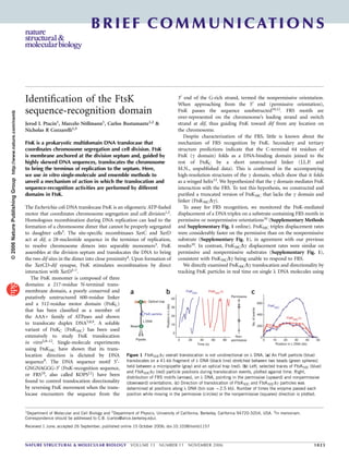
Optical trap
FtsK particle
Time (s)
FtsK50C
FtsK50C
∆γ
0
0
10
20
30
40
20 40 60 80
Permissive
Non-
permissivePipette
Bead
λ DNA
Position in λ DNA (kb)
Positioninλ(kb)
a b
FtsK50C
FtsK50C
∆γ
0
0
200
400
600
10 20 30 40 50
No.ofevents
c
Figure 1 FtsK50CDg overall translocation is not unidirectional on l DNA. (a) An FtsK particle (blue)
translocates on a 41-kb fragment of l DNA (black line) stretched between two beads (green spheres)
held between a micropipette (gray) and an optical trap (red). (b) Left, selected traces of FtsK50C (blue)
and FtsK50CDg (red) particle positions during translocation events, plotted against time. Right,
distribution of FRS motifs (arrows), on l DNA, pointing in the permissive (upward) and nonpermissive
(downward) orientations. (c) Direction of translocation of FtsK50C and FtsK50CDg particles was
determined at positions along l DNA (bin size B2.5 kb). Number of times the enzyme passed each
position while moving in the permissive (circles) or the nonpermissive (squares) direction is plotted.
Received 1 June; accepted 26 September; published online 15 October 2006; doi:10.1038/nsmb1157
1Department of Molecular and Cell Biology and 2Department of Physics, University of California, Berkeley, California 94720-3204, USA. 3In memoriam.
Correspondence should be addressed to C.B. (carlos@alice.berkeley.edu).
NATURE STRUCTURAL & MOLECULAR BIOLOGY VOLUME 13 NUMBER 11 NOVEMBER 2006 1023
BRIEF COMMUNICATIONS
©2006NaturePublishingGrouphttp://www.nature.com/nsmb
- 2. optical tweezers9 (Fig. 1a and Supplementary Methods). The dis-
tribution of FRS in l DNA promotes FtsK translocation preferentially
toward one end of the DNA10 (in the permissive direction), which
allowed us to quantify the directional movement of FtsK. We
characterized the movements of 81 FtsK50C particles on 16 l DNA
tethers and of 65 FtsK50CDg particles on 18 tethers. The mean
translocation velocities were 4.0 ± 1.5 kilobases (kb) s–1 for FtsK50C
particles and 4.1 ± 1.5 kb s–1 for FtsK50CDg, comparable to the 4.9
± 0.9 kb s–1 previously reported for FtsK50C
9. As previously observed9,
FtsK50C translocated in either direction (Fig. 1b, blue traces), but its
net movement was in the permissive direction in B90% of events. At
each position on the DNA, FtsK50C translocated more frequently in
the permissive than in the nonpermissive direction (Fig. 1c), and
B72% of the total distance traveled by FtsK50C was in the permissive
direction. We define burst size as the distance an FtsK particle traveled
in one direction without pausing or turning around. For FtsK50C, the
distribution of burst sizes was asymmetrically biased toward bursts in
the permissive direction (Supplementary Fig. 2 online), illustrating
quantitatively that FtsK50C is more likely to move without reversals in
the permissive than in the nonpermissive direction.
In contrast, FtsK50CDg particles translocated on l DNA in both
directions with equal frequency (Fig. 1b,c). The net distance trans-
located by FtsK50CDg was in the permissive direction in only B50%
of events, and the burst size distribution is symmetric. Long bursts in
the nonpermissive direction were more frequent than for FtsK50C
(Supplementary Fig. 2). Monte Carlo simulations assuming that
FtsK50CDg does not recognize FRS predicted translocation behaviors
of FtsK50C and FtsK50CDg that were congruent with our experimental
results (Supplementary Data online), suggesting FtsK50CDg does not
recognize FRS.
Finally, we used magnetic tweezers to examine the ability of FtsK50C
and FtsK50CDg to directly recognize FRS14 (Supplementary Methods).
In this assay, a single nicked DNA molecule is stretched between a
glass surface and a paramagnetic bead. Translocation events were
measured by monitoring changes in DNA extension induced by FtsK
looping9,14,15 (Fig. 2a and Supplementary Data). On a DNA lacking
FRS (DNAno-FRS), FtsK50C frequently looped the full length of the
DNA without pausing at 4.5 ± 1 kb s–1 (Supplementary Fig. 3 online).
We constructed a second DNA (DNAFRS,1/2) by inserting three over-
lapping FRS motifs at the center of DNAno-FRS
12. With DNAFRS,1/2, we
observed frequent reversals and pauses at extensions corresponding to
the locations of FRS repeats (Fig. 2b). We shortened one end of
DNAFRS,1/2, and the pause position shifted in agreement with the
change in the location of the FRS repeat (Supplementary Fig. 3).
FtsK50CDg translocated DNAno-FRS with translocation rates
(4.7 ± 1 kb s–1) and loop sizes similar to those of FtsK50C (Supple-
mentary Fig. 3). On DNAFRS,1/2, however, FtsK50CDg did not
specifically pause or reverse at the location of FRS (Fig. 2c).
The ability of FtsK50C and FtsK50CDg to interact with FRS was
quantified by compiling the distribution of occupancy times as
a function of DNA extension for all experiments (Fig. 2d). The
FtsK50C occupancy-time distribution peaks at the position of the
FRS repeat, whereas that of FtsK50CDg is flat, consistent with
FtsK50CDg having equal probability of interacting with any DNA
sequence on DNAFRS,1/2.
The g domain of FtsK specifically recognizes FRS, but it
may also interact nonspecifically with other DNA sequences,
affecting the processivity of FtsK even in the absence of FRS8.
In this study, we used the experimental FtsK50CDg distribution
of burst sizes to estimate the inherent processivity of the FtsK
motor to be B9 kb (Supplementary Data), considerably shorter
than distances FtsK translocates during chromosome dimer resolu-
tion16 (see below).
In summary, we show that deletion of the g domain disrupts the
ability of FtsK to respond to FRS sequences without affecting other
properties of its motor domain. Point mutations in the g domain have
recently been found to abrogate FtsK interactions with FRS13, in
support of our results. FRS recognition could be communicated
between the g and motor domains in two ways. FRS–g domain
interactions behind a translocating FtsK could communicate mechan-
ical strain to the complex that could switch the motor into reverse
translocation. Alternatively, FRS binding in front of a translocating
FtsK may result in a specific interaction that could lead to reversal of
the translocation direction allosterically. The latter mechanism may
also be used to stop the movement of FtsK upon an encounter with
DNA-bound XerD. In either model, the interaction between the
g domain–FRS complex and the motor could directly influence the
mechanochemical cycle of the motor.
FtsK does not track the DNA helix while translocating15, yet it
retains the ability to recognize specific DNA sequences via major
groove interactions10,12. The flexible linkage between the sequence-
recognition and motor domains could allow major groove interactions
without obligate groove tracking.
We found that the FtsK motor domain reverses direction stochas-
tically over distances considerably smaller than those FtsK must travel
in vivo (B250 kb16). This reversal behavior may have prompted
the evolution of a separate DNA-recognition module to exploit
the natural sequence polarity of the chromosome and ensure the
a
FtsK particle
Force
3× FRS
Objective Objective
Magnetic
bead
∆ extension
Glass
slide
b
3× FRS
Time (s)
FtsK50C
0
0
1
2
3
4
20 40 60 80 100
DNAextension(µm)
c
Time (s)
0
0
1
2
3
4
20 40 60 80 100
DNAextension(µm)
FtsK50C
∆γ
d
DNA extension (µm)
FtsK50C
0
0
2,000
1,500
1,000
500
2,500
3,000
1 2 3 4
No.ofevents
FtsK50C
∆γ
3× FRS
3× FRS
Figure 2 FtsK50CDg does not interact with FRS motifs. (a) A DNA molecule (black line) is tethered between a glass surface and a magnetic particle (green
sphere), and tension in the DNA is introduced using magnets (red triangles). FtsK (blue sphere) binds DNA and forms a loop, shortening the DNA extension.
Translocation-induced looping from FRSs (arrowheads) leads to FtsK pauses and reversals at characteristic extensions. (b,c) Representative traces of the
change in DNA extension versus time owing to FtsK50C (b) or FtsK50CDg (c) translocation on DNAFRS,1/2. (d) Occupancy-time distribution as a function of
DNA extension, for FtsK50C (blue) and FtsK50CDg (red). Regions corresponding to pauses at the bead and surface were omitted for clarity.
B R I E F COM M U N I CAT I O N S
1024 VOLUME 13 NUMBER 11 NOVEMBER 2006 NATURE STRUCTURAL & MOLECULAR BIOLOGY
©2006NaturePublishingGrouphttp://www.nature.com/nsmb
- 3. directional movement of FtsK. The limited mean burst size of the
FtsK motor (B9 kb) may explain the high density of FRS motifs along
the chromosome (every B 3.5 kb10), as frequent FRS motifs may
minimize nonproductive translocations after stochastic reversals.
Finally, the conservation of g domains in the FtsK/SpoIIIE family
of translocases and the stochastic reversibility of the FtsK motor
domain suggest a sequence-directed translocation mechanism for
other related translocases.
Note: Supplementary information is available on the Nature Structural & Molecular
Biology website.
ACKNOWLEDGMENTS
We dedicate this work to our friend and colleague Nick Cozzarelli, who passed
away during preparation of this paper. We thank J. Moffitt, J. Gore, P. Pease
and N. Crisona for critiques. US National Institutes of Health Grant GM31655
(J.L.P. and N.R.C.) and the Human Frontiers Science Organization (M.N.)
supported this work.
COMPETING INTERESTS STATEMENT
The authors declare that they have no competing financial interests.
Published online at http://www.nature.com/nsmb/
Reprints and permissions information is available online at http://npg.nature.com/
reprintsandpermissions/
1. Liu, G., Draper, G.C. & Donachie, W.D. Mol. Microbiol. 29, 893–903 (1998).
2. Iyer, L.M., Makarova, K.S., Koonin, E.V. & Aravind, L. Nucleic Acids Res. 32, 5260–
5279 (2004).
3. Blakely, G., Colloms, S., May, G., Burke, M. & Sherratt, D. New Biol. 3, 789–798
(1991).
4. Begg, K.J., Dewar, S.J. & Donachie, W.D. J. Bacteriol. 177, 6211–6222 (1995).
5. Aussel, L. et al. Cell 108, 195–205 (2002).
6. Yates, J. et al. Mol. Microbiol. 59, 1754–1766 (2006).
7. Yates, J., Aroyo, M., Sherratt, D.J. & Barre, F.X. Mol. Microbiol. 49, 241–249 (2003).
8. Saleh, O.A., Perals, C., Barre, F.X. & Allemand, J.F. EMBO J. 23, 2430–2439 (2004).
9. Pease, P.J. et al. Science 307, 586–590 (2005).
10. Levy, O. et al. Proc. Natl. Acad. Sci. USA 102, 17618–17623 (2005).
11. Massey, T.H., Aussel, L., Barre, F.X. & Sherratt, D.J. EMBO Rep. 5, 399–404 (2004).
12. Bigot, S. et al. EMBO J. 24, 3770–3780 (2005).
13. Sivanathan, V. et al. Nat. Struct. Mol. Biol. advance online publication 22 October
2006 (doi:10.1038/nsmb1158).
14. Bigot, S., Corre, J., Louarn, J.M., Cornet, F. & Barre, F.X. Mol. Microbiol. 54, 876–886
(2004).
15. Saleh, O.A., Bigot, S., Barre, F.X. & Allemand, J.F. Nat. Struct. Mol. Biol. 12, 436–440
(2005).
16. Corre, J. & Louarn, J.M. Mol. Microbiol. 56, 1539–1548 (2005).
B R I E F COM M U N I CAT I O N S
NATURE STRUCTURAL & MOLECULAR BIOLOGY VOLUME 13 NUMBER 11 NOVEMBER 2006 1025
©2006NaturePublishingGrouphttp://www.nature.com/nsmb