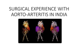
Management of aorto arteritis
•Download as PPTX, PDF•
0 likes•73 views

Deals with surgical management of takayasu diseases or aortoartiritis

Recommended
More Related Content
What's hot
What's hot (19)
Similar to Management of aorto arteritis
Similar to Management of aorto arteritis (20)
More from India CTVS
More from India CTVS (20)
Recently uploaded
Recently uploaded (20)
Management of aorto arteritis
- 1. SURGICAL EXPERIENCE WITH AORTO-ARTERITIS IN INDIA
- 2. In 1830, Rokushu Yamamoto described a peculiar condition of a 45 year old in his private practice record. Yamamoto found that there were no palpable pulses on the patient’s upper limbs and carotid.
- 3. In 1908, for the first time the Japanese ophthalmologist Mikito Takayasu (a) reported the case of a 21-year old woman with a peculiar optic fundus abnormality, characterised by arteriovenous anastomosis around the papilla (b).
- 4. In 1958, a symposium was organised on this subject. Dr. P. K.Sen, while chairing a session in the 4th Asian Pacific Congress of Cardiology, called it non specific aorto arteritis (NSAA) rather than Takayasus disease .
- 5. • Takayasu arteritis (TA) is a chronic, granulomatous, large-vessel panarteritis with preferential involvement of the aorta, its major branch arteries and the pulmonary artery. • It is an idiopathic inflammatory disease of the large elastic arteries occurring in the young and result in the occlusion or ectatic changes in vessels.
- 6. Synonyms • Takayasu’s Arteritis • Aortoarteritis • Pulseless Disease • Young female Arteritis • Occlusive thromboaortopathy • Aortic arch syndrome
- 7. Epidemiology • Worldwide incidence: 2.6 cases per million per year. • More frequent in Asian countries - Japan, Korea, China, India, Thailand, Singapore and Turkey. • Japanese patients with Takayasu arteritis - higher incidence of aortic arch involvement. • In contrast, series from India report higher incidences of abdominal involvement. Age: • Predominantly a disease of young females: 2nd or 3rd decades. • Mean age: • European study - 41yrs • Japan - 29yrs • India – 24yrs Sex: • F>M (~80% women) • India – F : M = 1.6 : 1
- 8. Pathophysiology • Predilection for the aorta and its branches. • Advanced lesions demonstrate a panarteritis with intimal proliferation, fibrosis, scarring and vascularisation of media. • Lesions :- stenotic, occlusive, or aneurysmal. • Vascular changes ->Complications :- • Hypertension - renal artery stenosis, stenosis of the suprarenal aorta; • Aortic insufficiency due to aortic valve involvement; • Pulmonary hypertension • Aortic or arterial aneurysm.
- 9. Other pathophysiologic consequences include: • Hypertensive ischemic retinopathy • Vertebrobasilar ischemia • Microaneurysms • Carotid stenosis • Hypertensive encephalopathy
- 11. Etiology • Exact etiology is unknown. • Underlying pathologic process is inflammatory. • Several etiologic factors having been proposed: • Spirochetes • Mycobacterium tuberculosis • Streptococcal organisms • Circulating antibodies due to an autoimmune process. • Genetic factors may play a role in the pathogenesis. • Raised ESR, leucocytosis, arthralgia and high titers of anti-aorta antibodies. • Rheumatic: A study showed some patients had raised ASO titre. Female predilection: Urinary estrogens elevated. Estradiol and progesterone (but not testosterones), enhance leucocyte adhesion to endothelial cells in the presence of TNF.
- 12. • (a) Active phase of TA in descending thoracic aorta showing a granulomatous inflammation at the medio-adventitial junction of aorta destroying the structure of an intercostal artery; (b) chronic phase of TA in descending thoracic aorta showing media (M) at the outer third with clusters of mononuclear cells. Note marked fibrosis of the adventitia (A) (Hematoxylin and Eosin, ×250). (c) The cells are lymphocytes, and few plasma cells and histiocytes (Hematoxylin and Eosin, ×250)
- 13. • Scanned images of the ascending aorta, arch, left subclavian artery, descending thoracic aorta (elastic van Gieson), and abdominal aorta (Hematoxylin and Eosin, and elastic van Gieson) in a diffuse pattern of TA. All segments show marked adventitial fibrosis, the major cause of wall thickening. Lymph nodes (arrows) are seen around abdominal aorta
- 14. (a) Multi-focal aortic disease with skip areas showing a large aneurysm in the abdominal aorta. The wall of the aneurysm when studied histologically also showed the presence of chronic dissection as seen in (b) (Hematoxylin and Eosin, ×100) and (c) (Elastic van Gieson, ×100). Arrow points to the intimal flap. The wall is represented only by thickened intima (I) and adventitia (A)
- 15. Major histocompatibility (MHC) class I– related chain A (MICA) Natural killer and T-cells Release of Proinflammatory cytokines Matrix metalloproteinases (MMPs) Inflammatory response Antigen Stimulates aortic tissue Expression of heatshock protein-65
- 17. Clinical Presentation • ~10% of patients are asymptomatic, with the disease detected based on abnormal vascular findings on examination. Constitutional symptoms: Headache (50%-70%) Malaise (35%-65%) Arthralgias (28%-75%) Fever (9%-35%) Weight loss (10%-18%)
- 18. Cardiac and vascular features: Bruit, with the most common location being the carotid artery (80%) Blood pressure difference of extremities (45%-69%) Claudication (38%-81%) Carotodynia or vessel tenderness (13%-32%) Hypertension (28%-53%; 58% with renal artery stenosis in one series) Aortic regurgitation (20%-24%) Raynaud’s syndrome (15%) Pericarditis (< 8%) Congestive heartfailure (< 7%) Myocardial infarction (<3%)
- 19. Neurologic features: Headache (50%-70%) Visual disturbance (16%-35%) - Strong association with common carotid and vertebral artery disease Stroke (5%-9%) Transient ischemic attacks(3%-7%) Seizures (0%-20%) Dermatologic manifestations: Erythema nodosum (6%-19%) Ulcerated subacute nodular lesions (< 2.5%) Pyoderma gangrenosum (<1%)
- 20. On ExaminationParticular attention to peripheral pulses. Blood pressure in all 4 extremities. Ophthalmologic examination. The most discriminatory finding is a systolic blood pressure difference (>10 mm Hg) between arms. Hypertension due to renal artery involvement (and sometimes leading to hypertensive encephalopathy) (~50% ofpatients).
- 21. Carotidynia may bepresent. Aortic regurgitation is a common finding. Absent or diminished pulses are the clinical hallmark of Takayasu arteritis. Pulses can be normal in many patients. Upper limbs are affected more often than lower limbs. When pulselessness occurs, patient monitoring can be difficult or impossible calf blood pressures must be obtained.
- 22. Ophthalmologic examination: Retinal ischemia, Retinal hemorrhages, Cotton-wool exudates, Venous dilatation and beading, Microaneurysms of peripheral retina, Optic atrophy, Vitreous hemorrhage, and Classic, wreathlike peripapillary arteriovenous anastomoses (extremely rare).
- 23. Dermatologic findings: resembling erythema nodosum or ulcerating nodular lesions may beseen. Other significant findings include the following: Vascular bruits – carotid, abdominal, subclavian and femoral arteries Amaurosis fugax Focal neurologic deficits consistent with cerebral infarction or TIA Eclampsia SAH, probably secondary to hypertension Myocardial infarction, DCM Pedal edema due to renal failure secondary to renal artery stenosis and glomerulonephritis
- 24. Differential DiagnosisTakayasu arteritis is rare and difficult to diagnose. Initially, symptoms arevague. Disease may have progressed considerably on presentation and diagnosis. Aortic Coarctation Atherosclerosis Buerger Disease (Thromboangiitis Obliterans) Giant CellArteritis Sarcoidosis Systemic Lupus Erythematosus Wegener Granulomatosis
- 25. Approach & Work UP Laboratory tests Nonspecific. ESR may be high (>50 mm/h) in early disease but normal later. TLC: normal orslightly elevated. Amoderate, normochromic anaemia may be present in individuals with active disease. Raised levels of soluble vascular cell adhesion molecule-1 (VCAM-1). Hypoalbuminemia is common. Urinalysis may be consistent with nephrotic syndrome.
- 26. Imaging studies CT scanning and MRI: patterns of stenosis or aneurysms of the arteries. Angiography: standard for diagnosis and evaluation of the extent of disease. Studies show that noninvasive imaging modalities - MRI, USG and 18F-FDG- PET allow diagnosis of Takayasu arteritis earlier in the disease than standard angiography and provide a means for monitoring disease activity. Angiography is used to evaluate only the appearance of the lumen and cannot be used to differentiate between active and inactive lesions.
- 28. Diagnostic Criteria Ishikawa criteria (1986) Obligatory: Age <40yrs; at the time of diagnosis, at onset of characteristic symptoms &signs of 1month duration. Major: 2 major Lesions in the left and right midsubclavian artery. The most severe stenosis or occlusion present in the mid portion of the artery from a 1cm point proximal to the left and right, respectively, of the vertebral artery orifices to a 3-cm distal point to the orifice as determined by angiography.
- 29. Minor: 9 minor High ESR Commoncarotid artery tenderness Hypertension Aortic regurgitation or annuloaortic ectasia Lesions of the pulmonary artery Left mid common carotid artery Distal brachiocephalic trunk Thoracic aorta Abdominal aorta Diagnosis: Obligatory criteria + 2 Major criteria or 1Major and ≥ 2 Minor criteria or ≥4 Minor criteria. Sensitivity of 60.4% and specificity of 95%.
- 30. Modified Ishikawa's Criteria for Takayasu's Arteritis (Modified According to Sharma et al.), 1996 The proposed modifications include: (a) removal of the obligatory criteria of age less than 40 years; (b) inclusion of characteristic signs and symptoms as a major criteria; (c) removal of age in defininghypertension; (d) deletion of the absence of aorto-iliac lesion, in defining abdominal aortic lesion; and (e) an addition of coronary artery lesion in absence of risk factors.
- 31. Three majorcriteria: 1. Left mid-subclavian artery lesion 2. Right mid-subclavian arterylesion 3. Characteristic signs and symptoms of at least 1month duration claudication, pulselessness or pulse differences in limbs, unobtainable or significant blood pressure difference (> 10 mmHg systolic blood pressure difference in limb), fever, neck pain, transient amaurosis, blurred vision, syncope, dyspnea or palpitations.
- 32. Ten minor criteria: 1.High ESR (>20 mm/h) 2. Carotid artery tenderness 3. Hypertension 4. Aortic regurgitation or annuloaortic ectasia 5. Pulmonary artery lesion 6. Left mid common carotid lesion 7. Distal brachiocephalic trunk lesion 8. Descending thoracic aorta lesion 9. Abdominal aorta lesion 10. Coronary artery lesion Diagnosis: (a) two major or (b) one major and two minor criteria or (c) four minor criteria. Sensitivity of 92.5% and specificity of 95%.
- 33. Criteria Definition 1.Age at disease onset in year Development of symptoms or findings related to Takayasu arteritis at age <40 years. 2. Claudication of extremities Development and worsening of fatigue and discomfort in muscles of one or more extremity while in use, especially the upperextremities. 3. Decreased brachial arterypulse Decreased pulsation of one or both brachial arteries. 4. BPdifference >10mmHg Difference of >10mmHg insystolic blood pressure between arms. 5. Bruit over subclavian arteriesor aorta Bruit audible on auscultation over one or both subclavian arteries or abdominal aorta. 6. Arteriogram abnormality Arteriographic narrowing or occlusion of the entire aorta, its primary branches, or large arteries in the proximal upper or lower extremities, not due arteriosclerosis, fibro- muscular dysplasia, or similar causes: changes usually focal or segmental. 1990 Criteria of American College of Rheumatology (ACR) for the Classification of Takayasu Arteritis
- 34. Diagnosis: 3 of these 6 criteria. (Sensitivity 77.4%, specificity 95%) >3 criteria yields a sensitivity of 90.5% and a specificity of 97.8%.
- 35. Takayasu arteritis can be divided into the following 6 types based on angiographic involvement:
- 37. Type I - Branches of the aortic arch.
- 38. Type IIa - Ascending aorta, aortic arch, and its branches.
- 39. Type IIb - Type IIa region plus thoracic descending aorta.
- 40. Type III - Thoracic descendingaorta, abdominal aorta, renal arteries, or a combination
- 41. Type IV - Abdominal aorta, renalarteries, or both.
- 42. Type V - Entire aorta and its branches.
- 43. Disease Activity New onset or worsening of 2 or more of the following features indicates active disease:
- 46. Corticosteroids Mainstay of therapyfor active disease. Some patients may require additional cytotoxic agents to achieve remission and taper of chronic corticosteroid treatment. Oral corticosteroids - 1mg/kg daily or divided twice daily and tapered over weeks to months as symptoms subside. IL-6 receptor inhibitor Humanized monoclonal antibody tocilizumab. IL-6 as a major component in the proinflammatory process of large-vessel vasculitis. Remission using tocilizumab as monotherapy. Then shifting to methotrexate for maintenance therapy. Medical Management
- 47. B-cell depletion Rituximab, a chimeric IgG1 antibody that binds to CD20 expressed on the surface of B cells, has shown to improve clinical signs and symptoms. Cytotoxic agents Used for patients whose disease is steroid resistant or relapsing. Continued for at least 1year after remission and are then tapered to discontinuation. Methotrexate (0.3 mg/kg/week), azathioprine (1-2 mg/kg/day), and cyclophosphamide (1-2mg/kg/day). Cyclophosphamide should be reserved for patients with the most severe and refractory disease states.
- 48. Anti-tumor necrosis factor agents Used in relapsingdisease. Initial dose of etanercept was 25 mg twice weekly (7 patients); infliximab (11patients [3 were switched from etanercept to infliximab]) was given at 3 mg/kg initially and at 2 weeks, 6 weeks, and then every 8 weeks thereafter. In 9 of the 14 responders, an increase in the anti-TNF dosage was required to sustain remission.
- 51. SURGERY
- 53. Cardiovascular procedures Bypass graft surgery: best long-term patency rate. Percutaneous balloon angioplasty: good outcomes for short lesions. Angioplasty and stenting: for recurrent stenosis. Conventional stents: high failure rates. Other procedures include aneurysm clipping and revascularization. PTCA is followed by restenosis at the angioplasty site within 1-2 years in a substantial number ofpatients.
- 54. Bypass graftsurgery Critical thoracic aortic arch arterial stenosis, upper and lower extremity ischemia, cerebrovascular accidents, and renal artery stenosis. Anastomotic stenosis or graft occlusion is a potential complication of surgery. Usually, the graft is a saphenous vein graft. Examples: Bypass of renal artery stenosis for renal salvage; Bypass of innominate or carotid artery; Bypass between subclavian-axillary and common carotid arteries; Extraintracranial bypass operations generally are performed for stenosis of the internal carotid or middle cerebral arteries.
- 55. Cardiovascular risk factors STRICT CONTROL of dyslipidemia, hypertension, and lifestyle factors that increase the risk of cardiovascular disease. These complications are the major cause of death in Takayasu arteritis. Aggressive therapy forhypertension. Low-dose aspirin may have a therapeutic effect in large vessel vasculitis. Antiplatelet agents and heparin may prove useful in preventing stroke. Warfarin also has been used. The literature reports a case of improvement in renal and systemic function with low-dose intravenous (IV) heparin therapy (10,000 U/d) followed by oral anticoagulant and antiplatelet agents.
- 62. Step 1: Exposure of both femoral arteries and the ascending aorta Step 2: Creation of pre-peritoneal tunnel Step 3: Placing the graft Step 4: Ascending aorta to graft anastomosis and femoral artery to graft anastomosis Surgical technique for Ascending Aorta- Bifemoral Bypass Grafting Surgical technique involves:
- 65. Results of Ascending Aorta-Bifemoral Bypass Grafting • Of the 56 patients undergoing this procedure (31 for bifurcation block and 25 for middle aortic syndrome) there were three deaths all due to renal failure, in one it appeared in the immediate post operative period and in two during the early follow up. • In the remaining patients there was marked improvement in limb ischemia, signs of claudication were relived, ischemic ulcers healed, hypertension was better controlled and peripheral pulses returned. • Long term results of this form of palliation too were gratifying. • All patients were maintained on anticoagulation and fared well except one patient who discontinued anticoagulants after 3 years and presented with a blocked graft. • Although the 10 year survival was only 48%, death in these patients was predominantly due to progression of the disease that resulted into end organ dysfunction. • A solitary incidence of graft block was salvaged by replacement of the blocked graft limb and embolectomy
- 66. Advantages of ascending aorta bifemoral graft: • In aortoarteritis, diffuse involvement of aorta is common as is hypertension, lower limb ischemia, impotence and renal involvement. • While attempting an abdominal aorta bi-femoral or thoracic aorta-bi-femoral bypass the graft it is often placed proximally on a diseased aortic segment. • Ascending aorta-bifemoral bypass graft avoids this problem. • It avoids a laparotomy and avoids adhesion specially in re do surgery. • In reoperation, change of limb of graft is easy as one has to work in superficial planes. • The technique is simple and is superior to axillo bifemoral graft as an emergency procedure as better blood flow through the graft is ensured. • While performing ascending aorta to bifemoral grafting, the surgeon avoids the planes of intense periarterial inflammation, adhesion, arterial inflammation, and calcification around the diseased aortic segments, thereby making the procedure very safe.
- 76. Prognosi s Substantial morbidity and mortality. Approximately 20% of patients have a monophasic and self-limited disease. ANational Institutes of Health (NIH) study of 60 patients with Takayasu arteritis: 20% of patients had a monophasic illness, self-limiting illness and therefore did not require immunosuppressive treatment. Remaining 80% of patients, who did not have a monophasic illness and who experienced 1exacerbation, immunosuppressive therapy resulted in remission in 60%. Of these, one half experienced relapse after immunosuppressive therapy was stopped.
- 77. Complications Stroke Intracranial haemorrhage Seizures Graft stenosis and/orocclusion Ischemia Organ failure Complications of hypertension Foetal injury Valvular heart disease Retinopathy Renovascular hypertension Long-term use of corticosteroids: infection, adrenal suppression, cataracts, hyperglycemia, hypertension (which complicates blood pressure control), osteoporosis, and asepticnecrosis.
- 78. Morbidity and mortality Overall 10-year survival rate is approximately 90%. Rate is reduced in the presence of major complications. 5- and 10-year survival rates are approximately 69% and 36%, respectively, in patients with 2 or more complications. 5- and 10-year survival rates associated with 1or fewer complications are 100% and 96%, respectively. Disease remission is the only factor that positively influences physical and mental quality of life.
Editor's Notes
- Potential role of pathogenic or commensal microbes in the pathogenesis of Takayasu arteritis. (A) Gut microbiota. A normal gut microbiota contributes to the development and education of the immune system, which is essential for the maintenance of physiologic homeostasis. Conversely, in the presence of dysbiosis, the number and function of regulatory T-cells are impaired and hyperactive cytotoxic T cells may grow without control, consequently the immune equilibrium is lost. The growing of pathogenic bacteria in the gut, likely harboring antigens that resemble antigens expressed by aortic or vascular tissues of the host, may promote the sensitization of the immune cells to the host own antigens, via molecular mimicry leading to autoimmunity. (B) Vascular microbiota. The colonization of pathogenic bacteria in aortic tissues may promote a local immune reaction against pathogen proteins and recruited immune cells may cross-react with antigens expressed in normal tissues leading to autoimmunity.