6. Here is an Aschoff nodule at high magnification. The most characteristic component is the Aschoff giant cell. Several appear here as large cells with two or more nuclei that have prominent nucleoli. Scattered inflammatory cells accompany them and can be mononuclears or occasionally neutrophils.
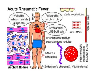
7. Microscopically, acute rheumatic carditis is marked by a peculiar form of granulomatous inflammation with so-called "Aschoff nodules" seen best in myocardium. These are centered in interstitium around vessels as shown here. The myocarditis may be severe enough to cause congestive heart failure.
8. Stenotic mitral valve seen from left atrium. Both commissures are fused; the cusps are severely thickened. The left atrium is huge. The valve is both incompetent and stenotic Opened stenotic mitral valve showing thickening distorted cusps, adherent commissures with calcification and thrombus deposition, and thickening, fusion and shortening of chordae tendinae
9. Aortic valve showing active valvulitis. The valve is slightly thickened and displays small vegetations – "verrucae" Stenotic mitral valve seen from left atrium, showing fusion of commissures, thickening and calcification of the cusps
10. Another peculiar cell seen with acute rheumatic carditis is the Anitschkow myocyte. This is a long, thin cell with an elongated nucleus.
16. Closer view of erythema marginatum Erythema marginatum on the trunk, showing erythematous lesions with pale centers and rounded or serpiginous margins
![RHEUMATIC HEART DISEASE ,[object Object],[object Object],[object Object],[object Object]](data:image/gif;base64,R0lGODlhAQABAIAAAAAAAP///yH5BAEAAAAALAAAAAABAAEAAAIBRAA7)