Call Girls Frazer Town Just Call 7001305949 Top Class Call Girl Service Avail...
Pedo.pdf
1. V,
u
1-
z
0
0
0
0
w
0.
I.I.
0
~
0
0
C0
I-
><
w
I-
410 SECTION 8 : PEDIATRIC ENDODONTICS
that there is a family of proteins that has bone induc-
tive properties and BMP is a generic term for this
family.
■ The effect of recombinant human BMP-2 and BMP-
4 when capped with inactivated dentin matrix on an
amputated canine pulp have been seen to imply that
recombinant human BMP-2 and BMP-4 induce dif-
ferentiation of adult pulp cell into odontoblast. The
recombinant human osteogenic protein 1 (hop- I )
when placed on artificially exposed dentin pulp of
four adult miniature pigs was seen to cause a sub-
stantial amount of hard tissue formation (osteoden-
tin and tubular dentin) thereby completely bridged
the defect. Hence, hop- I is a collagen carrier matrix
which appeared to be a suitable bio-active capping
agent.
■ We will be entering a new era, when commercially
available recombinant human BMPs will be made
available for experimentation and clinical trials. A
combination of BMPs may be necessary to ensure
maximal and predictable reparative dentinogenesis,
but there are details to be determined in logical steps.
If this material is proved to be fruitful in human clini-
cal trial this product could be an ideal material of
choice for vital tooth pulpotomy in primary teeth.
■ BMPs are osteogenic proteins capable which form a
part of TGF-beta. They are implicated in cell differ-
entiation, tissue morphogenesis, regeneration and re-
pair. Their genes are expressed during tooth devel-
opment and dentinogenesis. It has been proposed that
BMPs stimulate the induction and differentiation of
mesenchymal cells with varying degrees of dentinal
bridge formation.
■ Domingue AT (2003) compared MTA, calcium hy-
droxide and adhesive dentin bonding agent in pul-
potomy. He stated that MTA forms crystals of cal-
cium oxide in an amorphous structure of:
Table 35.9 Deciduous tooth pulpectomy
Indications
Strategically important tooth (e.g., in case of the
deciduous second molar where the permanent
first molar has not erupted)
Irreversible pulpitis
· Minimal periapical changes with sufficient bone support
Al least 213rd of the root length available
· Internal resorption without any obvious perforation
33% Ca
- 49% PO4
2% Carbon
3% Chloride
6% Silica
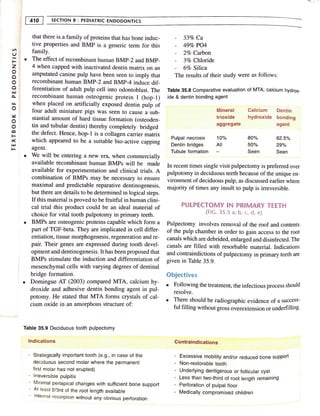
The results of their study were as follows:
Table 35.8 Comparative evaluation of MTA, calcium hydrox-
ide & dentin bonding agent
Mineral Calcium Dentin
trioxide hydroxide bonding
aggregate agent
Pulpal necrosis 10% 80% 62.5%
Dentin bridges All 50% 29%
Tubule formation Seen Seen
In recent times single visit pulpectomy is preferred over
pulpotomy in deciduous teeth because of the unique en-
vironment of deciduous pulp, as discussed earlier where
majority of times any insult to pulp is irreversible.
PULPECTOMY IN PRIMARY TEETH
(FIG. 35.5 a, b, c, d, e)
Pulpectomy involves removal of the roof and contents
of the pulp chamber in order to gain access to the root
canals which are debrided, enlarged and disinfected. The
canals are filled with resorbable material. Indications
and contraindictions of pulpectomy in primary teeth are
given in Table 35.9.
Objectives
■ Following the treatment, the infectious process should
resolve.
■ There should be radiographic evidence of a success-
ful filling without gross overextension or underfilling.
Contraindications
· Excessive mobility and/or reduced bone support
- Non-restorable tooth
· Underlying dentigerous or follicular cyst
- Less than two-third of root length remaining
- Perforation of pulpal floor
Medically compromised children
2. -
• The treatment should permit resorption of the pri-
mary _
root _structures and filling materials at the ap-
propriate time to permit normal eruption of succeda-
neous tooth.
• There should be no radiographic evidence of further
breakdown of the supporting tissues.
• Treatment should alleviate and prevent further sen-
sitivity, pain or swelling.
■ There should be no internal orexternal root resorption
or other pathology.
Technique (Fig. 35.6 A-F)
•:lccess Opening for Pulpectomy in Primary Teeth
Although the apical forarnen must be sealed by endo-
dontic therapy, the root canal is what provides the path-
way to apex. Therefore it is important for the pedodon-
tist to be familiar with the various paths that the root
takes in getting to the apex. These are the typical con-
figurations of the root canals. After the establishment
of a diagnosis and treatment plan the first part of treat-
ment directly applied to the tooth is the access cavity
preparation, also known as the endodontic entry. The
canal preparation can be divided into the coronal phase
and the radicular phase.
Apical moisture proof seal, the first essential for suc-
cess, is not possible unless the space to be filled is care-
fully prepared and debrided to receive the final restora-
tion. The coronal phase must give direct access to the
root canals and the apical foramina so that these areas
may be properly cleaned and shaped by the intraradicular
phase. Therefore all the treatment hinges on the accu-
racy and correctness of the entry. If the access is im-
properly prepared as to position, depth, or extent, it will
be difficult to reach the optimal result.
R
_
ules for Proper Access Preparation .
Endodontic Dogma: "Careful cavity preparatwn and
rout canal obturation are the keystones to successful
root canal therapy."
I. The objective of entry is to gain direct access _to the
apical foramina and not merely to the canal orifices.
2· Access cavity preparations are different from typical
occ.:Jusal preparations and are not guided by topog~a-
phy of the occlusaJ grooves, pits, and fissures avoid-
ing the underlying pulp.
3 ·t'h · th nder treat-
. e likely interior anatomy of the too u
f17t;nt must be determined for access opening. Each
1001'1 ha.., a different length number and configura-
CH. 35 : TREATMENT MODALITIES 413
l!on of root canals and so radiographs taken from dif-
ferent angles must be considered.
4. Endodontic entries are prepared always through the
occlusal or the lingual surface and never the proxi-
mal or the gingival surface. When proximal or
gingival tooth destruction occurs, affected area should
be excavated and restored with either temporary or
permanent restorative material and the normal access
is prepared.
5. As a part of the access preparation, the unsupported
cusps of posterior teeth must be reduced to avoid
weakening of the tooth structure.
To achieve optimal preparation three factors of internal
anatomy must be considered:
l. Size of the Pulp Chamber
In young patients the preparations are more exten-
sive than in the older patients with receeded pulps
and smaller pulp chambers. This is quite apparent in
preparing the anterior teeth ofyoungsters whose large
root canals require larger instruments and filling
materials.
2. Shape of the Pulp Chamber
The finished outline form should accurately reflect
the shape of the pulp chamber, e.g., the floor of the
pulp chamber in a molar is usually triangular owing
to the triangular position of the canal orifices. This
shape is so extended to the walls of the cavity onto
the occlusal surface, hence the final outline form is
usually triangular.
3. Number, position and curvature of the root canals
Primary Root Canal Anatomy
To complete endodontic treatment on primary teeth suc-
cessfully, the clinician must have a thorough knowledge
of the anatomy of the primary root canal systems and
the variations that normally exist.
■ Primary anteriors
The form and shape of the root canals of the primary
anterior teeth resembles the form and shape of the
exteriors of the teeth.
■ Maxillary incisors
The root canals of the primary maxillary central and
lateral incisors are almost round but somewhat com-
pressed. Normally, these teeth have one canal with-
-t
m
X
-t
c,,
0
0
~
0
'Tl
"ti
m
C
0
C
0
z
-t
l"'I
V,
3. V,
u
1-
z
0
0
0
0
UJ
c..
u..
0
::-=:
0
0
C0
I-
x
UJ
I-
414 SECTION 8 : PEDIATRIC ENDODONTICS
out a bifurcation. Apical ramifications or accessory
canals and lateral canals are rare, but do occur.
■ Mandibular incisors
The root canals of the primary mandibular centraJ
and lateral incisors are flattened on the mesial and
distal surfaces and sometimes grooved, pointing to
an eventual division into two canals. The presence
of two canals is seen less than 10% of the time. Oc-
casionally, lateral or accessory canals are observed.
■ Maxilla,y and mandibular canines
The root canals of the maxillary and mandibular ca-
nines correspond to the exterior root shape, a rounded
triangular shape with the base toward the facial sur-
face. Sometimes, the lumen of the root canals is com-
pressed in the mesial-distal direction. Bifurcation of
the canal does not normally occur. Lateral canals and
accessory canals are rare.
■ Primary molars
The primary molars normally have the same number
of roots and positions of the roots as the correspond-
ing permanent molars. The maxillary molars have
three roots, two facial and one palatal; the mandibu-
lar have two roots, mesial and distal. The roots of the
primary molars are long, slender compared with the
crown length and width, and they diverge to allow
for a permanent tooth bud formation.
■ Maxillary first primary molars
The maxillary first primary molars have from two to
four canals that roughly correspond to the exterior
root form with much variation. The palatal root is
often round; it is often longer than the two facial roots.
Bifurcation of the mesial-facial root into two canals
occurs in approximately 75% of the maxillary first
primary molar.
■ Maxillary second primary molars
The maxillary primary molar has two to five canals,
roughly corresponding to the exterior root shape.The
mesial-facial root usually bifurcates or contains two
distinct canals. This occurs in approximately 85% to
95% of the maxillary second primary molars.
■ Mandibular first primary molars
The mandibular first primary molar usually has three
canals, roughly corresponding to the external root
anatomy but may have two to four canals. It is re-
ported that approximately 75% of the mesial roots
contain two canals, whereas only 25% of the distal
roots contain more than one canal.
■ Mandibular second primary molars
The mandibular second primary molar may have two
to five canals but usually has three. The mesial root
has two canals approximately 85% of the time,
whereas the distal root contains more than one canal
only 25% of the times.
Access Opening for Primary Anterior Teeth
Access opening for endodontic treatment on primary or
permanent anterior teeth have traditionally been through
the lingual surface. This continues to be the surface of
choice except for the maxillary primary incisors. Be-
cause of the problems associated with the discoloration
of endodontically treated primary incisors, it has been
recommended to use a facial approach followed by an
acid etch composite restoration to improve esthetics.
The anatomy of maxillary primary incisors is such that
access may successfully be made from the facial sur-
face. The only variation to the opening is more exten-
sion to the incisal edge than with the normal lingual
access in order to give as straight an approach as pos-
sible into the root canal.
Access Opening for Primary Posterior Teeth
Access opening into the posterior primary root canals is
essentially the same as those for the permanent teeth.
Important differences between the primary and perma-
nent teeth are the:
■ Length and the bulbous shape of the crowns
■ A very thin dentinal wall at the pulpal floor and the
root.
■ The depth necessary to penetrate into the pulp cham-
ber is quite less than that in the permanent teeth.
■ Likewise, the distance from the occlussal surface to
the pulp floor of the pulp chamber is much less than
in permanent teeth. In the primary molars, care muSt
be taken not to grind on the pulpal floor since perfo·
ration is likely.
When the roof of the pulp chamber is perforated a11
<l
the pulp chamber is identified, the entire roof sho~ld,
1
~~
removed with a bur. Since the crowns of the P1
,rn. )
. ·ds the cxt.:-
teeth are more bulbous, less extenswn towai ·
4. ~ - - - - - - - - - - - - - - - - - - - - - - -- -Cn . ~
,.:' · TRi:/·.1MENT MODALITIES ~
•or ofthe tooth is necessary to uncover the openings of
n h .
the root canals t an m the permanent teeth.
canal Cleaning and Shaping
Isolation (Fig. 35.1)
1
Use of the rubber dam is essential in any endodontic
procedure as it is the best method of isolating the
toothfrom the oral cavity. First introduced by Barnum
(1864), it is useful in providing a clean, dry and
sterilizable field (refer isolation).
Oebridement
1
Canal cleaning and shaping is one of the most im-
portant phases of primary endodontic therapy. The
main objective of the chemico--mechanical prepara-
tion of the primary tooth is debridement of the ca-
nals. Although an apical taper to the canals is desir-
able, it is not necessary to have an exact shape to the
canals. The biomechanical preparation in the primary
teeth can be said to be different enough to warrant
the following considerations:
Fig. 35.7 Isolated primary maxillary first molar during
PUlpectomy
1. Relative pulpectomy: Due to tortuous course of
root canal coupled with the numerous accesso1?'
canals, the complete removal of pulp in the p~-
mary teeth may often be difficult, if not impossi-
ble. Thus, all such procedures can be regarded as
Partial pulpectomy procedures.
2 s · · the primary teeth
· elective filling: Resorptton m
may have started at the time of treatment. Also,
the slender roots with thin apical ends may pre-
d. ' f t re in cases of
1
spose the tooth to a root rac u
. . h h procedure of se-
exccss,ve preparation. T us t e
lcc:Live filing of the canals should be followed.
■ It is important to establish ihe working length to pre-
vent overextension through the apical foramen. It is
suggested that the working length be shortened, 2-3
nun short of the radiographic root length, especially
in the teeth showing signs of apical root resorption.
Working length can also be determined by electronic
apex locator.
Electronic Apex Locator for Working Length Determi-
nation (Fig. 35 BJ
Apex Locator (Root ZX)
Root ZX is a third generation electronic apex locator
manufactured by J. Morita Corp. It is a device which
can be used for measuring the working length. It con-
sists of a LCD monitor, a file holder tip and a contrary
electrode to complete the circuit.
Fig. 35.8 Electronic Apex Locator (Root ZX)
Working Principle of Root ZX
The meter in the display indicates the position of the
file tip. As the file approaches the apex, the audible alarm
will beep slowly when the meter reaches '2', then the
bar indicating the apical constriction of the root canal
flashes on and off. A meter reading of 0.5 indicates that
the tip of the file is at the apical constriction. At this
point the image of the root canal will start flashing and
the sound of the alarm will change. It is essential that
the file be taken to the anatomic apex (the major fora-
men) and then returned to the apical constriction (the
minor foramen). This ensures that all the constrictions
that can occur in the canal have been negotiated. If the
file reaches the major foramen (meter reading 0), the
alarm will change to a single sustained beep and the
word 'APEX' will flash.
-I
rn
><
-I
=
0
0
~
0
'Tl
"C
rn
C
0
C
0
z
-I
("'
VI
5. Vl
u
1-
z
0
0 ,
0
0
u.J
C.
~
0
0
cc
I-
><
u.J
I-
416 SECTION 8 : PEDIATRIC ENDODONTICS
Operator Instructions for Root ZX
The operating instructions for the Root ZX states, "The
working length of the canal used to calculate the length
of the filling material is actually somewhat sh01ter. Find
the length of the apical seat (i.e., the end point of the
filling material) by subtracting 0.5-1.0 mm from the
working length indicated by the 0.5 reading on the
meter." They suggested that the Root ZX should be used
with the 0.0 or APEX increment mark as the most accu-
rate apical reference point. The clinician should then
adjust the working length on the endodontic instrument
according to the margin of safety that is desired (i.e., I
mm short).
■ Instruments should be gently curved to help negoti-
ate the canals. This helps in maintaining the original
shape of the canal and thus lessens the risk of perfo-
ration. Shaping of the canals proceeds in much the
same manner as is done to receive a gutta-percha fill-
ing. The canals are enlarged several file sizes past
the first file that fit snugly into the canal, with a mini-
mum size of 30 to 35.
■ Since many of the pulpal ramification cannot be
reached mechanically, copious irrigation during
cleaning and shaping must be maintained. Debride-
ment of the primary root canal is more often accom-
plished by chemical than mechanical means. The use
of sodium hypochlorite to digest organic debris and
RC-prep to produce effervescence must play an im-
portant part in removal of the tissue from the inac-
cessible area of the root canal system.
■ If the inflammation is beyond the coronal pulp with
only interradicular but no periapical radiolucency, a
single-visit pulpectomy is preferred. On the other
hand, if the pulp is necrotic with periapical inv~lve-
ment filling procedure is delayed until a later time.
Afte; canal debridement, the canals are again copi-
ously flushed with sodium hypochlorite and are the_n
dried with sterile paper points; a pellet of cotton 1s
barely moistened with camphorated ~arachlorophenol
and sealed into the pulp chamber with temporary ce-
ment. At a subsequent appointment the canal is re-
lered. As lono as the patient is free of all signs and
en t:,
• • •
symptoms of inflammation, th~ canals a~e agarn im-
oated with sodium hypochlonte and dned prepara-
t:,
tory to filling.
ROTARY INSTRUMENTS IN PEDIATRIC
ENDODONTICS (FIG. 35 .9)
The rotary Ni-Ti files are specially designed to provide
superior flexibility and unmatched efficiency. The
bl 1
. . . •.i,
1 Yen.
a e c 1111cians to create unt1orm y tapered shapes ·
1
' naria
tomically difficult and curved canals. ·
These files are made of Ni-Ti, allowing for tlexibilit
. d y~
be used smoothly even m curve canals. The latch ty e
design of the files allows attachment to a handpiec/
Advantages
■ Provide a more consistently dense fill due to uniform
debridement.
■ Allow for greater apical enlargement.
■ Prevent apical exposure.
■ Provide better shape than traditional hand-filing.
■ Significantly reduce instrumentation time.
Disadvantages
■ Skill is required for practice for a beginner.
■ Resorption ofroots in primary teeth may causeaprob-
lem.
■ Problem of breakage of files in canals.
■ Repeated use increases risk of fracture, specially in
curved canals.
Fig. 35.9 Use of rotary files in pediatric endodontics
FILLING OF THE PRIMARY ROOT CANALS
Root Canal Filling Materials
. d.fferences
Developmental, anatomic and physiologic I
d'f.
h II for 1
between the primary and permanent teet ca . Is
-1
1- 11a1ena ·
ferences in the criteria for root canal ft 111g 1
' ., Is
. t'·11· o rna1en,1
The ideal requirements of a root canal I inc
for the primary teeth are as follows:
/deal Requirement5 . , ·y root.
■ Should resorb al a similar rate as the prunai
6. •
•
Should be hannless to the periapical tissue:, unJ 10
(he pennanent tooth germ; ifpressed beyond the apex
it should resorb readily.
It should have a stable disinfecting power.
It should be inserted easily into the root canal and be
removed easily if necessary.
1
Should adhere to the walls of the canal and should
not shrink.
1
It should not be soluble in water.
1
Should be radiopaque and not discolor the tooth.
No material currently available meets all these criteria.
The filling materials most commonly used for primary
pulp canals are zinc oxide-eugenol paste, iodofonn paste
and calcium hydroxide.
Zinc Oxide-Eugenol Paste
Zinc oxide - eugenol paste (ZOE) is probably the most
commonly used filling material for primary teeth. Camp
in 1984 introduced the endodontic pressure syringe to
overcome the problem of underfilling, a relatively com-
mon finding when tbkk mix of ZOE is employed.
Underfilling, however, is frequently clinically accept-
able. Overfilling, on the other hand, may cause a mild
foreign body reaction. Another disadvantage of ZOE
paste is the difference between its rate of resorption and
that of the tooth root.
lodoform Paste
Several authors have reported the use of KRI paste; It
resorbs rapidly and has no undesirable effects on suc-
cedaneous teeth when used as a pulp canal medicament
in abscessed primary teeth. Further, KRI paste that ex-
trudes into the periapical tissue is rapidly replaced with
< -I .[,HMENT MODALITIES 417
.1 n,.. ~1;,1, ·, ! ·· J fu11 nd 111 have a long lasting
b;1c1eric1Jal p0 L
ent1,tl. S 1u: 1odnform paste does not set
into a hard mass, it can be removed if re-treatment is
required. KRI was found to have a success rate of 84%
as compared to ZOE, which showed a success rate of
only 65%.
■ A paste developed by Maisto has been used clini-
cally for many years, and good results have been re-
ported with its use. This paste has the same composi-
tion as the KRI paste with additions.
Composition of commonly used root canal materials are
given in Table 35.10 for primary teeth.
Calcium Hydroxide
■ This material is generally not used in pulp therapy
for primary teeth. However, several clinical and his-
topathologic investigations of calcium hydroxide and
iodoform mixture (Vitapex, Neo Dental Chemical
Products Co., Tokyo) have been published by Fuchino
and Nishina (1980). This material was found to be
easy to apply and resorbs at a slightly faster rate than
that of the root. It has no toxic effects on permanent
successor and is radiopaque. For these reasons, the
calcium hydroxide-iodoform mixture can be consid-
ered to be a nearly ideal primary tooth root canal fill-
ing material. Other preparations with a similar com-
position are available in the United States with the
trade name of Endoflas (Sanlon Laboratories, A.A.
7523 Cali, Colombia S.A).
■ Chawla et al (1998) carried out a pilot study in the
mandibular primary molars using calcium hydroxide
paste as a root canal filling material and found it to
be a success.
Table 35.10 Composition of commonly used root canal materials for primary teeth
Walkhotf KRI paste Maisto paste Vitapex Endoflas Colla cote Guedes-Pinto
paste paste
·-
Parachloro- lodoform 80.8% Zinc oxide 14 g Calcium Zinc oxide Synthetic 0.30 g
Phenol Camphor 4.86% lodoform 42 g hydroxide 56.5% collagen iodoform
Camphor Parachlorophenol Thymol 2 g lodoform Barium 0.25 g calcium
Menthol 2.025% Chlorphenol Oily sulfate 1.63% hydroxide 0.1 ml
Menthol 1.215% Camphor 3 cc additives lodoform 40.6% Camphorated
Lanolin 0.5 g Calcium paramonochl-
hydroxide 1.07% orophenol
Eugenol
Pentachloro-
phenol
-f
m
><
-f
c:,
0
0
~
0
-n
"ti
m
0
0
0
0
z
-f
f"I
V,
7. V,
u
1-
z
0
C
0
C
w
0..
u.
0
~
0
0
co
I-
><
w
I-
418 -~S~EC~T~l~O~N~8'.:...:_:~P~ED~l~A~T~Rl~C~E~N~D~O~D~O~N~T~l~C~S----------------------
.___~
--
• We have also observed almost a 100% clinical suc-
cess in 10 endodontically treated primary molars
which were filled with vitapex (calcium hydroxidised
idoform).
Colla Cote
It is a soft, white, pliable, biocompatible sponge obtained
from bovine collagen. It can be applied to moist or bleed-
ing canals. It is an absorbable collagen barrier which
prevents or diminishes extravasation of root canal fill-
ing material during primary molar pulpectomies. Apart
from its use in endodontic therapy (surgical or non sur-
gical) it also provides a scaffold for bone orowth and so
e,
it can be applied on wounds.
Endoflas
Endoflas is a root canal sealer material, which is com-
posed of zinc oxide, barium sulfate, iodoform, calcium
hydroxide, eugenol and pentachlorophenol. It can be
used as an alternative root canal filling material for pri-
mary teeth. One condition for success of Endoflas is the
prevention of microleakage. A permanent restoration
should be placed as soon as possible after clinical signs
and symptoms of inflammation are eliminated. A retro-
spective study done on primary teeth using endoflas has
shown 70% success rate.
Gutta-Percha (Not indicated for primary teeth)
Since gutta-percha is not a resorbable material, its use
is contraindicated in the primary teeth.
Comparison of materials used for pulpectomy in pri-
mary teeth is given in Table 35.11
Obturation Techniques
• Several techniques have been used for the filling of
materials into the deciduous teeth canals.
1. The primary teeth with their larger canals can be
filled with the thin mix coating the walls of the
canal with the help of a reamer in an anti-clock
wise direction while taking it out slowly followed
by the placement of the thicker mix which is then
pushed manually.
2. Pastes can also be filled by means of a Lentulo
spiral mounted on the micromotor hand piece.
Lentulospiral mounted on the slow speed hand-
piece has shown success rate of 96% and 92%
when hand held. The direction of rotation needs
to be checked for the material to properly flow
into the canal (Figs. 35.11, 35.12).
3. The endodontic pressure syringe is also effective
for placing the ZOE into the canals. The Vitapex
system also uses a syringe with the material in it
(Fig 35.10). The syringe is introduced upto 1/5'h
the distance from the apex of the canal and the
material is slowly injected as the syringe is with-
drawn from the canal.
■ Regardless of the method adopted to fill the canals,
care should be used to prevent extrusion of the mate-
rials into the periapical tissues. The adequacy of the
obturation is checked by radiographs. In case a small
amount of the ZOE is inadvertently forced through
the apical foramen, it is left alone since the material
is resorbable (Fig. 35.13).
• When the canals are satisfactorily obturated, a fast-
set temporary cement is placed in the pulp chamber
Table 35.11 Comparison of materials used for pulpectomy in primary teeth
Properties
1. Resorbs at the same rate as the tooth
2. Harmless
3. Overfill resorbs
4. Antiseptic
5. Easily applied
6. Adheres to the wall
7. Easily removed
8. Radiopaque
9. No discoloration
ZOE
y
y
y
y
y
Ca(OH)2
with
lodoform (VITAPEX)*
y
y
y
y
y
y
y
y
'f-Yes ' Vi/apex - Neo Dental Chemical Products Co. Ltd., Tokyo, Japan (2000)
KAI
paste
y
y
y
y
y
y
y
8. ~---------------------------~C~
H
~3
~S_::_:_
T
~R~E~A'.:1:T~M~
E
~
N~T~M~O~O~A~L~IT~l~E~S- 419
Fig. 35.10 Vitapex material for obturating root canals in
primary teeth
Fig. 35.12 Endodontic instrument holder
to seal over the ZOE canal filling. The primary tooth
is restored with a stainless steel crown.
Follow-up After Primary Pulpectomy
• The rate of success following primary pulpectomy is
high. However, these teeth should be periodically
checked for the success of the treatment and to inter-
cept any problem associated with failure. While
resorbing normally without interference with erup-
tion of the pemrnnent tooth, the primary tooth should
remain asymptomatic, firm in the alveolus and free
from pathosis. Ifevidence of pathosis is detected, ex-
traction and conventional space maintenance are rec-
ommended.
• It has been pointed out that pulpally treated primary
teerh may occasionally present a problem of over-
rctention. After normal physiologic resorption of the
-
Fig. 35.11 Lentulo spiral used to obturate root canals
Fig. 35.13 Overobturation seen following ZOE obtura-
tion of mandibular second molar (distal root)
root reaches the pulp chamber, the large amount of
ZOE present may impair the resorption and lead to
prolonged retention of the crown. Treatment usually
consists of simple removal of the crown and allow-
ing the permanent tooth to erupt.
Management of Acute Alveolar Abscess
Incision and drainage of abscess followed with antibi-
otics along with anti-inflammatory analgesics. Access
opening can also be done to facilitate drainage. After
acute phase subsides (in 24-48 hr), pulpectomy or ex-
traction of offending tooth can be planned.
Young Permanent Tooth
Endodantic Management
The completion of root development and closure of apex
occurs up to 3 years after eruption of the teeth.
-I
rn
X
-I
c::,
0
0
;:,::
0
'Tl
,,
rn
0
0
0
0
z
-I
n
VI