
Recommended
Recommended
More Related Content
Similar to skin part 2.pptx
Similar to skin part 2.pptx (20)
More from HiraKhan218459
More from HiraKhan218459 (17)
Recently uploaded
Recently uploaded (20)
skin part 2.pptx
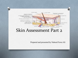
- 1. Skin Assessment Part 2 Prepared and presented by Naheed Feroz Ali
- 2. Vascular lesions Petechiae Ecchymosis Hematoma Cherry Angioma Spider angioma
- 3. Vascular lesions O Ecchymosis – purplish blue bruises O Petechia - pin point red capillary bleeding
- 4. Vascular lesions O Hematoma: A collection of blood outside of blood vessels. Most commonly, hematomas are caused by an injury to the wall of a blood vessel.
- 5. Vascular lesions O Cherry Angioma: A cherry angioma is classified as a bright, cherry-red or purple spot, which is due to the dilated capillaries they’re made up of.
- 6. Vascular lesions O Spider angiomas are caused by small arteries that travel to the surface of the skin and branch out, resembling a small red spider with tiny legs
- 7. Burns O A burn is an injury to the skin or other organic tissue primarily caused by heat or due to radiation, radioactivity, electricity, friction or contact with chemicals. O Degrees of Burns: First-degree (superficial) burns Second-degree (partial thickness) burns Third-degree (full thickness) burns Fourth-degree burns
- 8. First-degree (superficial) burns. O First-degree burns affect only the outer layer of skin, the epidermis. O The burn site is red, painful, dry, and with no blisters. O Mild sunburn is an example.
- 9. Second-degree (partial thickness) burns. O Second-degree burns involve the epidermis and part of the lower layer of skin, the dermis. O The burn site looks red, blistered, and may be swollen and painful.
- 10. Third-degree (full thickness) burns O Third-degree burns destroy the epidermis and dermis. O They may go into the innermost layer of skin, the subcutaneous tissue. O The burn site may look white or blackened and charred. O Involves all layers of skin O Dry surface O Often leathery O Tight swelling O Relatively painless
- 12. Fourth-degree burn O Fourth-degree burns go through both layers of the skin and underlying tissue as well as deeper tissue, possibly involving muscle and bone. There is no feeling in the area since the nerve endings are destroyed.
- 13. Pressure ulcers O Pressure ulcers are localized areas of skin breakdown that occur as a result of prolonged pressure. Necrotic tissue develops because the vascular supply to the area is diminished. O Staging pressure ulcers Stage I Stage ii Stage iii Stage iv
- 15. Stage I O Intact skin that doesn’t blanch O May differ in color from surrounding area in people with darkly pigmented skin O Usually over a bony prominence O May be painful, firm or soft, and warmer or cooler than surrounding tissue
- 16. Stage II O Superficial partial-thickness wound O Presents as a shallow, open ulcer without slough and with a red and pink wound bed
- 17. Stage III O Involves full-thickness wound with tissue loss and possibly visible subcutaneous tissue but no exposed muscle, tendon, or bone
- 18. Stage IV O Involves full-thickness skin loss, with exposed muscle, bone, and tendon O May be accompanied by eschar, slough, undermining, and tunneling
- 19. Unstageable O Involves full-thickness tissue loss, with base of ulcer covered by slough and yellow, tan, gray, green, or brown eschar O Can’t be staged until enough slough and eschar are removed to expose the wound base
- 20. Common skin disorders O Contact dermatitis: Contact dermatitis is an inflammatory disorder that results from contact with an irritant. Primary lesions include vesicles, large oozing bullae, and red macules that appear at localized areas of redness. These lesions may itch and burn.
- 22. Psoriasis O Psoriasis is a chronic disease of marked epidermal thickening. Plaques are symmetrical and generally appear as red bases topped with silvery scales. The lesions, which may connect with one another, occur most commonly on the scalp, elbows, and knees.
- 23. Psoriasis
- 24. Urticaria (hives) O Occurring as an allergic reaction, urticaria appears suddenly as pink, edematous papules or wheals (round elevations of the skin). O Itching is intense. O The lesions may become large and contain vesicles
- 26. Scabies O Cause by Mites. O The lesions appear in a straight or zigzagging line about 3/8 (1 cm) long with a black dot at the end. O Commonly seen between the fingers, at the bend of the elbow and knee, and around the groin, abdomen, or perineal area O Scabies lesions itch and may cause a rash.
- 27. Scabies
- 28. Herpes zoster O Herpes zoster appears as a group of vesicles or crusted lesions along a nerve root. The vesicles are usually unilateral and appear mostly on the trunk. These lesions cause pain but not a rash.
- 29. Tinea corporis (ringworm) O Tinea corporis is characterized by round, red, scaly lesions that are accompanied by intense itching. O These lesions have slightly raised, red borders consisting of tiny vesicles. Individual rings may connect to form patches with scalloped edges. O They usually appear on exposed areas of the body
- 31. Hair Assessment O •Quality and Quantity O •Color -black, grey, white, dyed O •Texture -thick, fine, smooth, silky, dry , dull, shiny, lustrous O –Hypothyroidism & malnutrition = dull, dry hair O •Cleanliness –Clean, oily, dandruff, infested lice/ nits, scaliness -dermatitis
- 32. Hair Assessment O Distribution –amount & density of hair O –alopecia localized patch, –aging, malnutrition, chemotherapy O –Hirsutism –Excessive facial hair – steroids, Cushing syndrome O –Pattern of loss –hair falling, male pattern balding O –Gen hair loss –infection, Nut def, hormonal, thyroid, liver, drugs or radiation.
- 34. Hair abnormalities O Hair abnormalities can cause patients emotional distress. Among the most common hair abnormalities are alopecia and hirsutism. O Hair abnormalities may be caused by certain drugs or endocrine problems.
- 35. Alopecia O Alopecia occurs more commonly and extensively in men than in women. O Diffuse hair loss, may occur as a result of pyrogenic infections, chemical trauma, ingestion of certain drugs, and endocrinopathy and other disorders. O Tinea capitis, trauma, and full-thickness burns can cause patchy hair loss.
- 37. Hirsutism O Excessive hairiness in women, or hirsutism, can develop on the body and face, affecting the patient’s self-image. O Localized hirsutism may occur on pigmented nevi. O Generalized hirsutism can result from certain drug therapy or from such endocrine problems as Cushing’s syndrome, polycystic ovary syndrome, and acromegaly
- 39. Nails PE ( Inspect / palpate) O •Color O –Pink, pallor, cyanosis, reddish, yellowish cuticle O –Capillary refill –brisk, sluggish due to circulation O –Yellow fungal infections, raised sides - Paronychia
- 40. Contour O –Convex, smooth trimmed edges O –Curved nails with normal angle O –Edges bitten, broken –biting or injury O –Spoon shaped nails –concave -Iron deficiency anemia O –Lines / Ridges / depression –Beau’s lines in acute illness O –Clubbing >180 degrees -hypoxia O Splinter hemorrhages caused by trauma
- 42. Consistency -Thickness O –Thick , brittle , soft nails O –Due to Excessive water contact / detergent use O –Illness, trauma, Iron , calcium, Mg deficiency O –Thin nails –nutritional deficiency anemia O –Thick nails -decreased circulation
- 44. Cleanliness O –Clean trimmed and manicured O –Dirty, broken or pointed fingernails
- 45. Nail abnormalities O Nail abnormalities include : Clubbed fingers, splinter hemorrhages of the nail bed Muehrcke’s lines.
- 46. Clubbed fingers O Clubbed fingers can result from chronic tissue hypoxia. O Normally, the angle between the fingernail and the point where the nail enters the skin is about 160 degrees. O Clubbing occurs when that angle increases to 180 degrees or more.
- 48. Splinter hemorrhages O Splinter hemorrhages are reddish brown narrow streaks under the nails. O They run in the same direction as nail growth and are caused by minor trauma. O They can also occur in patients with bacterial endocarditis.
- 49. Muehrcke's lines O Muehrcke's lines or leukonychia striata are longitudinal white lines that can indicate trauma but may also be associated with metabolic stress, which impairs the body from using protein
- 50. Onycholysis O Onycholysis is the medical term for when your nail separates from the skin underneath it.
- 51. Paronychia Fungal infection of nails
- 52. Benign versus cancerous lesions O Lesions may be benign, such as a benign nevus, or mole. O However, changes in an existing growth on the skin or a new growth that ulcerates or doesn’t heal could indicate cancer or a precancerous lesion.
- 53. Neoplastic Lesions O Pre cancerous O Seborrheic keratosis O Actinic keratosis O Cancerous O •Melanoma O •Basal cell Carcinoma O •Squamous cell carcinoma O •Kaposi sarcoma
- 54. Types of skin cancer OBenign nevus : Symmetrical, round, or oval shape Sharply defined borders Uniform, usually tan or brown color Less than 6 mm in diameter Flat or raised
- 55. Precancerous actinic keratosis O Abnormal changes in keratinocytes O Can become squamous cell carcinoma
- 56. A seborrheic keratosis is a common noncancerous (benign) skin growth
- 57. Dysplastic nevus O Abnormal growth of melanocytes in a mole O Can become malignant melanoma
- 59. Kaposi sarcoma O Kaposi's sarcoma is a type of cancer that forms in the lining of blood and lymph vessels. O The tumors (lesions) of Kaposi's sarcoma typically appear as painless purplish spots on the legs, feet or face. O The underlying cause of Kaposi's sarcoma is infection with a virus called human herpes virus
- 60. Kaposi sarcoma
- 61. Skin Assessment (PRACTICE) INSPECTION AND PALPATION O COLOR O TEMPERATURE O TEXTURE O TURGOR O MOBILITY O MOISTURE O SENSATION O INTEGRITY
- 62. Nail Assessment O Color O Contour O Consistency O Cleanliness
- 63. Hair Assessment O Color O Cleanliness O Distribution O Pattern of hair loss
- 64. LESION O Color Of The Lesion O Odor (If Any) O Location – Exposed Areas , Folds, Localized, Generalized O Discharge – Dry, Oozing, Clear Fluid, Blood, Pus, Serum O Size – Describe In Cms, Mm, <0.5 Cms / > 0.5 Cms O Arrangement – Discrete , Linear, Clustered, Annular, Circular, & Irregular. O Type- Primary, Secondary & Vascular